

 PDF
PDF ePub
ePub Citation
 Print
Print


Abstract
The purpose of this in vitro study was to compare the effects of root canal debridement following rotary Ni-Ti instruments with positive versus negative rake angle. Seventy sound, extracted human anterior teeth & premolars were randomly divided into four groups. The used rotary instruments were Ni-Ti HERO 642(Micro-Mega in France, 20 specimen), Ni-Ti ProFile(Maillefer, Ballaigues, Switzerland, 20 specimen), stainless steel engine reamer(Mani, Matsutani Seisakusho Co.,Japan, 20 specimen) and negative control group(10 specimen) was only extirpated with barbed broach(Mani, Matsutani Seisakusho Co.,Japan)
Group 1 & 2 teeth were prepared to a #40 at the apex followed by 1 mm using crown-down technique. Group 3 teeth were instrumented from a #15 to a #40 in sequential order. After preparation and final irrigation, the roots split longitudinally into a bucco-lingual direction. Root halves were cross-sectioned in apical third portion again. all root specimens were prepared for SEM investigation & photographed. Separate evaluations were undertaken for smear layer on prepared walls with a five score-index for each using reference photograph in root halves. the penetration depth of smear layer into dentinal tubules was also estimated in the other halves. the following results were obtained :
1. Smear layer was observed on all the prepared walls with three experimental groups except negative control group
2. Smear layer characteristics
1) HERO 642 groups showed snowy & dusty appearance & were observed only few some dentinal tubuli open on the prepared walls, and the penetration depth of it into dentinal tubules may be 1-2 µm thick.
2) ProFile groups showed shiny & burnished appearance & complete root canal wall covered by a homogenous smear layer with no open dentinal tubuli and penetration depth of it into dentinal tubules may be 1-2 µm thick.
3) Engine reamer groups showed obviously file's passed tracks on the prepared walls & were observed complete root canal wall covered by a homogenous smear layer with no open dentinal tubuli.
The results revealed that a completely clean root canal could not be achieved regardless of positive & negative rake angle, which is in accordance with the majority of studies on root canal cleanliness.
In conclusion, throughout irrigation with antibacterial solutions or chelating agents is recommended to remove the smear layer on prepared canal walls.
Figures and Tables
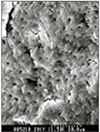
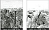
 | Fig. 3Scanning electron micrograph of canal wall prepared with stainless-steel engine reamer(×1,500).
|
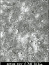
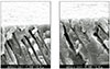
 | Fig. 4Scanning electron micrograph of canal wall extirpated with barbed broach but not instrumented(×1,500).
|
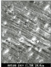
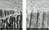
 | Fig. 5the penetration depth of smear layer into dentinal tubules was observed on canal wall prepared with Ni-Ti HERO 642(×3,000, ×5,000).
|
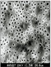
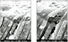
 | Fig. 6the penetration depth of smear layer into dentinal tubules was observed on canal wall prepared with Ni-Ti ProFile(×3,000, ×5,000).
|
 | Fig. 7the penetration depth of smear layer into dentinal tubules was observed on canal wall prepared with stainless-steel engine reamer(×3,000, ×5,000).
|

References
1. Williams S, Goldman M. Penetrability of the smeared layer by a strain of Proteus vulgaris. J Endod. 1985. 11:385–388.

2. Kennedy WA, Walker WA. Smear layer removal effects on apical leakage. J Endod. 1986. 12:21–27.
3. Pitt Ford TR, Roberts GJ. Tissue response to glass ionomer retrograde root fillings. Int Endod J. 1990. 23:233–238.
4. Baker NA, Eleazer PD, Averbach RE, Seltzer S. Scanning electron microscopic study of the efficacy of variious irrigating solutions. J Endod. 1975. 1:127–135.
5. Yamada RS, Armas A, Goldman M, Lin PS. A scanning electron microscopic comparison of a high volume final flush with several irrigating solutions: Part III. J Endod. 1983. 9:137–142.
6. Orstavik D, Haapasalo M. Disinfection by endodontic irrigants and dressings of experimentally infected dentinal tubules. Endod Dent Traumatol. 1990. 6:142–149.
7. Haikel Y, Gasser P, Allemann C. Dynamic fracture of hybrid endodontic hand instruments compared with traditional files. J Endod. 1991. 17:217–220. Camps JJ, Pertot WJ. : Torsional and stiffness properties of nickel-titanium K files. Int Endod J 1995; 28: 239-43.
8. Vulcain JM, Calas P. The three wave concept of Hero 642. Endod Prac. 1999. 4:20–30.
9. Thomson SA, Dummer PMH. Shaping ability of Profile.04 Taper Series 29 rotary nickel-titanium instruments in simulated root canals. Part 1. Int Endod J. 1997. 30:1–7.
10. Hülsmann M, Rümmelin C, Schäfers F. Root canal cleanliness after preparation with different endodontic handpieces and hand instruments: a comparative SEM investigation. J Endod. 1997. 23:301–306.
11. Brännström M, Nyborg H. : Cavity treatment with a microbicidal fluoride solution: growth of bacteria and effect on the pulp. J Prosthet Dent. 1973. 30:303–310.
12. Uitto VJ, Haapasalo M, Laakso T, Salo T. Degradation of basement membrane(Type IV) collagen by protease from some anaerobic microorganism. Oral Microbiol Immunol. 1988. 3:97–102.
13. Heard F, Walton RE. Scanning electron microscopic study comparing four root canal preparation techniques in small curved canals. Int Endod J. 1997. 30:323–331.
14. Cameron JA. Factors affecting the clinical efficiency of ultrasonic endodontics: a scanning electron microscopy study. Int Endod J. 1995. 28:47–53.
15. Peters OA, Barbakow F. Effects of irrigation on debris and smear layer on canal walls prepared by two rotary techniques : a scanning electron microscopic syudy. J Endod. 2000. 26:6–10.
16. Bechelli C, Zecchi Orlandini S, Colafranceschi M. Scanning electron microscopic stydy on the efficacy of root canal wall debridement of hand versus lightspeed instrumentation. Int Endod J. 1999. 32:484–493.
17. Liolios E, Economides N, Parissis-Messimeris S. The effectiveness of three irrigating solutions on root canal cleaning after hand and mechanical preparation. Int Endod J. 1997. 30:51–57.
 XML Download