

 PDF
PDF ePub
ePub Citation
 Print
Print


I. Introduction
Pulpal and periradicular tissues react to bacterial infections as do other connective tissues elsewhere in the body. The degree of pulpal and periapical response to bacterial irritants varies from slight tissue inflammation to complete pulpal necrosis or acute periradicular osteomyelitis with systemic signs and symptoms of severe infection. As a results of presence of microorganisms in the dentin, a variety of immunocompetent cells can be recruited to the dental pulp. The pulp is initially infiltrated by chronic inflammatory cells, such as macrophages, lymphocytes, and plasma cells. Pulp studies have shown the presence of immnocompetent cells and cells that recognize foreign antigens1). As a results of the interaction of microorganisms and their by-products, various mediators of inflammation, such as neuropeptides, vasoactive amines, kinins, complement component, and arachidonic acid metabolites, are released2).
It was shown that cytokines play important roles and regulate the intensity and duration of the immune response against potentially pathogenic agents. The occurrence of Interleukin(IL)-1 and IL-1 producing cells has been demonstrated in human inflamed pulps3). The roles of IL-2 and IL-6 have also been studied in healthy and inflamed dental pulps4,5).
In one study on periodontal disease, the amount of IL-6 and IL-10 were significantly higher in the inflamed gingival tissues than in the peripheal blood from the healthy subjects6). IL-6 was detected in human pulps, periapical lesions5) and odontogenic cysts7).
Interleukin-6 is produced by mononuclear phagocytes, vascular endothelial cells, fibroblasts, and other cells in response to IL-1 and TNF8). One of the best described actions of IL-6 is on B lymphocytes. IL-6 serves as a growth factor for activated B lymphocytes late in the sequence of B cell differentiation. Activated B lymphocytes differentiate into plasma cells and produce antibody against antigens or infective microorganisms. IL-6 may serve as a cofactor of T lymphocytes and thymocytes activation. Activated T lymphocytes can produce cytokines including IL-10 that mediate inflammatory reactions. IL-6 also acts as a cofactor with other cytokines for the growth of early bone marrow hematopoietic stem cells. In previous studies of pulpal and periapical pathology, IL-6 is known to be one of pro-inflammatory cytokines9,10).
Interleukin-10 is produced by the TH2 subset of CD4+ helper cells8,11). One of the major activities of IL-10 is to inhibit cytokine (i.e.. TNFα, IL-1, chemokine, and IL-12) production by macrophages. It inhibits the production of IFN-γ which contribute to pathologic bone resorption in periapical lesion12). And It also suppresses the production of IL-6 by T-lymphocytes13). The effect of these actions is to inhibit T cell-mediated immune response. In addition to its inhibitory effects on macrophages, IL-10 has stimulatory actions on B lymphocytes.
Kakehashi et al. have shown that pathogenesis of pulpal and periapical lesion is closely related to microorganisms14). In their study, dental pulps of conventional and germ-free rats were exposed to their own flora. Pulpal and periradicular lesions developed in conventional rats but failed to develop in germ-free rats.
The purpose of this study was to determine IL-6 and IL-10 in human pulpal inflammation and to investigate their roles in the progress of pulpal inflammation.
II. Materials and Methods
1. Preparation of tissue samples and Protein assay
Total 60 teeth were extracted. The experimental group consist of 30 teeth with inflamed pulps. The control group consist of 30 teeth with healthy pulps. The extracted teeth were removed from the liquid nitrogen tank, and allowed to thaw for 10 minutes. After the teeth were cracked open, pulp tissues were carefully removed from the teeth. Half of the pulp tissue from one tooth was used for detection of IL-6 and the other half was used for IL-10. Pulp tissues were homogenized in buffer(0.1 M potassium chloride, 0.02 M TRIS; pH = 7.6) in glass homogenizer and centrifuged (2000 RPM, 4C, 10 min). Supernatants were collected. The concentrations of protein in tissue sample were measured by protein assay kit. The concentrations were measured in g/ml tissue sample(BCA protein assay kit, Pierce, USA).
2. ELISA
The concentrations of IL-6 and IL-10 were measured using ELISA kits (R & D System Inc., USA).
IV. Discussion
The concentrations of IL-6 and IL-10 were higher in the experimental group than those in the control group. This findings may suggest that IL-6 and IL-10 might be involved in developing pulpal inflammation stimulated by specific bacteria. Matsushima et al demonstrated that Gram-negative bacteria, such as L. casei, from carios lesions, might be involved in developing pulpitis through the stimulation of IL-6 production15).
The results of this study were comparable to the study of Barkhordar's5), which examined the level of interleukin-6 in inflamed human dental pulps and periapical lesion. In their study, the inflamed pulpal tissues exhibited significantly higher levels of IL-6. In the study of Nakanishi16), differences between normal and inflamed pulp were found in the levels of IL-6, but the difference were not statistically significant.
One investigator17) examined the production of various cytokines including IL-6 and IL-10 in murine periapical inflammation. The production of both cytokines increased beginning on day 7 and increased to day 14. This results indicate that a cytokine network is activated in response to bacterial irritation and IL-6 and IL-10 played a role in the progress of periapical pathogenesis.
Another study18) have shown that expression of IL-6 mRNA was significantly higher in diseased periodontal tissues compared to healthy contols. They also have shown the correlation between extent of tissue damage and bone destruction. These results were comparable to our study and support the findings that IL-6 and IL-10 was produced and released to have a role in the process of pulp inflammation.
There are a few studies on the role of IL-6 and IL-10 in pulpal inflammation. But the the role of IL-6 and IL-10 in the development of pulpal and periapical pathogenesis were not clearly identified.
A study of Opal SM et al., have shown that IL-6 is one of anti-inflammatory cytokine19). On the contrary, in the study of Panichi et al. IL-6 was known to be one of three pro-inflammatory cytokines9). IL-10 is known to be anti-inflammatory cytokine that suppresses the production of IL-6.
In one study it was suggested that IL-6 produced by dental pulp cells is involved in the metabolism of extracellular matrix and the destruction of dental pulp tissue15). Ishimi et al have shown that IL-6 induces bone resorption both alone and in concert with other bone-resorbing agents20). Although Lowik et al have shown that IL-6 may be a mediator in PTH-stimulated osteoclastic bone resorption21).
At the early stage of pulpal inflammation, IL-6 was produced and it stimulated B lymphocyte to produce antibody and activate T lymphocyte to produce their own cytokines including IL-10. And in the advanced stage of pulpal inflammation, IL-10 was produced and inhibited the production of cytokines, including IL-1 and IL-612).
Further studies are necessary to elucidate the roles of IL-6 and IL-10 in developing irreversible inflammation in the dental pulp.
V. Conclusion
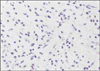
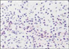
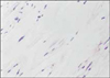
Total 60 teeth were extracted. Pulp tissues were carefully removed from the teeth. The concentrations of IL-6 and IL-10 were measured using ELISA kits. Pulp tissues were stained and examined under light-microscope.
The results were as follows:
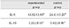
The concentrations of interleukin-6 in the experimental group were higher than those in the control group (P<0.05).
The concentrations of interleukin-10 in the experimental group were higher than those in the control group (P<0.05).
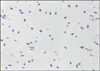
In the histoloical study, the dental pulp tissues in the experimental group showed intense infiltration of polymorphonuclear leukocytes.
 XML Download