

 PDF
PDF ePub
ePub Citation
 Print
Print


Abstract
The purpose of this study is to identificate root canal system including ideal access placement, root curvature, canal configuration, incidence of isthmus in mandibular incisors for success of endodontic treatment.
200 mandibular incisors were selected. The ideal access placement was determimed as follows. The teeth were radiographed from mesiodistal and buccolingual views using intraoral dental film. The image was divided into coronal, middle and apical third using the proximal film. Straight line access was determined by measuring the faciolingual canal width and placing points at midway point between the buccal and lingual wall at the junction of the middle and apical third and at the juntion of coronal and middle third of the root canal. A line was drawn connecting these two points extending through the crown of the tooth. The point at which the line crossed the external crown surface was recorded as facial, incisal, lingual. Degree of root curvature was determined by Schneider Protractor Method. Both section method and clearing method were used in this study. By section method, 100 mandibular incisors were embedded in clear resin and transeverse serial sectioned at 0.5, 1.0, 2.0, 3.0, 4.0, 5.0mm level from root apex. The resected surfaces were stained by methylene blue and examined under ×40 magnification with a stereomicroscope. By clearing method, 100 mandibular incisors were cleared in methysalicylate after decalcification with 10% nitric acid and evaluated under ×18 magnification with a stereomicroscope.
The results were as follows ;
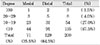
1. 29% had the center of the plotted straight-line access facial to incisal edge, whereas 71% had straight-line access at the incisal edge. When incisal wear classified as extensive, the straight-line access was plotted on the incisal edge 95.5%. When incisal wear classified as slight/none, the straight-line access was plotted on the facial 65.9%.
2. Degree of curvature of main canal was straight or almost straight, and only 10% in buccolingual direction had a degree of curvature greater than 20 degrees and 5.5% in mesiodistal direction had.
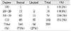
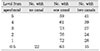
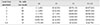
3. In section method, canal configuration analysis showed that 51% of the specimen classified as type I, 27% as type II, 12% as type III, 10% as type IV. For thoses setions with two canals, the incidence of an isthmus was 26.7%, 64.3%, 79.2%, 96.3%, 97.4%, 97.6% at each level and highest in 3~5mm sections.
4. In clearing method, canal configuration analysis showed that 74% of the specimen classified as type I, 11% as type II, 6% as type III, 9% as type IV.
These results suggested that traditional access from lingual should be moved as far toward the incisal as possible to locate and debride the lingual canal and root canal system should be cleaned, shaped completely and obturated three dimensionally for successful endodontic treatment.
Figures and Tables
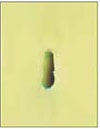
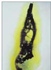
Fig. 3
Representative Photograph of Type IV root canal at 1.0mm from root apex by section method (×40).

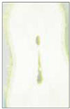
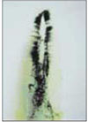
Fig. 4
Representative Photograph of Type IV root canal at 2.0mm from root apex by section method (×40).
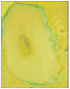
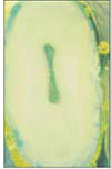
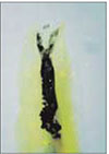
Fig. 5
Representative Photograph of Type IV root canal at 3.0mm from root apex by section method (×40).
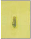
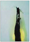
Fig. 6
Representative Photograph of Type IV root canal at 4.0mm from root apex by section method (×40).


References
1. Ingle JI, Barkland LK. Endodontics. 1994. 4th ed. Williams & Wilkims;95–96.
2. Miyashita M, Kasahara E, Yasuda E, et al. Root Canal System of the Mandibular Incisor. J Endod. 1997. 23(8):479–484.

3. Benjamin KA, Dowson J. Incidence of two root canals in human mandibular incisor teeth. Oral Surg. 1974. 38(1):122–126.
4. Pineda F. Roentgenographic investigation of the mesiobuccal root of the maxillary first molar. Oral Surg. 1973. 36(2):253–260.
5. Weller RN, Niemczyk SP, Kim S. Incidence and Position of the Canal Isthmus. Part 1. Mesiobuccal Root of the Maxillary First Molar. J Endod. 1995. 21(7):380–383.
6. Ingle JI, Barkland LK. Endodontics. 1994. 4th ed. Williams & Wilkims;116–119.
7. Beer R, Baumann MA, Kim S. Endodontology. 2000. 1th ed. Thieme;47.
8. Schneider SW. A comparison of canal preparations in straight and curved root canals. Oral Surg. 1971. 32(2):271–275.
9. Weine FS. Endodontic Therapy. 1989. 4th ed. Mosby;239–243.
10. Mauger MJ, Waite RM, Alexander JB, Schindler WG. Ideal Endodontic Access in Mandibular Incisors. J Endod. 1999. 25(3):206–207.
11. Janik JM. Access cavity preparation. Dent Clin North Am. 1984. 28:809–818.
12. Zillich RM, LaTurno SAL, Arbor A. Straight- line endodontic access to anterior teeth. Oral Surg. 1985. 59:418–419.
13. Weine FS, Kelly RF, Lio PJ. The effect of preparation procedure on original canal shape and on apical foramen shape. J Endod. 1975. 1:255–262.
14. Zmener O, Balbachan L. Effectiveness of nikel- titanium files for preparing curved root canals. Endod Dent Traumatol. 1995. 11:121–123.
15. Hession RW. Endodontic Morphology II - A Radiographic Analysis. Oral Surg. 1977. 44:610.
16. Baumann MA, Doll GM. Spatial Reproduction of the Root Canal System by Magnetic Resonance Microscopy. J Endod. 1997. 23(1):49–51.
17. Robertson D, Leeb J, Mckee M, Brewer E. A clearing technique for the study of root canal system. J Endod. 1980. 6(1):421–424.
18. Hasselgren G, Nellestam P, Bynum-Hasselgren RM. Teeth with Transparent Roots - An Improved Teaching Aid for Preclinical Endodontics. J Endod. 1987. 13(3):126–127.
19. Kartal N, Yanikoglu FC. Root Canal Morphology of Mandibular Incisors. J Endod. 1992. 18(11):562–564.
20. Rankien-Wilson RW, Herry P. The bifurcated root canal in lower anterior teeth. J Am Dent Assoc. 1965. 70:1162–1165.
21. Green D. Double canals in single roots. Oral Surg. 1973. 35(5):689–696.
22. Vertucci FJ. Root canal anatomy of the human permanent teeth. Oral Surg. 1984. 58(5):589–599.
23. Hsu YY, Kim S. The resected root surface: The issue of canal isthmus. Dent Clin North Am. 1997. 41(3):529–540.
24. Senia ES, Marshall FJ, Rosen S. The solvent action of sodium hypochlorite on pulp tissue of extracted tissues. Oral Surg. 1971. 31:96.
25. Szajkis S, Tagger M. Periapical healing in spite of incomplete root canal debridement and filling. J Endod. 1983. 9:203–209.
 XML Download