

 PDF
PDF ePub
ePub Citation
 Print
Print


Abstract
The aim of this study is to compare the adaptability of thermoplasticized injectable gutta-percha technique to the canal walls in ribbon-shaped canals.
Thirty resin models simulated ribbon-shape canals were instrumented to #40 using .06 taper Profile systems. Three groups of each 10 resin models were obturated by the lateral condensation technique(LC) and the two thermoplasticized injectable gutta-percha technique; Ultrafil Endoset+Obtura II(EO) and Ultrafil Firmset(UF), respectively.
After resin model were kept at room temperature for 4 days, they were resected horizontally with microtome at 1, 2, 3, 4 and 5mm levels from apex. At each levels, image of resected surface were taken using CCD camera under a stereomicroscope at ×40 magnification and stored. Ratio of the area of gutta-percha was obtained by calculating area of gutta-percha cone to the total area of canal using digitized image-analyzing program. The data were collected then analyzed statistically using One-way ANOVA.
The results were as follows.
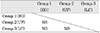
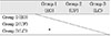
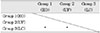
1. At 1mm levels, there was no statistically significant difference in the mean ratio of gutta-percha among the groups.
2. At 2mm level, EO showed the highest mean ratio of gutta-percha (p<0.05) and there was no significant difference between LC and UF.
3. At 3, 4, 5mm levels, EO and UF had significantly greater mean ratio of gutta-percha than LC(p<0.05) and there was no significant difference between EO and UF.
In conclusion, the thermoplasticized injectable gutta-percha techniques demonstrated relatively favorable adaptability to canal walls than lateral condensation technique in ribbon-shaped canals except for 1mm level.
Figures and Tables
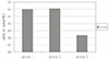
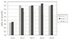
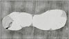
Fig. 3
Representative photograph of Ultrafil Endoset+Obtura II group at the 1mm level showing the incomplete obturation of isthmus.(orginal magnification ×40)
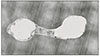
Fig. 4
Representative photograph of Ultrafil Firmset group at the 1mm level showing the incomplete obturation of isthmus.(orginal magnification ×40)
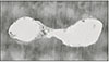
Fig. 5
Representative photograph of Lateral condensation group at the 1mm level showing the incomplete obturation of isthmus.(orginal magnification ×40)
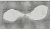
Fig. 6
Representative photograph of Ultrafil Endoset+Obtura II group at the 2mm level showing the complete obturation of isthmus.(orginal magnification ×40)
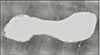
Fig. 7
Representative photograph of Ultrafil Firmset group at the 2mm level showing the complete obturation of isthmus.(orginal magnification ×40)
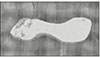
Fig. 8
Representative photograph of Lateral condensation group at the 2mm level showing the complete obturation of isthmus.(orginal magnification ×40)
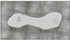
Fig. 9
Representative photograph of Ultrafil Endoset+Obtura II group at the 3mm level showing the good adaptability to the canal wall.(orginal magnification ×40)
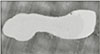
Fig. 10
Representative photograph of Ultrafil Firmset group at the 3mm level showing the good adaptability to the canal wall.(orginal magnification ×40)
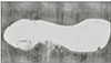
Fig. 11
Representative photograph of Lateral condensation group at the 3mm level showing the gaps between gutta-perchas.(orginal magnification ×40)
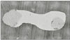
Fig. 12
Representative photograph of Ultrafil Endoset+Obtura II group at the 4mm level showing the good adaptability to the canal wall.(orginal magnification ×40)
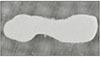
Fig. 13
Representative photograph of Ultrafil Firmset group at the 4mm level showing the good adaptability to the canal wall.(orginal magnification ×40)
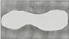
Fig. 14
Representative photograph of Lateral condensation group at the 4mm level showing the gaps between gutta-perchas and canal wall.(orginal magnification ×40)
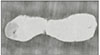
Fig. 15
Representative photograph of Ultrafil Endoset+Obtura II group at the 5mm level showing the good adaptability to the canal wall.(orginal magnification ×40)
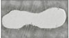
Fig. 16
Representative photograph of Ultrafil Firmset group at the 5mm level showing the good adaptability to the canal wall.(orginal magnification ×40)
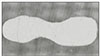
Fig. 17
Representative photograph of Lateral condensation group at the 5mm level showing the gaps between gutta-perchas and canal wall.(orginal magnification ×40)


References
1. Nguyen NT. Cohens S, Burns RC, editors. Obturation of the root canal system. Pathways of the pulp. 1984. 3rd ed. St. Louis: CV Mosby Co.;205–259.
2. Ingle JI, Dow PR. Isotope determination of root canal failure. Oral Surg. 1955. 8:1100–1104.

3. Norman WR, Niemczky SP, Kim S. Incidence and position of the canal isthmus Part I Mesiobuccal root of the maxillary first molar. J Endod. 1995. 21:380–383.
4. Pineda F. Roentgenograhic investigation of the mesiobuccal root of the maxillary first molar. Oral Surg Oral Med Oral Pathol. 1973. 36:253–260.
5. Green D. Double canals in single roots. Oral Surg. 1973. 35:689–696.
6. Cambruzzi JV, Marshall FJ. Molar endodontic surgery. J Can Dent Assoc. 1983. 49:61–65.
7. Vertucci FJ. Root canal anatomy of the human permanent teeth. Oral Surg Oral Med Oral Pathol. 1973. 35:226–231.
8. Baryton SM, Davis SR, Goldman M. Gutta-percha root canal fillings. Oral Surg Oral Med Oral Pathol. 1973. 35:226–231.
9. Weller RM, Kimbrough WF, Anderson RW. A comparison of thermoplastic obturation techniques : Adaptation to the canal walls. J Endod. 1997. 23:703–706.
10. Yee FS, Marlin J, Gron P. Three-dimensional obturation of the root canal using injection-molded thermoplasticised dental gutta-percha. J Endod. 1977. 3:168–174.
11. Torabinejad M, Skobe Z, Trombly PL, Krakow AA, Gron P, Marlin J. Scanning electron microscopic study of root canal obturation using thermoplasticized gutta-percha. J Endod. 1978. 4:245–250.
12. Marlin J, Krakow AA, Desilets RP, Gron P. Clinical use of injection-molded thermoplasticized gutta-percha for obturation of the root canal system : a preliminary report. J Endod. 1981. 7:277–281.
13. Michanowicz A, Czonstkowsky M. Sealing properties of an injection thermoplasticized low-temperature gutta-percha: a preliminary study. J Endod. 1984. 10:563–566.
14. ElDeeb ME. The sealing ability of injection-molded thermoplasticized gutta-percha. J Endod. 1985. 11:84–86.
15. LaCombe JS, Campbell AD, Hicks ML, Pelleu GB. A comparison of the apical seal produced by two thermoplasticized injectable gutta-percha techniques. J Endod. 1988. 14:445–450.
16. Beaty RG, Baker PS, Haddix J, Hart F. The efficacy of four root canal obturation techniques in preventing apical dye penetration. J Am Dent Assoc. 1989. 119:633–637.
17. Michaileso PM, Valcarcel J, Grieve AR, Levallois B, Lerner D. Bacterial leakage in endodontics : an improved method for quantification. J Endod. 1996. 22:535–539.
18. Czonstkowsky M, Michannowicz A, Vazquez J. Evaluation of an injection of thermoplasticized low temperature gutta-percha using radioactive isotopes. J Endod. 1985. 11:71–74.
19. Eguchi DS, Peters DD, Hollinger JO, Lorton L. A comparison of the area of the canal space occupied by gutta-percha following four gutta-percha obturation techniques using Procosol sealer. J Endod. 1985. 11:166–175.
20. Giani O, Visvisian C, Caso C. Quality of apical in curved canals using three types of spreaders. J Endod. 2000. 26:581–585.
21. WU MK, Wesselink PR. Endodontic leakage studies reconsidered. Part I. Methodolgy, application and relevance. Int Endod J. 1993. 26:37–43.
22. Pitt Fort TR. Relation between seal of root fillings and tissue response. Oral Surg. 1983. 55:291–294.
23. McSpadden J. Multiphase gutta-percha obturation technique. Dent Econ. 1993. 83:95–97.
24. Dulac KA, et al. Comparison of the obturation of lateral canals by six techniques. J Endod. 1999. 25:376–380.
25. Hsu YY, Kim S. The resected root surface. The issue of canal isthmus. Dent Clin North Am. 1997. 41:529–540.
 XML Download