ABSTRACT
Microleakage at the occlusal margins was not evident in group 1 and group 2, but it showed in group 3 (p<0.05).
Microleakage in group 1 and group 3 was significantly lower than in group 2 at gingival margins (p<0.05).
Microleakage at gingival margins was greater than at occlusal margins in group 1 and group 2, but microleakage at occlusal margins was greater than at gingival margins in group 3 (p<0.05).
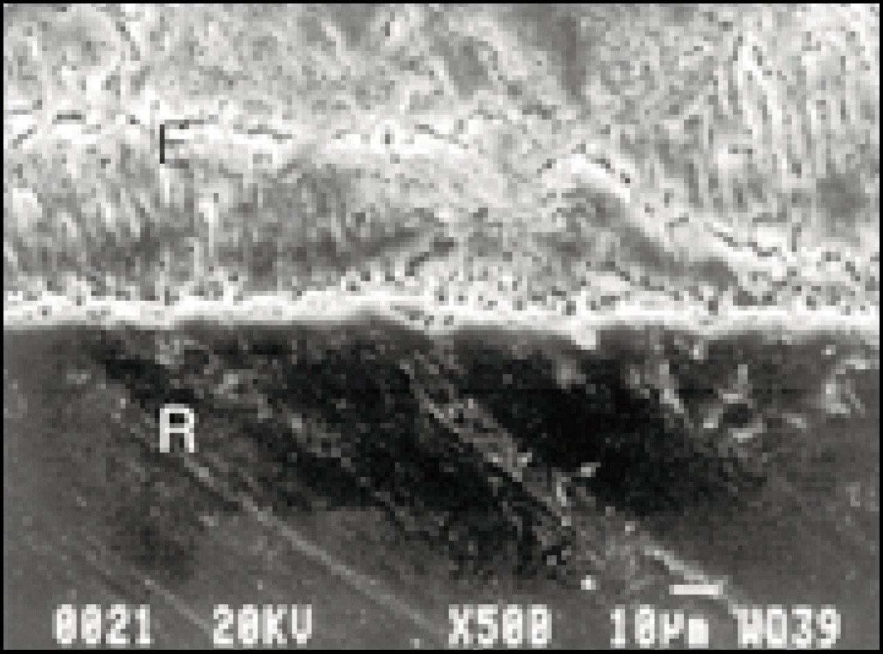
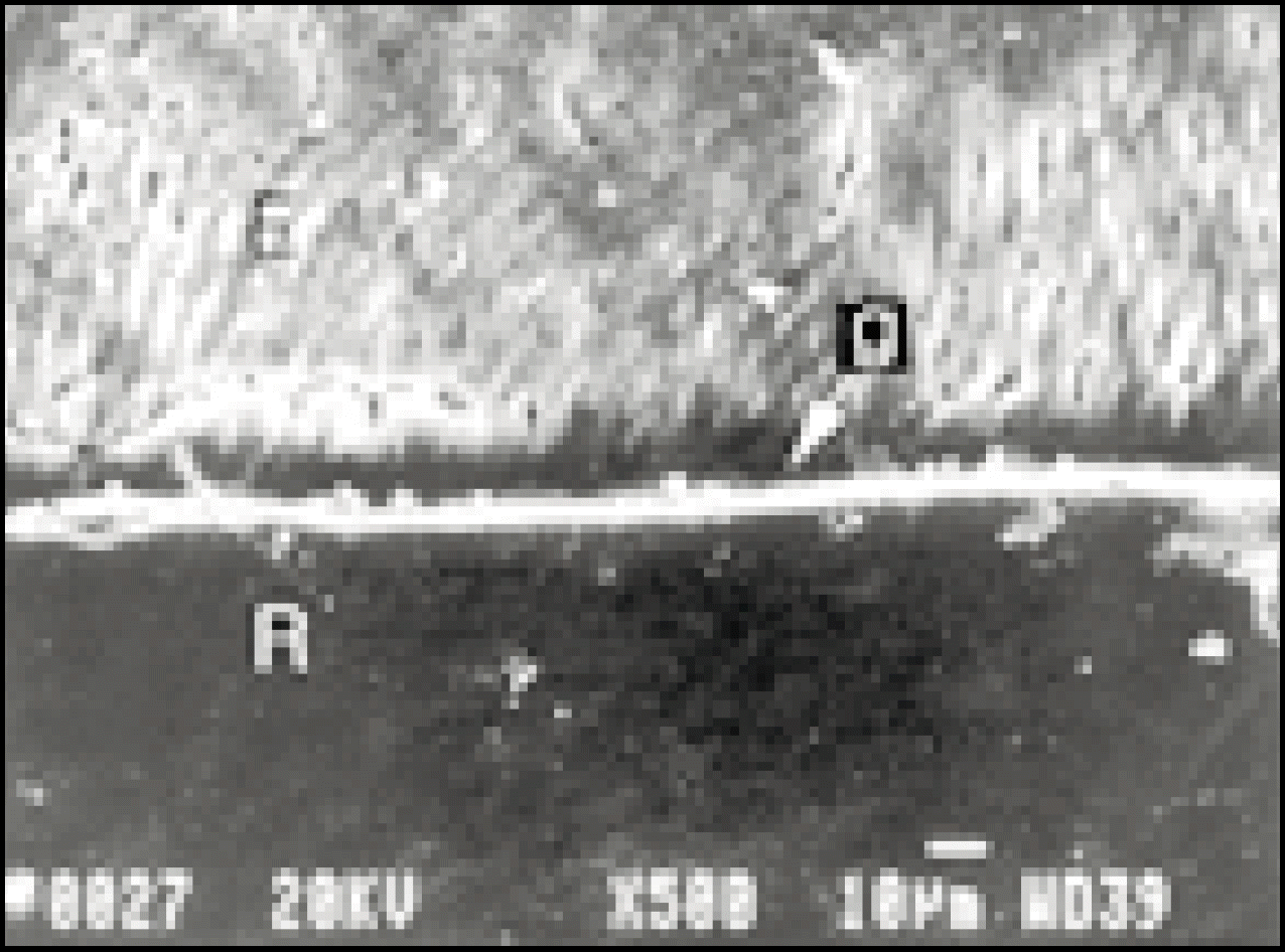
In group 1 and group 2, no gaps at occlusal margins showed. But gaps showed in group 3. Occlusal margins were free from a hybrid layer in all groups.
The thickness of the marginal hybrid layers was 2.5∼5 μm thick in group 5 μm thick in group 2 and 1.5 μm thick in group 3.
There was no corelation between microleakage and thickness of marginal hybrid layer.
References
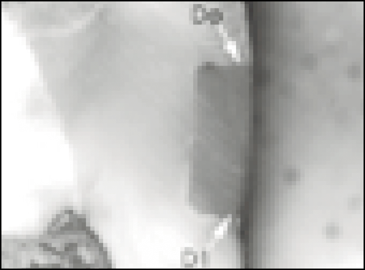 | Fig. 1.Microleakage in group 1 (Single Bond/Filtek Z-250)
Microleakage did not showed at occlusal margin(Do).
Microleakage of degree 1(D1) was showed at gingival margin (× 15).
|
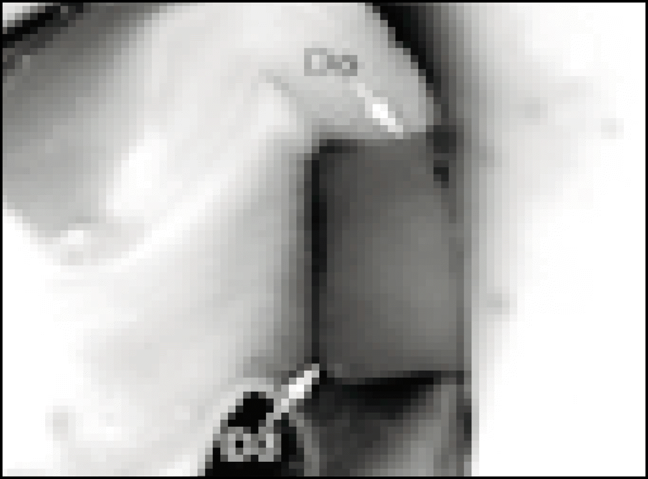 | Fig. 2.Microleakage in group 2 (Prime&Bond NT/Esthet∙X)
Microleakage did not showed at occlusal margin(Do).
Microleakage of degree 3(D3) showed at gingival margin (× 15).
|
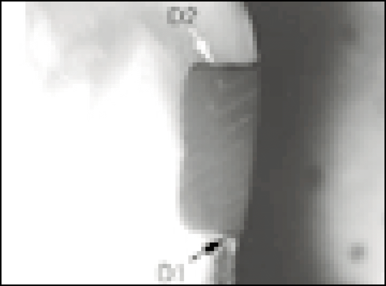 | Fig. 3.Microleakage in group 3 (UniFil Bond/UniFil F)
Microleakage of degree 2(D2) showed at occlusal margin.
Microleakage of degree 1(D1) showed at gingival margin (× 15).
|
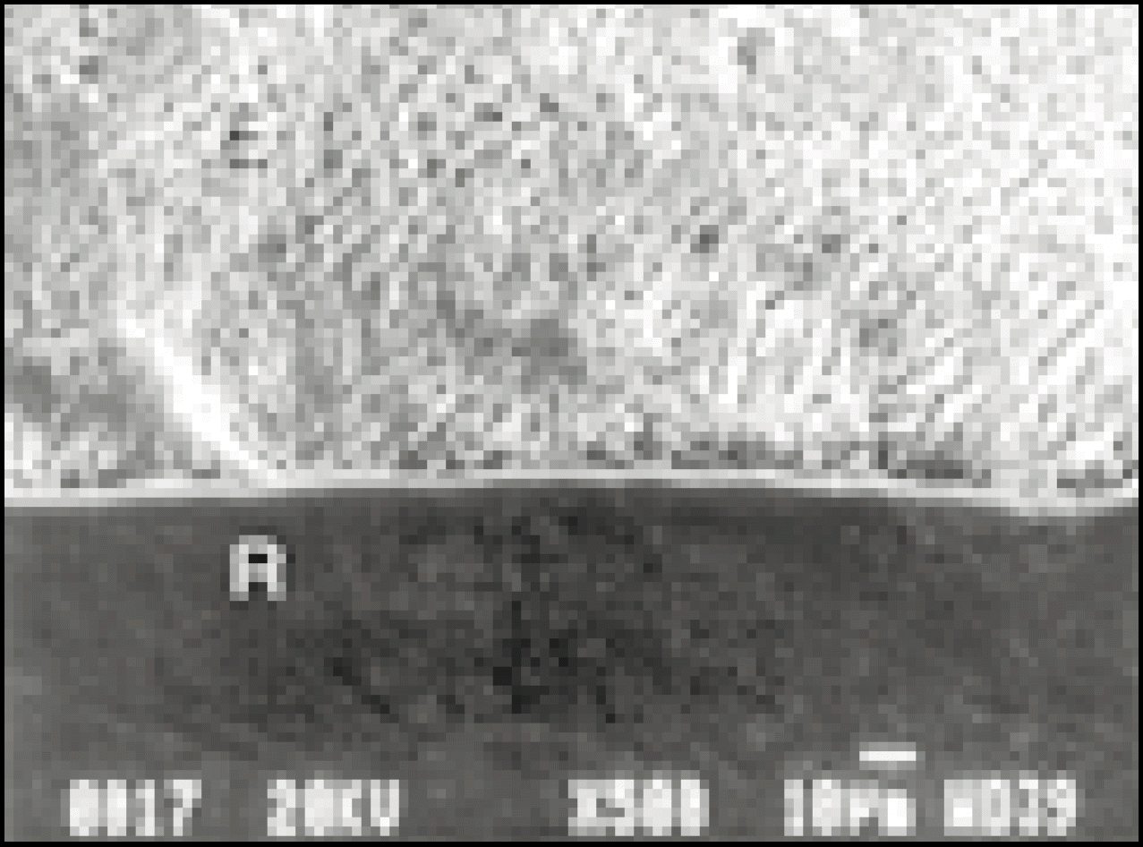 | Fig. 4.SEM of occlusal margin in group 1 (× 500)
Close adaptation between interface of resin(R) and enamel(E) was evident and no gaps showed at occlusal margins.
Hybrid layer was free.
|
 | Fig. 5.SEM of occlusal margin in group 2 (× 500)
Close adaptation between interface of resin(R) and enamel(E) was evident and no gaps showed at occlusal margins. Hybrid layer was free.
|
 | Fig. 6.SEM of occlusal margin in group 3 (× 500)
Tiny gaps(g) between interface of resin(R) and enamel(E) showed. Hybrid layer was free.
|
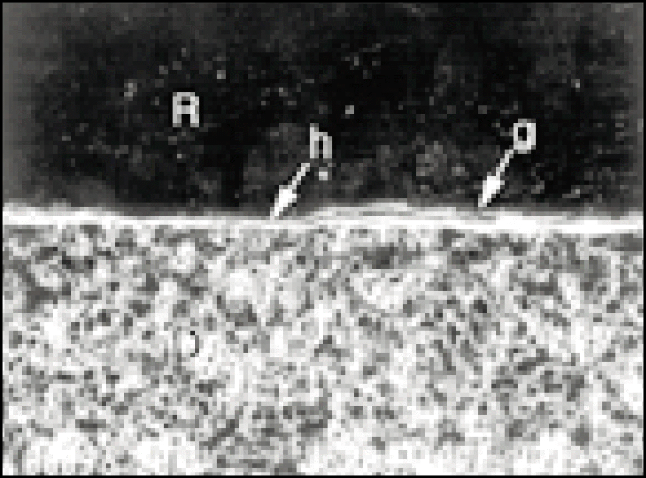 | Fig. 7.SEM of gingival margin in group 1 (× 500)
Thickness of the marginal hybrid layers(h) was 2.5∼5μm and tiny gaps(g) between interface of resin(R) and detin(D) showed.
|
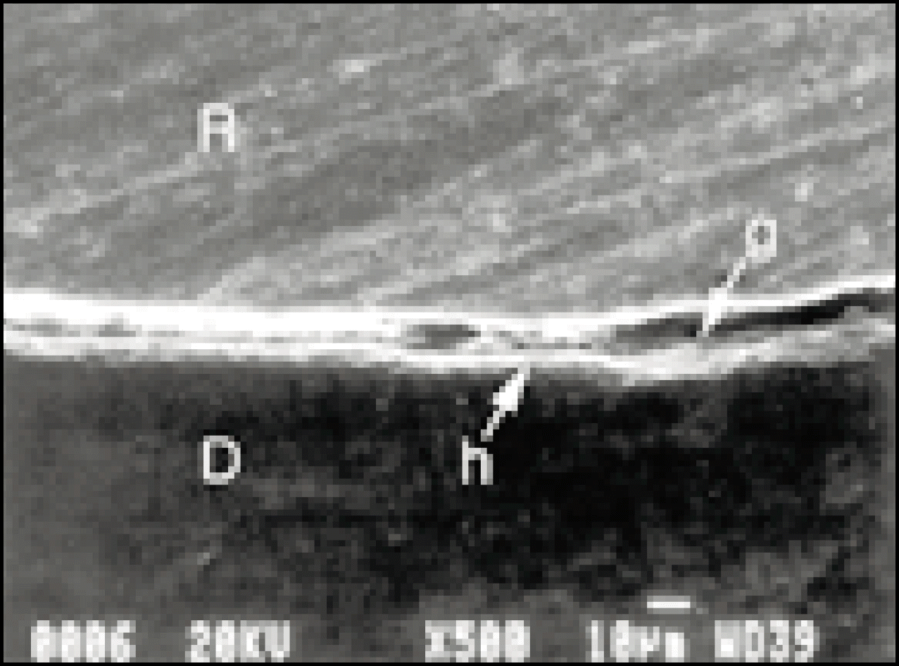 | Fig. 8.SEM of gingival margin in group 2 (× 500)
Thickness of the marginal hybrid layers(h) was 5μm and wide gaps(g) between interface of resin(R) and dentin(D) showed.
|
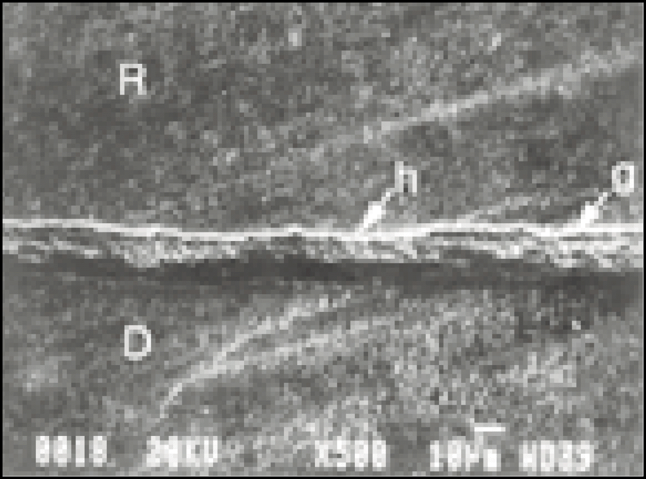 | Fig. 9.SEM of gingival margin in group 3 (× 500)
Thickness of the marginal hybrid layers(h) was 1.5μm and tiny gaps(g) between interface of resin(R) and dentin(D) showed.
|
Table 1.
| Group | Adhesive | Composite resin | Manufacturer |
|---|---|---|---|
| 1 | Single Bond | Filtek Z-250 | 3M Dental Products |
| 2 | Prime&Bond NT | Esthet. X | Dentsply/Caulk |
| 3 | UniFil Bond | UniFil F | GC Co. |
Table 2.
Table 3.
| Score\Group | Occlusal Margin | Gingival Margin | ||||||||
|---|---|---|---|---|---|---|---|---|---|---|
| 0 | 1 | 2 | 3 | n | 0 | 1 | 2 | 3 | n | |
| 1 | 10 | 0 | 0 | 0 | 10 | 7 | 1 | 0 | 2 | 10 |
| 2 | 10 | 0 | 0 | 0 | 10 | 0 | 1 | 1 | 8 | 10 |
| 3 | 1 | 3 | 5 | 1 | 10 | 4 | 6 | 0 | 0 | 10 |
Table 4.

|
Table 5.

|


 PDF
PDF ePub
ePub Citation
 Print
Print


 XML Download