

 PDF
PDF ePub
ePub Citation
 Print
Print


I. INTRODUCTION
For good endodontic obturation, a canal system that has been cleaned and shaped in all its dimensions is necessary. However, once this has been accomplished, the complete seal of the root canal system is an essential component in assuring longterm clinical success as it is this seal that maintains the barrier between the oral environment and the periradicular tissues. The essentials of an excellent barrier begin with the assurance that the apical preparation has been properly sealed. This must not only be possible theoretically but also be accomplished in such a manner by the clinician19). Ingle14) reported that 59% of endodontic failures are due to incomplete obturation of the root canal system. Naidorf25) indicated that improper obturation allows fluids to enter the root canal space, that may become infected as a result.
Throughout the years, many studies on obturation techniques and filling materials of root canals have been developed for hermetic sealing of root canal system.
Lateral gutta-percha condensation is currently the most accepted canal obturation method. Advantages of this technique include its predictability, relative ease of use, conservative preparation, and controlled placement of materials. But disadvantages include a lack of homogeneity of the gutta-percha mass, increased number of voids and sealer pools, and less adaptation to canal walls and irregularities9).
Therefore, a number of different filling techniques based on preheated gutta-percha have been introduced with the aim of enhancing three-dimensional filling of the root canal.
Johnson(1978)16) demostrated a simple method of carrying thermoplasticized gutta-percha to the extent of the prepared canal. A flexible metal or plastic carrier the same size as the final apical instrument is coated with alpha-phase gutta-percha. Recently, this method of obturation was commercialized under the name of Thermafil Endodontic Obturators(Tulsa Dental Product, U.S.A).
Schilder(1967)31) popularized the use of warm vertical compaction of gutta-percha and sealer. Based on this techniques, Buchanan (1996)5) developed a new method of vertical compaction of warm gutta-percha and called it, "continuous wave of condensation technique"
Recently, MicroSeal endodontic obturation technique(TYCOM, USA) is based on the combination of different types of gutta-percha ; α-, β-phase gutta percha. Sealer and master cone is placed in the root canal, and then remaining space of root canal is filled with MicroSeal condenser coated with warm thermoplasticized gutta-percha.
A study by Beatty et al.3) showed Thermafil was more effective than laterally condensed gutta-percha techniques in restricting apical dye penetration. In contrast, studies by Lares and ElDeeb23) and Chohayeb7) reported that leakage was significantly less with the lateral condensation technique than with Thermafil. In Scott's study33), there was no significant difference between them.
Gutmann12) showed that Thermafil and continuous wave technique were not significantly different in the overall canal obturation and in the apical third adaptation.
As appears from the above, the apical sealing ability of these obturation techniques was different in each report. Therefore many studies have been accomplishing. However, it has not been decided yet and is still controversial.
These different obturation techniques still needs to be performed in combination with root canal sealer. With the development of obturation techniques, many sealers have been developed. And the sealing ability of root canal sealer have been investigated. There have been, however, few study reported on the sealing ability of different root canal sealer used in conjunction with various obturation techniques. Also, the evaluation of relationship between the obturation quality and sealing ability has been confined to a few studies.
Therefore, the purpose of this study is to evaluate and compare the apical sealing capacity of four different obturation techniques (MicroSeal™, Thermafil®, Continuous wave technique, and Cold lateral condensation) in conjunction with root canal sealer (Sealapex, AH26®). Criteria of evaluation include linear dye leakage, presence or absence of material extrusion
II. MATERIALS AND METHODS
Eighty recently extracted human permanent anterior teeth with straight single canal and complete apex were used in this study. Adherent soft tissue on root surface were removed by periodontal currete.
Uniform access preparation were made with high speed burs. After access, the coronal and middle third of the canals were enlarged with size ISO 050, 070 or 090 Gates Glidden burs.
A #15 K-file (Dentsply-Maillefer, Ballaigues, Switzerland) was introduced into the canal and advanced until it appeared at the apical foramen. The working length of each root was established by substacting 1mm from this measurement.
The apical preparation was then completed with ProFile® instruments(Dentsply-Maillefer, Ballaigues, Switzerland). First, yellow 0.04 taper ProFile® was used at the working length, and then yellow 0.06 taper ProFile® was used at the same length. Red 0.04, 0.06 taper, blue 0.04, 0.06 taper and green 0.04, 0.06 taper ProFile® were used sequently at the working length. According to the initial apical file size of each teeth, final apical instrument was determined. Each ProFile® instrument was introduced into the canal at a constant speed of 300 rpm with gentle push-pull motion.
During canal cleaning and shaping, RCPREP ™(Premier, Philadelphia, U.S.A.) was used as lubricant with each size instrument. After using each size of instrument, canals were irrigated with 5.25% NaOCl. Patency of apical foramen was determined and maintained by passing a size 15K-file through the apex.
Root canals were dried with an endodontic irrigation probe(Max-i-Probe®, 30 gauge/dark blue, Dentsply) and sterile standardized paper points.
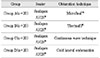
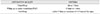
Eighty teeth were randomly divided into 4 groups of 20 teeth each, according to obturation techniques. 10 teeth of each group used Sealapex(KERR®, Sybron, U.S.A) and in 10 other teeth of each group, AH 26® sealer(Dentsply, Konstanz, Germany) was used(Table 1).
A. Canal Obturaion
Group A : MicroSeal™ obturation (n=20)
First, MicroSeal™ customized master cone was inserted into the root canal with sealer. Then MicroSeal™ spreader was inserted 1mm short from the working length. The gutta-percha-coated MicroSeal™ condenser was immediately carried to the void previously created in the canal by spreader and then rotation of the condenser started at a speed of 6000rpm. After approximately 2 seconds, the MicroSeal™ condenser was removed slowly from root canal.
Group B: Thermafil® obturation (n=20)
Before obturation, the walls of the canal were coated with a small amount of sealer using a file. Thermafil® obturators of the same size as the master apical file were selected. The Thermafil® obturator was heated by ThermaPrep Plus oven according to manufacturer's recommendations, and the Thermafil® obturator was inserted in the canal to the full working length. The carrier was left in place. After obturation, an inverted cone bur was used to cut through the shank of each carrier at the level of the root canal orifice. The gutta-percha around the carrier shaft then was vertically condensed with a hand plugger.
Group C: Continuous wave technique (n=20)
A Buchanan's plugger, a Schilder's plugger, and Obtura II tip all to 3mm short from the working length were pre-fitted. A nonstandardized master gutta-percha cone was selected and cut at its apical third to the size of the master apical file of each preparation, using a gutta-percha gauge(Dentsply-Maillefer, Ballaigues, Switzerland) and a sharp scalpel.
Following placement of the master gutta-percha cone and sealer, the pre-fitted Buchanan's plugger was activated by the System B™(Analytic Tec., U.S.A) and inserted into the root canal for obturating the apical 3mm according to the Continuous wave obturation technique. Then the Schilder's plugger was inserted steadily at the 3mm level for vertical condensation. Next, the Obtura II(Obtura Co., U.S.A) was used for backfill at 200℃.
Group D: Cold lateral condensation (n=20)
This group was the control. A standard-sized gutta-percha cone of the same size as the master apical file was coated with the sealer and seated in the root canal. A finger spreader and accessory gutta-percha cones were used for lateral condensation. Cones were added until the spreader would not penetrate beyond the coronal third of the canal.
In all groups, the access cavity were restored with Ariston pHc(Vivadent, Liechtenstein) following the obturation. The teeth were taken a digital radiograph(Biomedysis CO., Korea) in mesiodistal and buccolingual directions to study the quality of the obturation. The teeth were kept in an incubator at 37℃ at 100% humidity for 48 hrs to ensure that AH26® sealer had set. The teeth were coated with two layers of nail varnish, except the apical 2mm that were left exposed. The roots were immersed in 2% methylene blue for 48hrs at 37℃. Afterward, the teeth were rinsed under running tap water to remove the dye on the external root surface. The nail varnish was gently removed with a scalpel blade. Samples were then decalicified in 5% nitric acid for 48 hr(changing the solution every 24 hr), dehydrated in ascending concentrations of alcohol(80, 90, 100%), and rendered transparent with methyl salicylate.
B. Evaluation
Linear Dye Penetration Measurement
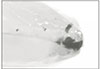
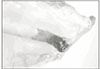
The cleared specimens were examined independently by two observers at 40 magnification with a stereomicroscope(Olympus, Japan). A grid calibrated in millimeters was used to determine the extent of leakage. To minimize possible bias in measurements, two examiners measured the linear dye leakage for each sample and the average was recorded. Dye impregnation was measured for each tooth from all four surfaces(mesial, distal, buccal and lingual-palatal) and scored the most coronal extent of dye visible along the gutta-percha filling material according to the criteria in Table 2.
Presence or absence of material extrusion
Any extrusion of gutta-percha or sealer during obturation was noted.
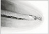
On digital image of all teeth bucco-lingually and mesio-distally, length between end of gutta-percha obturated and radigraphic apex was measured. After this measurement, underfilling, filling up to apical constriction(FAC), and overfilling were evaluated according to the criteria in Table 3.
III. RESULTS
A total of 72 teeth were tested. eight teeth were eliminated during evaluation because, vertical root fracture or apical perforation were noticed and determined to have occurred before exposure to the ink; 72 teeth remained.
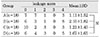
The linear dye penetration measurements for each group are listed in Table 4. The mean leakage scores weve as follows; 1.11±1.52 in Microseal™, 2.10± 1.82 in Thermafil®, 1.21 ±1.40 in Continuous wave technique, and 1.67±1.65 in Cold lateral condensation group.
Statistical analysis was carried out using the Kruskal-Wallis test for nonparametric data to determine whether there were significant difference among the groups. There were no significant difference in linear dye penetration among four obturation techniques.
But Microseal™ produced the smallest amount of mean linear dye penetratin score followed order from smallest to largest by Continuous wave technique, Cold lateral condensation, and Thermafil®.
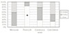
From the statistical analysis using Kruskal-Wallis test, it revealed significant difference in the degree of dye penetration between the two sealers. The mean leakage score of all samples using AH26®(0.92±1.42) was lower than Sealapex(2.14±1.60)(p<0.05).
The mean leakge scores for each sealer in four group, were shown in Fig. 1. In MicroSeal™ group, samples using AH26® were less leakage than Sealapex. This revealed statistically significant difference by Wilcoxon signed rank test(p<0.05).
The relationship between obturation quality(underfilling, FAC, overfilling) and mean linear dye penetration analyzed statistically using Kruskal-Wallis test. There was no correlation between the obturation quality and microleakage.
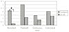
Fig. 2 showed frequency of underfilling, FAC, and overfilling for each obturation technique. The Thermafil® group showed the higher frequency of overfilling compared with the other obturation by Chi-square test(p<0.05).
IV. DISCUSSION
Various methods have been used for evaluating the apical sealing property of root canal filling materials and associated obturation techniques. Examples of such methods are dye penetration tests34), radioactive isotope studies4), electrochemical leakage tests27), bacterial penetration tests8), and scanning electron microscopic analysis37). Among these methods, dye penetration studies are the most common, although published findings reveal large standard deviations9).
Dye penetration studies are vertical, cross sectioning and clearing of the roots. Among these methods, clearing method is much time to consume, difficult to know the decalcification degree, and uncleared area can be appeared by incomplete dehydration. But, the examiner is able to evaluate dye penetration level and adaptation of the filling material in a three-dimensional manner20,26) unlike the other methods1). In this study, the leakage patterns around the tooth were observed by rotating the tooth21).
On measurement of the apical leakage using dye penetration tests, the maintenance of patency is important. Adams et al.13) reported that dentinal plug can influence the apical leakage. Therefore, not dentinal plug but filling material must seal apical stop2). In this study, dye penetration was only assessed when extending coronally beyond the end of gutta-percha obturated and not at the apical end6).
Matloff et al.24) showed that methylene blue penetrates in greater depth than other tracing dyes. Whereas, Edmund et al10) reported other inks that might provide better contrast showed that they dissolved during the decalcification and clearing processes. Also, he reported that there was no evidence of this problem with india ink. In this study, methylene blue was used as dye. The dissolution of methylene blue during the decalcification and clearing processes might happen and affect the dye penetration level.
Pitt Ford(1983)30) reported that the sealing effect in vitro have not relation to tissue reaction in vivo. In order to evaluate the sealing ability, several in vitro methods have been designed. Among these methods, the results of dye penetration studies, however, are sometimes confusing and often result in variable conclusion. The lack of agreement has been discussed by Wu & Wesslink39) who questioned the validity of leakage studies and recommended that more research should be devoted to leakage methodology.
It is known that most root canal sealers shrink during setting21), and dissolve with time28). The seal can be destroyed by dissolution of sealer components. In this study, we used Sealapex and AH26 as root canal sealer in conjunction with four obturation techniques. AH26 was originally developed as a sole obturating material or for single-cone techniques32). It is, however, commonly used for more complex obturation techniques such as lateral and vertical condensation. We found the excellent adherence of the sealer to the canal walls and gutta-percha in AH26 group. This finding is agreement with Limkangwalmongkol23), Schroeder32). Tagger and coworkers36) reported no leakage of the dye when α-acting gutta-percha or lateral condensation with AH26® sealer were used. Sealapex may posses significant solubility, which accounts for its biological activity at the root apex35). AH26& expand initially, Which might result in the reduction of microleakage 17). One main disadvantage observed with blue dye penetration studies was the dark color that resulted form the oxidized silver content. This may somtimes mistakenly be interpreted as leakage9).
The Thermafil obturation technique showed a predisposition for material extrusion beyond the apical foramen in 84.2% of the specimens(Fig 2), and It showed the higher frequency of overfilling compared with the other obturation techniques(p<0.05). This finding is in aggrement with other studies(Haddix et al.15), Gutmann et al.12), Pathomvanich & Edmunds28)). This can increase postoperative pain.
There are many studies about the apical sealing ability according to the obturation techniques. LaCombe et al.21) found that laterally condensed gutta-percha showed less linear leakge than low-temperature and high-temperature thermoplasticized gutta-percha. But, Hata et al.13) and Goldberg et al.11) founded that thermoplasticized filling techniques and cold lateral condensation were not significant difference.
This study suggests that all four obturation techniques evaluated here may be equally effective in obturation of well-instrumented root canals under ideal conditions, and sealed well with no statistically significant differences between them. This result were agreement with study showed above.
In this study, we used single root with straight canal. Lares and ElDeeb22) speculated that there was the difference between single rooted and multi-rooted teeth. Therefore, further work is also needed to determine the apical sealing ability between singlerooted and multi-rooted teeth, curved root and straight root.
V. CONCLUSION
This study evaluated the apical sealing ability of four obturation techniques. In this study, eighty teeth were instrumented using ProFile® and randomly divided into 4 groups of 20 teeth each, according to obturation techniques.
The canals were obturated by four obturation techniques: Group A- Microseal™. Group B- Thermafil®, Group C- Continuous wave technique, Group D-Cold lateral condensation. 10 teeth of each group used Sealapex and in 10 other teeth of each group, AH26® sealer, was used.
After obturation, the teeth were taken a digital radiograph in mesiodistal and buccolingual directions to study the quality of the obturation (underfilling, filling up to apical constriction, or overfilling). Afterward, the teeth were coated with two layers of nail varnish, and immersed in 2% methylene blue and subsequently cleared. The most coronal extent of dye penetration was measured using stereomicroscope (X40).
Results obtained from the study above were as follows:
There was no statistically significant difference among groups. All four obturation techniques showed the similar results in the apical sealing ability.
The mean leakage score of all samples using AH26® were lower than Sealapex(p<0.05). In Micro Seal™ group, samples using AH26® were less leakage than Sealapex(p<0.05).
There was no corelation between the quality of obturation and microleakage.
The Thermafil® group showed the higher frequency of overfilling compared with the other obturation(p<0.05).
As a result, there are no significant difference for apical sealing ability among four obturation techniques. Therefore, the other advantages must be considered such as operator's experience, obturation time, the ease of retreatment etc. It is reasonable to choose the obturation method with careful consideration about these points.

 XML Download