

 PDF
PDF ePub
ePub Citation
 Print
Print


Abstract
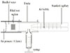
Leakage studies have been performed frequently, since a fluid-tight seal provided by various dental filling materials has been considered clinically important. The leakage of the various root-end filling materials has been widely investigated mostly dye penetration method. These dye studies cannot offer any information about the quality of the seal of a test material over a long period of time The purpose of this study was to evaluate the microleakage of root end cavities in blood contamination filled amalgam, intermediate restorative material(IRM), light cured glass ionomer cement(GI) and mineral trioxide aggregate(MTA) by means of a modified fluid transport model. Fifty standard human root sections, each 5mm high and with a central pulp lumen of 3mm in diameter, were and filled with our commonly used or potential root end filling materials after they were contaminated with blood. At 24h, 72h, 1, 2, 4, 8, and 12 weeks after filling, leakage along these filling materials was determined under a low pressure of 10KPa(0.1atm) using a fluid transport model.
The results were as follows:
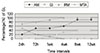
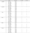
1. MTA group showed a tendency of decreasing percent of gross leakage (20ml/day) in process of time, whereas the other materials showed a tendency of increasing in the process time.
2. At the all time interval, GI group leaked significantly less than amalgam group and IRM group (p<0.05).
3. At the 4 weeks, the percentage of gross leakage in MTA group decreased to 0% thereafter, the low percentage of gross leakage was maintained in MTA group until the end of the experiment, whereas the percentage in IRM group increased to 100%.
4. At the 12 weeks, percentage of gross leakage was significantly low in MTA group(0%), comparison with GI group(40%), amalgam group(90%) and IRM group(100%), but there was no significant difference between latter two materials.
Figures and Tables
References
1. Sjögrem U, Hägglund B, Sundqvist G, Wing K. Factors affecting the long term results of endodontic treatment. J Endod. 1990. 16:498–504.
2. Ingle JI, Backland LK. Endodontics. 1994. 4th ed. Philadelphia: Lea & Febiger Book;689–763.
3. Baumgartner JC, Galkler WA. Bacteria in the apical 5mm of infected root canals. J Endod. 1991. 17:380–383.
4. Harty FJ, Parkins BJ, Wengrat AM. The success rate of Apicoectomy, A retrospective study of 1016 case. Br Dent J. 1970. 129:407–418.

5. Gartner A, Dorn S. Advances in endodontic surgery. Dent Clin North Am. 1992. 36:357–378.
6. Cho YB, Hong CU, Shin DH. Effects of some root end filling materials on the repair of periradicular tissue. J Korean Acad Conserv Dent. 1995. 20(1):17–32.
7. Rud J, Andreason JO, Jensen JEM. Radiographic criteria for the assessment of healing after endodontic surgery. Int J Oral Surg. 1972. 1:195–214.
8. Dorn SO, Gartner A. Retrofilling materials : a retrospective success-failure study of amalgam, EBA, and IRM. J Endod. 1990. 16:391–393.
9. Frank AL, Click DH, Paterson SS, Weine FS. Long-term evaluation of surgically placed amalgam fillings. J Endod. 1992. 18:391–398.
10. Rapp EL, Grown CE, Newton CW. An analysis of success and failure of apicoectomies. J Endod. 1991. 17:508–512.
11. Smee G, Bolanos OR, Morse DR, Furst MF, Yesilsoy C. A comparative leakage study of P-30 resin bonded ceramic, Teflon, amalgam, and IRM as retrofilling seals. J Endod. 1987. 13:117–121.
12. Bondra DL, Hartwell GR, Macpherson MG, Portell FR. Leakage in vitro with IRM, high copper amalgam, and EBA cement as retrofilling materials. J Endod. 1989. 15:157–160.
13. Wilson AD, Kent BE. A new translucent cement for dentistry. Br Dent J. 1972. 132:133–135.
14. Tobias RS, Brows RM, Plant CG, INgram DW. Pulpal response to a glass ionomer cement. Br Dent J. 1978. 144:345–350.
15. Pitt Ford TR, Robers GJ. Tissue response to glass ionomer retrograde root fillings. Int Endod J. 1990. 23:233–238.
16. Abdal Ak, Retief DH. The apical seal via the retrosurgical approach 1 A Preliminary study. Oral Surg. 1982. 53:614–621.
17. Ruyter IE, Sjøvik IJ. Monomer composition of dental composites and sealants. J Dent Res. 1978. 57(spec. iss):249. abstr. 700.
18. Rueggeberg FA, Craig RG. Correlation of parameters used to estimate monomer conversion in a light-cured composite. J Dent Res. 1988. 67:932–937.
19. Asmussen E. Restorative resins: hardness and strength vs quantity of remaining double bonds. Scand J Dent Res. 1982. 90:484–489.
20. Blackman R, Gross M, Seltzer S. An Evaluation of the biocompatibility of a glass ionomer-silver cement in rat connective tissue. J Endod. 1989. 15:76–79.
21. Pissiotis E, Sapounas G, Spangberg LSW. Silver glass ionomer cement as a retrograde filling material : a study in vitro. J Endod. 1991. 17:225–229.
22. Bates CF, Carnes DL, del Rio CE. Longitudinal sealing ability of mineral trioxide aggregate as a root-end filling material. J Endod. 1996. 22(11):575–578.
23. Torabinejad M, Watson TF, Pitt Ford TR. Sealing ability of a mineral trioxide aggregate when used as a root end filling material. J Endod. 1993. 19:591–595.
24. Torabinejad M, Dby Wc, Naidorf IJ. Inflammatory and immunological aspects of the pathogenesis of human periapical lesions. J Endod. 1985. 11:479–488.
25. Torabinejad M, Hong CU, Pitt Ford TR, Kettering JD. Cytotoxiciy of four root end filling materials on mouse L929 cells. J Endod. 1993. 19:203.
26. Hong Cu, McKendry DJ, Pitt Ford TR, Torabinejad M. Healing of furcal lesions repaired by amalgam or mineral trioxide aggregate. J Endod. 1994. 29:197.
27. Torabinejad M, Hong CU, Kettering JD. Antibacterial effects of some root end filling materials. J Endod. 1995. 21:403–406.
28. Torabinejad M, Higa RK, McKendry DJ, Pitt Ford TR. Dye leakage of four root end filling materials: Effects of blood contamination. J Endod. 1994. 20(4):159–163.
29. Pashley DH, Thompson SM, Stewart FP. Dentin permeability: effects of temperature on hydraulic conductance. J Dent Res. 1983. 62:956–959.
30. Wu MK, De Gee AJ, Wesselink PR. Leakage of four root canal sealers at different thicknesses. Int Endod J. 1994. 27:304–308.
31. Derkson GD, Pashley DH, Derkson ME. Microleakage measurement of selected restorative materials: a new in vitro method. J Prosthet Dent. 1986. 56:435–440.
32. King KT, Anderson RW, Pashley DH, Pantera EA. Longitudinal evaluation of the seal of endodontic retrofillings. J Endod. 1990. 16:307–310.
33. Trowbridge HO, Kim S. Pulp development, structure and function. Pathways of the pulp. 1994. 6th ed. St. Louis: Mosby;322–325.
34. Wu MK, Kontakiotis EG, Wesselink PR. Long-term seal provided by some root-end filling materials. J Endod. 1998. 24(8):557–560.
35. Wu MK, Kontakiotis EG, Wesselink PR. Decoloration of 1% methylene blue solution in contact with dental filling materials. J Dent. 1998. 26:585–589.
36. Philips RW. Skinner's science of dental materials. 1973. 7th ed. Philadelphia: WB b Saunders.
37. Callis PD, Mannan G. Microleakage of retrograde fillings: assessment using a novel measurement system. J Endod. 1994. 20:555–557.
38. Inose S, Yoshimura M, Tinkle JS, Marshall FJ. A 24-week study of the microleakage of four retrofilling materials using a fluid filtration method. J Endod. 1991. 17:369–375.
39. Kent BE, Lewis BG, Wilson AD. The properties of glass ionomer cement. Br Dent J. 1973. 135:322–326.
40. Barkhordar RA, Pelzner RB, Stark MM. Use of glass ionomers as retrofilling materials. Oral Surg Oral Med Oral Pathol. 1989. 67:734–739.
41. Roth S. A laboratory study of glass ionomer cement as a retrograde filling material. Aust Dent J. 1991. 36(5):384–390.
42. Chong BS, Pitt Ford TR, Watson FF, Wilson RF. Sealing ability of potential retrograde root filling materials. Endod Dent Traumatol. 1995. 11:264–269.
43. Jesslén P, Zetterqvist L, Heimdahl A. Long-term results of amalgam versus glass ionomer cement as apical sealant after apicectomy. Oral Surg Oral Med Oral Pathol Oral Radiol Endod. 1995. 79:101–103.
44. Torabinejad M, Rastegar AF, Kettering JD, Pitt Ford TR. Bacterial leakage of mineral trioxide aggregate as a root-end filling material. J Endod. 1995. 21:109–112.
45. Kettering JD, Torabinejad M. Investigation of mutagenicity of mineral trioxide aggregate and other commonly used root-end filling materials. J Endod. 1995. 21:537–542.
46. Torabinejad M, Hong CU, Lee SJ, Monsef M, Pitt Ford TR. Investigation of mineral trioxide aggregate for root-end filling in dogs. J Endod. 1995. 21:603–608.
47. Torabinejad M, Pitt Ford TR, McKendry DJ, Abedi HR, Miller DA, Kariyawasam SP. Histologic assessment of mineral trioxide aggregate as a root-end filling in monkeys. J Endod. 1997. 23:225–228.
48. Torabinejad M, Hong CU, Pitt Ford TR, Kariyawasam SP. Tissue reaction to implanted SuperEBA and mineral trioxide aggregate in the mandible of guinea pigs: a preliminary report. J Endod. 1995. 21:569–571.
49. Torabinejad M, Pitt Ford TR, Abedi HR, Tang HM. Tibia and mandible reaction to implanted root-end filling material. J Endod. 1997. 23:263.
50. Koh ET, Pitt Ford TR, McDonald F. Mineral trioxide aggregate stimulates cytokine production in human osteoblasts. J Bone Miner Res. 1995. 10S:S406.
51. Koh ET, McDonald F, Pitt Ford Ford, Torabinejad M. Cellular response to Mineral trioxide aggregate. J Endod. 1998. 24:543–547.
 XML Download