

 PDF
PDF ePub
ePub Citation
 Print
Print


Abstract
Dental caries is a chronic disease that causes the destruction of tooth structure by the interaction of plaque bacteria, food debris, and saliva.
There has been attempts to induce remineralization by supersaturating the intra-oral environment around the surface enamel, where there is incipient caries.
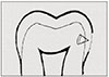
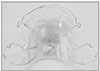
In this study, supersaturated remineralized solution "R" was applied to specimens with incipient enamel caries, and the quantitative ananlysis of remineralization was evaluated using microradiography. Thirty subjects volunteered to participate in this study. Removable appliances were constructed for the subjects, and the enamel specimen with incipient caries were embedded in the appliances. The subjects wore the intra-oral appliance for 15 days except while eating and sleeping.
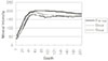
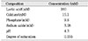
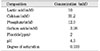
The removable appliance were soaked in supersaturated solution "R", saline, or Senstime® to expose the specimen to those solutions three times a day, 5 minutes each time. After 15 days, microradiography was retaken to compare and evaluate remineralization.
The results were as the following:
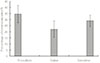
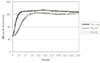
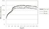
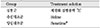
1. The ratio of remineralized area to demineralized area was significantly higher in the supersaturated solution "R" and Senstime® than in the saline. (p<0.05)
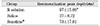
2. Remineralization in the supersaturated buffer solution "R" occurred in the significantly deeper parts of the tooth, compared to the Senstime® group containing high concentration of fluoride.(p<0.05)
As in the above results, the remineralization effect of remineralized buffer solution "R" on incipient enamel caries has been proven. For clinical utilization, further studies on soft tissue reaction and the effect on dentin and cementum are necessary.
In conclusion compared to commercially available fluoride solution, remineralization solution "R" showed better remineralization effect on early enamel caries lesion, so it is considered as effecient solution for clinical application.
Figures and Tables
References
1. Arends J, Shuthof J, Jongebloed WL. Lesion depth and microhardness indentations on artificial white spot lesions. Caries Res. 1980. 14:190–195.

2. Backer Dirks O. Posteruptive changes in dental enamel. J Dent Res. 1966. 45:503–511.
3. Brudevold F, McCann HG, Gron P. Caries resistant teeth as related to the chemistry of enamel. Wolstenholm. 1965. London: 121 Churchill.
4. Chow LC, Takagi S. Remineralization of Root Lesions with Concentrated Calcium and Phosphate Silutions. Dent Mater J. 1995. 14:31–36.
5. Clarkson BH, Krell D, Wefel JS, Crall J, Feagin FF. In vitro caries-like lesion produced by Streptococcus mutans and Actinomyces viscosus using sucrose and starch. J Dent Res. 1987. 66:795–798.
6. Dawes C, Weatherell JA. Kinetics of fluoride in the oral fluids. J Dent Res. 1990. 69:638.
7. Exterkate RAM, Damen JJM, Ten Cate JM. A single-section model for enamel de- and remineralization suties 1 The effects of different Ca/P ratios in Remineralization solutions. J Dent Res. 1993. 72:1599–1603.
8. Hay DI, Smith DJ, Schluckebier , Moreno EC. Relationship between concentration of human salivary statherin and inhibition of calcium phosphate precipitation in stimulated human parotid saliva. J Dent Res. 1984. 63:857–863.
9. Heilman JR, Wefel JS. Effect of remineralization on demineralized root surfaces. J Dent Res. 1989. 68:351.
10. Herkstr ter FM, Noordmans J, ten Bosch JJ. Wavelength-independent microradiography: Its use to measure mineral changes in curved and thick samples. Caries Res. 1990. 24:399.
11. Johnson M, Richardson CF, Bergey EJ, Levine MJ, Nancollas GH. The effect of human salivary crystatins and statherin on hydroxyapatite crystallization. Arch Oral Biol. 1978. 23:993–996.
12. Koulourdes T. Implication of remineraliztion in the tratment of dental caries in processing symposium on current topics in dental caries in commemoration of the decenial anniversary of Hihon university school of dentistry at Matsudo. 1982. Matsudo, Japan: 198.
13. Larsen MJ. Cemically induced in vitro lesions in dental enamel. Scand J Dent Res. 1974. 82:496–509.
14. Larsen MJ. Degrees of saturation with respect to apatite in parotid saliva at various pH values. Scand J Dent Res. 1975. 83:7–12.
15. Margolis HC, Murphy BJ, Moreno EC. Development of caries-like lesions in partially saturated lactate buffers. Caries Res. 1985. 19:36–45.
16. Margolis HC, Moreno EC, Murphy BJ. Effect fo low levels of fluoride in solution on enamel deminalization. J Dent Res. 1986. 65:23–29.
17. Mellberg JG, Ripa LW, Leske GS. Fluoride in preventive dentistry, theory and clinical application. Quintessence. 1983. 151–179.
18. Moreno EC, Zahradnik RT. Chemistry of enamel subsurface demineralization in vitro. J Dent Res. 1974. 53:226–235.
19. Moreno EC, Zahradnik RT. Chemistry of enamel subsurface demineralization in vitro. J Dent Res. 1974. 53:226–235.
20. Ogaard B, Rolla G, Helgeland K. Alkali soluble and alkali insoluble fluoride retention in demineralized enamel in vivo. Scand J Dent Res. 1983. 91:200.
21. Silverstone LM. Remineralization and enamel caries new concepts. Dent Update. 1983. 10:261–273.
22. Silverstone LM. Observations on the dark zone in early enamel caries and artificial caries-like lesions. Caries Res. 1967. 1:260–274.
23. Silverstone LM. Structure of carious enamel, including the early lesion. Oral Sci Rev. 1973. 3:100–160.
24. Sperber GH, Bunnocore MG. Enamel surface in whitw spot formation. J Dent Res. 1963. 42:724–731.
25. Ten Bosch JJ, van der Mei HC, Borsboom PCF. Optical monitor of in vitro caries. Caries Res. 1984. 18:540–548.
26. Ten Cate JM, Jongebloed WL, Arends J. Remineralization of artificial enamel lesions in vitro IV. Influence of and diphonates in short and long term remineralization. Caries Res. 1981. 15:60–69.
27. Ten Cate JM, Arends J. Remineralization of artificial enamel lesions in vitro III A study of the deposition mechanism. Caries Res. 1980. 14:351–358.
28. Ten Cate JM, Arends J. Remineralization of artificial enamel lesions in vitro. Caries Res. 1977. 11:277–286.
29. Theuns HM, Van Dijk JWE, Driessens FCM, Groeneveld A. Effect of time, degree of saturation, pH and acid concentration of buffer solutions on the rate of in-vitro demineralization of human enamel. Arch Oral Biol. 1985. 30:37–42.
30. Thylstrup A, Fejershov O. Textbook of clinical cariology. 1994. Second edition. 231.
31. Thylstrup A, Fejershov O. Textbook of clinical cariology. 1994. Second edition. 240.
32. Wefel JS. Effects of fluorides on careis development and progression using intraoral models. J Dent Res. 1990. 69:626.
33. Wei SHY. Clinical uses of fluorides. 1985. Lea & Febiger;16–24.
34. Kum KY, Lee CY. The effect of four kinds of acid and concentration on the formation of artificial carious lesion in human tooth enamel. J Korean Acad Conserv Dent. 1996. 21:470–488.
35. Kim MK, Kum KY, Lee CY. The influence of ph on remineralization of artificial dental caries. J Korean Acad Conserv Dent. 1997. 22:193–208.
36. Kim SR, Lee CY, Kum KY. The remineralizing effects of early enamel car10us lesion by supersaturated buffer solution under ph cycling model. J Korean Acad Conserv Dent. 2001. 26:341–349.
37. Kim JB, Choi YJ, Back DI, Shin SC, Kim DK. Clinical Preventive Dentistry. 1991. Iuchulpansa;193–218.
38. Park SH, Lee CY, Lee CS. The effect of acid concentration and ph of lactate buffer solution on the progress of artificial caries lesion in human tooth enamel. J Korean Acad Conserv Dent. 1993. 18:277–290.
39. Park JW, Hur B, Lee CY. The effects of the degree of saturation of acidulated buffer solutions in enamel and dentin remineralization and afm observation of hydroxyapatite crystals. J Korean Acad Conserv Dent. 2000. 25:459–473.
40. Oh HS, Kum KY, Ro BD, Lee CY. The influence of ph on the formation of artificial root caries in acid buffer solution. J Korean Acad Conserv Dent. 1999. 24:495–502.
41. Lee CY. Artificial caries formation in acid buffering solution. Yonsei Dent J. 1992. 7:34–41.
42. Han WS, Kum KY, Lee CY. The influence of fluoride on remineralization of artificial dental caries. J Korean Acad Conserv Dent. 1996. 21:161–173.
 XML Download