Abstract
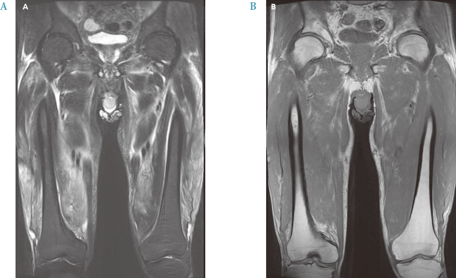
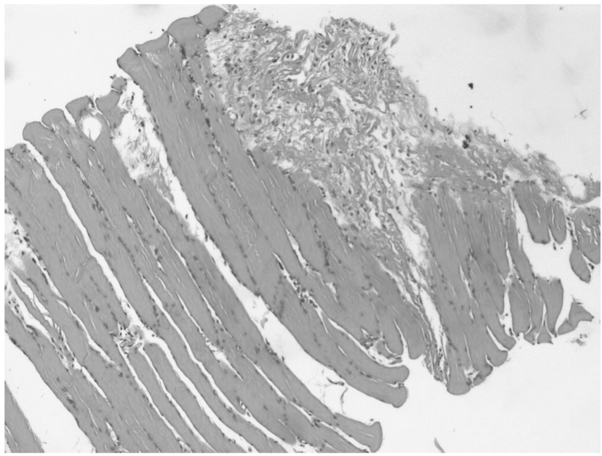
Diabetic muscle infarction (DMI) is a rare condition that usually occurs in diabetic patients who have longstanding microvascular complication. The typical presentation is a painful swelling with abrupt onset in the lower limbs, particularly involving hyper-intense signals in T2-weighted magnetic resonance imaging (MRI) images. The treatment consists of bed rest, analgesics, and physical therapy. The authors encountered a case of DMI with bilateral tender swelling on the anteromedial aspect of the thighs. DMI is less likely to develop in patients with good glycemic control. Recently, however, a few cases demonstrated that DMI can also develop in patients with good glucose control. However, diffuse and extensive infarction of muscle, such as in our case, is rare. It is important to consider differential diagnoses in order to avoid misdiagnosis and non-essential treatment such as overuse of antibiotics or steroid treatment. In this case, we diagnosed the patient using MRI, muscle biopsy, and electromyography and successful treatment involved bed rest and analgesics. We herein report a case of 76-year-old man with very extensive and diffuse DMI in spite of well-controlled type 2 diabetes.
Go to : 
References
1. Rocca PV, Alloway JA, Nashel DJ. Diabetic muscular infarction. Semin Arthritis Rheum. 1993; 22:280–7.

2. Umpierrez GE, Stiles RG, Kleinbart J, Krendel DA, Watts NB. Diabetic muscle infarction. Am J Med. 1996; 101:245–50.

3. Banker BQ, Chester CS. Infarction of thigh muscle in the diabetic patient. Neurology. 1973; 23:667–77.

4. Trujillo Santos AJ, Alcalá Pedrajas JN, Moreno García MM, García de Lucas MD, García Sánchez JE. A 70-year-old woman with right calf pain and inflammation. Rev Clin Esp. 2001; 201:153–4.
6. Kapur S, Brunet JA, McKendry RJ. Diabetic muscle infarction: case report and review. J Rheumatol. 2004; 31:190–4.
7. Trujillo-Santos AJ. Diabetic muscle infarction: an underdiagnosed complication of long-standing diabetes. Diabetes Care. 2003; 26:211–5.
8. Bjornskov EK, Carry MR, Katz FH, Lefkowitz J, Ringel SP. Diabetic muscle infarction: a new perspective on pathogenesis and management. Neuromuscul Disord. 1995; 5:39–45.

9. Kapur S, McKendry RJ. Treatment and outcomes of diabetic muscle infarction. J Clin Rheumatol. 2005; 11:8–12.

10. Yu HM, Jin HY, Park TS. Case of recurrent diabetic muscle infarction related to strict blood glucose control. Korean J Med. 2013; 84:737–41.

11. Hinton A, Heinrich SD, Craver R. Idiopathic diabetic muscular infarction: the role of ultrasound, CT, MRI, and biopsy. Orthopedics. 1993; 16:623–5.

12. Morcuende JA, Dobbs MB, Crawford H, Buckwalter JA. Diabetic muscle infarction. Iowa Orthop J. 2000; 20:65–74.
13. Wang SI, Park JH, Lee JH. Acute compartment syndrome in association with spontaneous muscle infarction. J Korean Orthop Assoc. 2012; 47:75–8.

14. Hwang JH, Jeong ST, Ra YJ, Jung JY. Diabetic muscle infarction in diabetes; three cases report. J Korean Acad Rehabil Med. 2003; 27:803–7.
Go to : 
 | Fig. 1.Magnetic resonance imaging of the patient's thighs shows extensive muscle edema, unevenly and bilaterally involving all visualized muscles of the thighs. Overlying soft tissue and skin thickening are seen prominently on the right. (A) Coronal T2-weighted image demonstrating increased signal intensity in both involved muscles. (B) Coronal T1-weighted image showing decreased signal intensity in both involved muscles. |


 PDF
PDF ePub
ePub Citation
 Print
Print


 XML Download