

 PDF
PDF ePub
ePub Citation
 Print
Print


Abstract
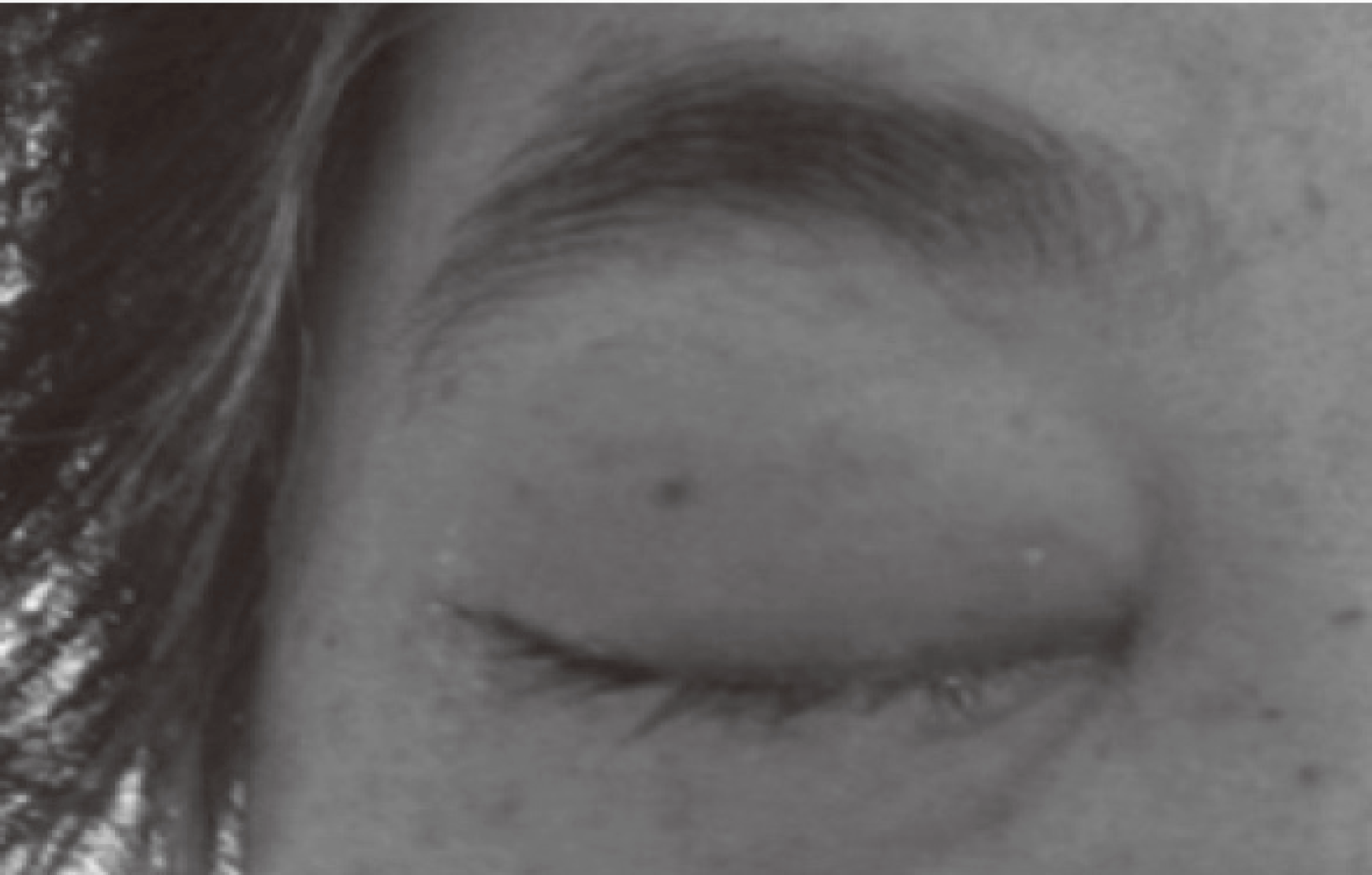
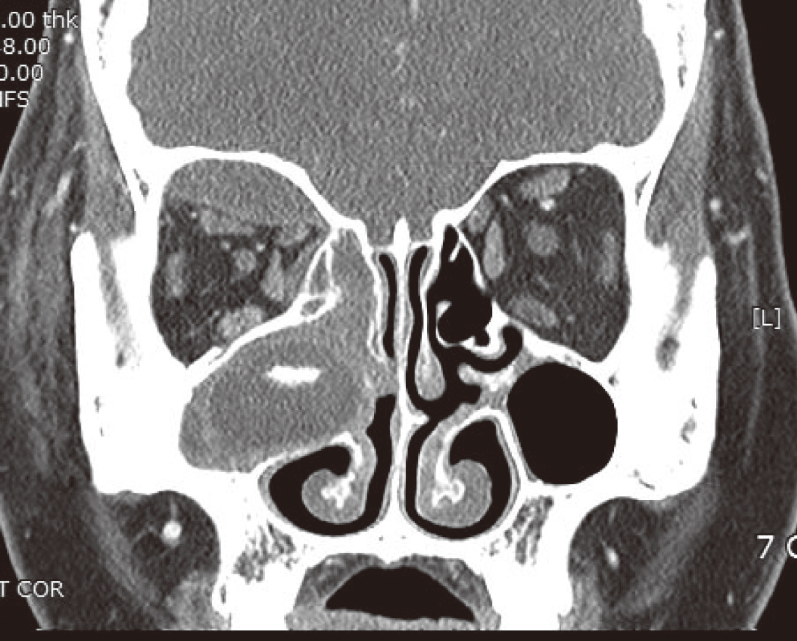
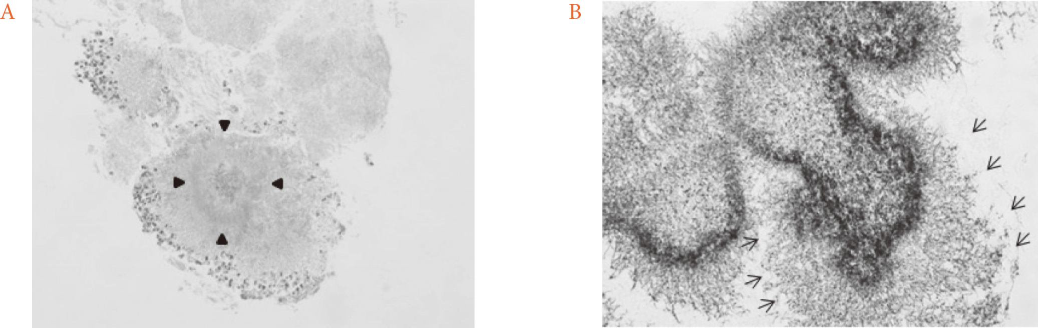
Orbital and paranasal actinomycosis have not been commonly reported. We report a case of this uncommon infection, which was improved after endonasal endoscopic drainage and antibiotics. A 53-year-old woman with type 2 diabetes mellitus complained of inability to lift her right upper eyelid and painful swelling over the preceding two days. Broad-spectrum antibiotics did not resolve her lesion. In ophthalmic examination, decreased visual acuity, upper and medial gaze limitation, and a relative afferent pupillary defect of her right eye were observed. Computed tomography of the orbit showed aggravated orbital cellulitis, preseptal cellulitis, subperiosteal abscess, and maxillary and ethmoid sinusitis. After endonasal endoscopic drainage and systemic antibiotics, her clinical symptoms dramatically improved. Microbiological analysis of the maxillary excisional biopsy showed Actinomycosis. This case is of interest due to the rare orbital presentation of actinomycosis infection and the importance of appropriate surgical drainage and long-term antibiotics treatment in such cases. Because delayed diagnosis and treatment of rhino-orbital actinomycosis can cause permanent vision loss or intracranial abscess, it requires careful clinical attention.
References
1. Moon SW, Lee YJ, Lee YS, Jung JH. A case of simultaneous orbital cellulitis and intracranial complication. J Korean Ophthalmol Soc. 2009; 50:467–70.

2. Jung HM, Hwang YJ, Ahn YS, Youn JS, Lee MG, Kim WJ, Lee EW. Two cases of endobronchial actinomycosis that were cured by bronchoscopic removal and short-term antibiotic therapy. Korean J Med. 2010; 79:563–8.
3. de Andrade AL, Novaes MM, Germano AR, Luz KG, de Almeida Freitas R, Galvão HC. Acute primary actinomycosis involving the hard palate of a diabetic patient. J Oral Maxillofac Surg. 2014; 72:537–41.
4. Cocuroccia B, Gubinelli E, Fazio M, Girolomoni G. Primary cutaneous actinomycosis of the forehead. J Eur Acad Dermatol Venereol. 2003; 17:331–3.
5. Melgarejo Moreno PJ, Hellín Meseguer D, Gil Vélez M, Ruiz Macia JA. Primary laryngeal actinomycosis. Acta Otorrinolaringol Esp. 1997; 48:237–8.
6. Samant S, Sandoe J, High A, Makura ZG. Actinomycosis mimicking a tonsillar neoplasm in an elderly diabetic patient. Br J Oral Maxillofac Surg. 2009; 47:417–8.
7. Ramos JM, Pacho E, Esteban J, Ponte MC. Paramandibular mass in a diabetic woman. Enferm Infecc Microbiol Clin. 1993; 11:105–6.
8. Davanos E, Rahman SM, Nogid B. Treatment of Eikenella corrodens and Actinomyces odontolyticus foot abscess in a penicillin-allergic patient. Ann Pharmacother. 2008; 42:1706–10.
9. Chandler JR, Langenbrunner DJ, Stevens ER. The pathogenesis of orbital complications in acute sinusitis. Laryngoscope. 1970; 80:1414–28.
10. Botting AM, McIntosh D, Mahadevan M. Paediatric pre- and post-septal periorbital infections are different diseases. A retrospective review of 262 cases. Int J Pediatr Otorhinolaryngol. 2008; 72:377–83.
11. Chung SW, Cho JG, Lee SH, Lee SH, Hwang SJ, Lee HM. Clinical analysis of orbital subperiosteal abscess. J Rhinol. 2005; 12:50–4.
12. EyeNet. Evaluation and management of orbital subperiosteal abscess. Available from:. http://www.aao.org/eyenet/article/evaluation-management-of-orbital-subperiosteal-abs. (updated 2009 Jul 1).
13. Weese WC, Smith IM. A study of 57 cases of actinomycosis over a 36-year period. A diagnostic ‘failure’ with good prognosis after treatment. Arch Intern Med. 1975; 135:1562–8.
14. Mallika OU, Sujatha N, Smitha Narayan, Sinumol S. Orbital and preseptal cellulitis. Kerala J Ophthalmol. 2011; 23:10–4.
15. Garcia GH, Harris GJ. Criteria for nonsurgical management of subperiosteal abscess of the orbit: analysis of outcomes 1988–1998. Ophthalmology. 2000; 107:1454–6. discussion 1457–8.
 XML Download