

 PDF
PDF ePub
ePub Citation
 Print
Print


Abstract
Spontaneous pneumomediastinum (SPM) is defined as the presence of extraluminal gas in the mediastinal space without any clear traumatic cause. It has been reported in association with asthma exacerbation, emesis, childbirth, seizure, excessive shouting, drug inhalation and diabetic ketoacidosis (DKA). SPM complicated by DKA is infrequently accompanied with chest pain and DKA can lead to changes in respiratory rate and depth; this complication might be underestimated. Here, we report a 21-year-old male with throat pain on swallowing due to SPM complicated by DKA. Clinicians need to consider this complication in differential diagnoses.
Go to : 
References
1. Chaithongdi N, Subauste JS, Koch CA, Geraci SA. Diagnosis and management of hyperglycemic emergencies. Hormones (Athens). 2011; 10:250–60.

2. Mondello B, Pavia R, Ruggeri P, Barone M, Barresi P, Monaco M. Spontaneous pneumomediastinum: experience in 18 adult patients. Lung. 2007; 185:9–14.
3. Pauw RG, van der Werf TS, van Dullemen HM, Dullaart RP. Mediastinal emphysema complicating diabetic ketoacidosis: plea for conservative diagnostic approach. Neth J Med. 2007; 65:368–71.
4. Hamman L. Spontaneous mediastinal emphysema. Assoc Am Phys. 1937; 52:311–9.
5. Pooyan P, Puruckherr M, Summers JA, Byrd RP Jr, Roy TM. Pneumomediastinum, pneumopericardium, and epidural pneumatosis in DKA. J Diabetes Complications. 2004; 18:242–7.
6. Kim YW, Kwon YB, Jin HG, Ryu KH, Oh HY, Park SW. A case of subcutaneous emphysema and pneumomediastinum associated with diabetic ketoacidosis. Korean J Med. 1990; 39:429–33.
7. Jung KS, Jo UH, Lee JW, Park HK, Kim YS, Lee MK, Park SG. A case of subcutaneous emphysema and pneumomediastinum associated with diabetic ketoacidosis. Korean J Med. 2002; 63:198–9.
8. Abolnik I, Lossos IS, Breuer R. Spontaneous pneumomediastinum. A report of 25 cases. Chest. 1991; 100:93–5.
9. Bullaboy CA, Jennings RB Jr, Johnson DH, Coulson JD, Young LW, Wood BP. Radiological case of the month. Pneumomediastinum and subcutaneous emphysema caused by diabetic hyperpnea. Am J Dis Child. 1989; 143:93–4.
10. Takada K, Matsumoto S, Hiramatsu T, Kojima E, Watanabe H, Sizu M, Okachi S, Ninomiya K. Management of spontaneous pneumomediastinum based on clinical experience of 25 cases. Respir Med. 2008; 102:1329–34.
11. Bejvan SM, Godwin JD. Pneumomediastinum: old signs and new signs. AJR Am J Roentgenol. 1996; 166:1041–8.
12. Kaneki T, Kubo K, Kawashima A, Koizumi T, Sekiguchi M, Sone S. Spontaneous pneumomediastinum in 33 patients: yield of chest computed tomography for the diagnosis of the mild type. Respiration. 2000; 67:408–11.
13. Banki F, Estrera AL, Harrison RG, Miller CC 3rd, Leake SS, Mitchell KG, Khalil K, Safi HJ, Kaiser LR. Pneumomediastinum: etiology and a guide to diagnosis and treatment. Am J Surg. 2013; 206:1001–6.
14. Cummings RG, Wesly RL, Adams DH, Lowe JE. Pneumopericardium resulting in cardiac tamponade. Ann Thorac Surg. 1984; 37:511–8.
15. Meeking DR, Krentz AJ. Pneumomediastinum complicating diabetic ketoacidosis. Diabet Med. 1996; 13:587–8.
16. Nessan VJ. Recurrent pneumomediastinum in diabetic ketoacidosis. Postgrad Med. 1974; 55:139–40.
17. Ruttley M, Mills RA. Subcutaneous emphysema and pneumomediastinum in diabetic ketoacidosis. Br J Radiol. 1971; 44:672–4.
18. Caceres M, Ali SZ, Braud R, Weiman D, Garrett HE Jr. Spontaneous pneumomediastinum: a comparative study and review of the literature. Ann Thorac Surg. 2008; 86:962–6. S.
Go to :
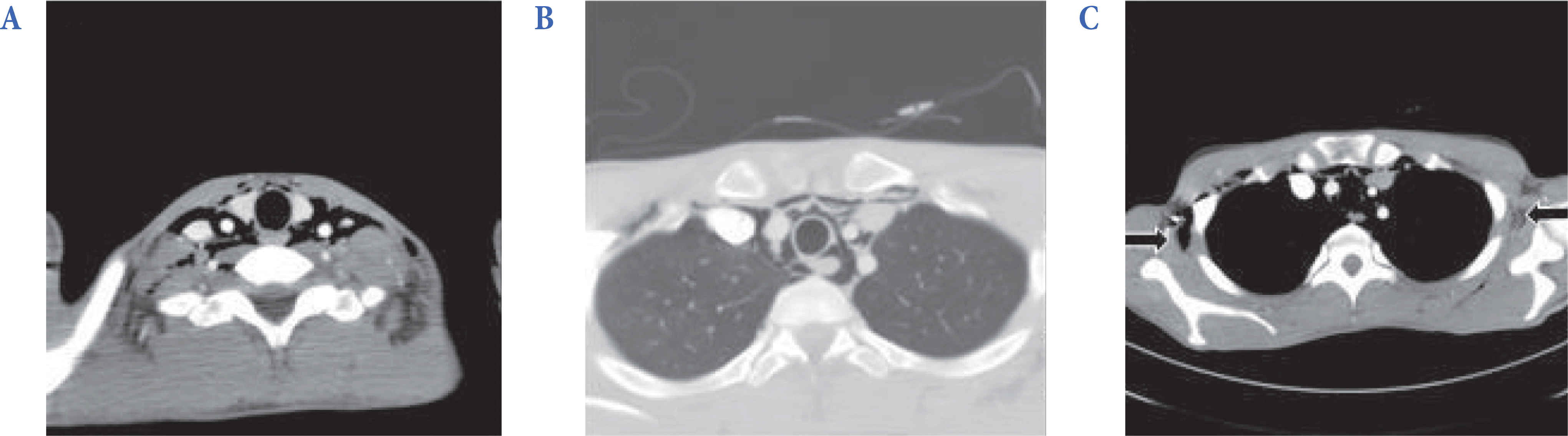
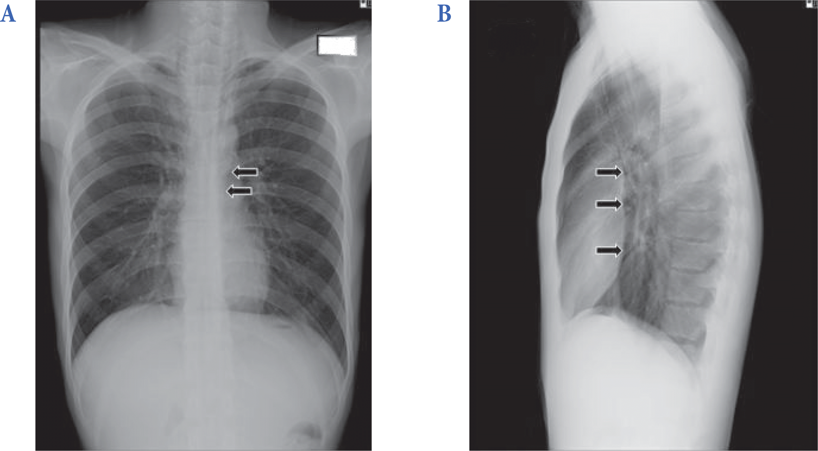 | Fig. 1.(A) Postero-anterior view and (B) left-lateral view chest radiograph. (A) The radiolucent lines beside the aorta indicate the mediastinal emphysema (arrows). (B) The presence of air dissecting the posterior cardiac border and mediastinal structures is demonstrated by left-lateral view (arrows). |
 XML Download