Abstract
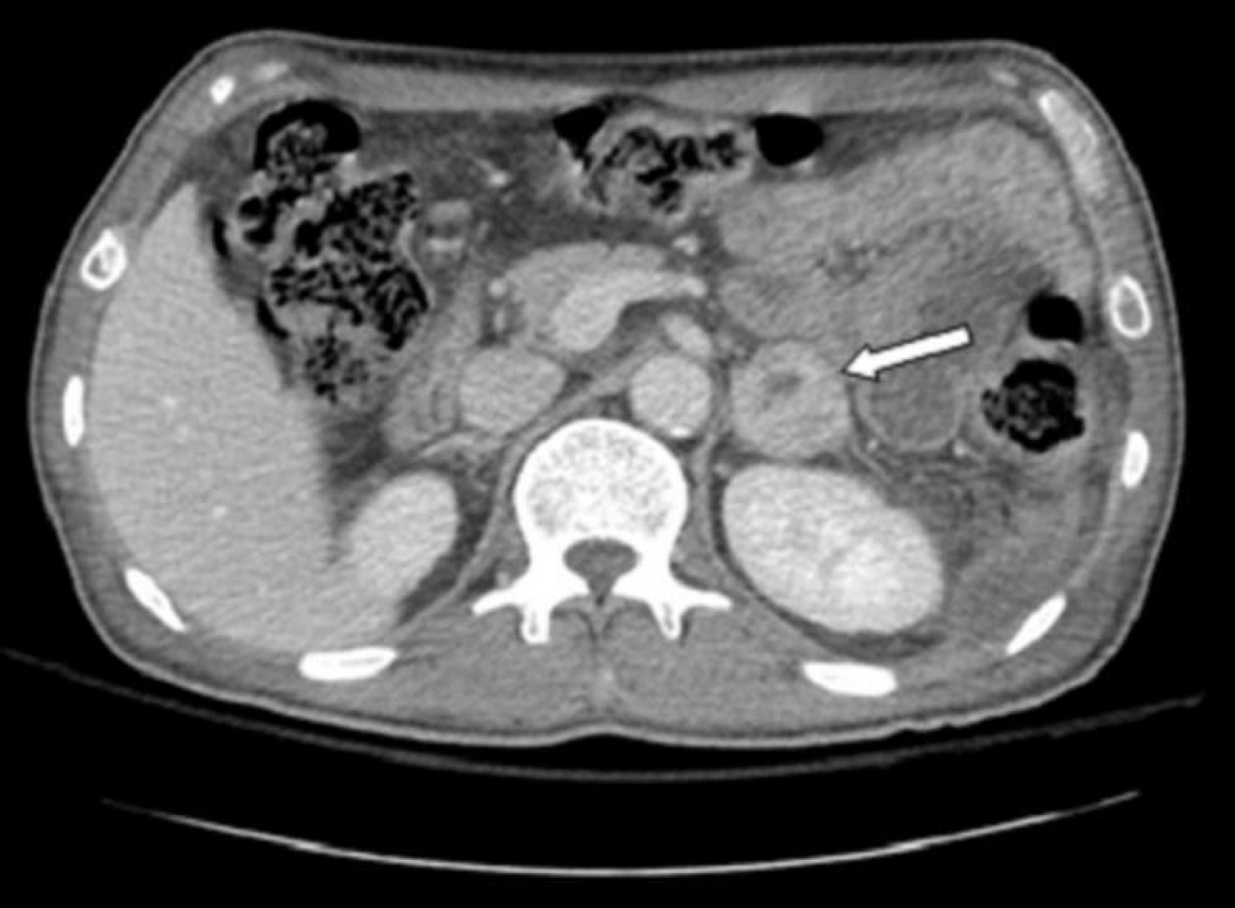
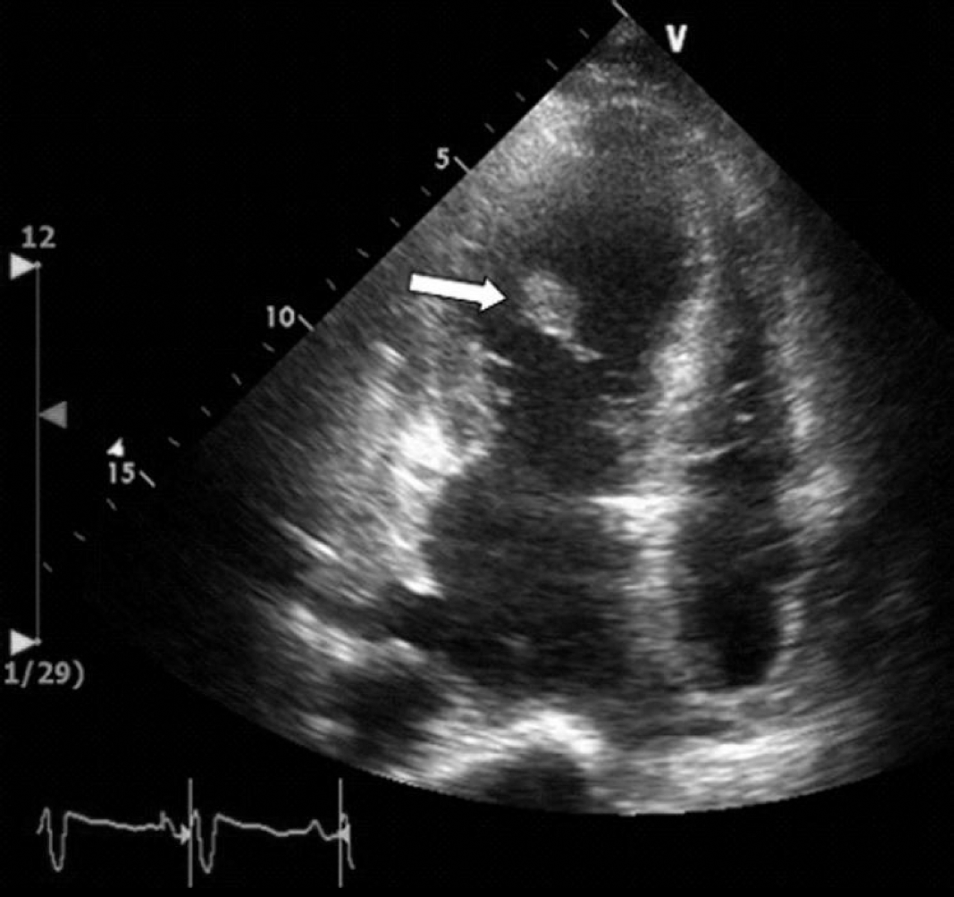
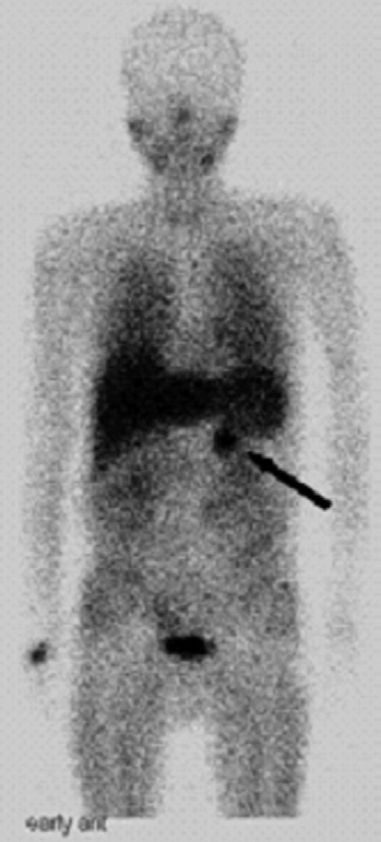
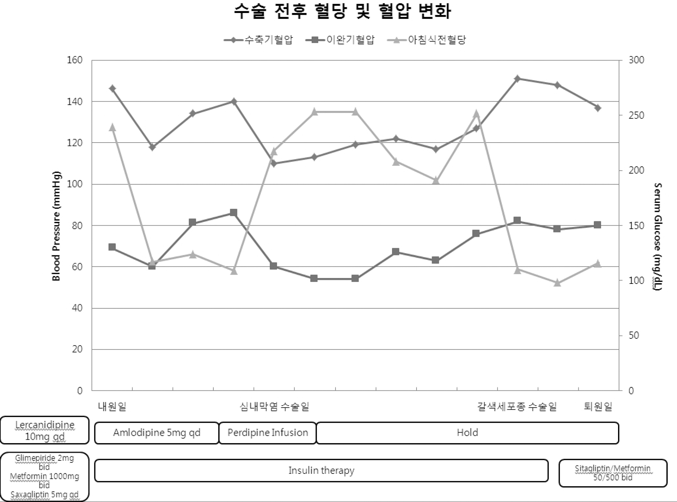
Pheochromocytoma is a rare neuroendocrine tumor that is usually derived from adrenal medulla or chromaffin cells along with sympathetic ganglia. In Western countries, the prevalence of pheochromocytoma is estimated to be between 1:6,500 and 1:2,500, compared with an incidence in the United States of 500 to 1,100 cases per year. Despite this low incidence, pheochromocytoma should always be considered for differential diagnoses because previous studies have shown that this condition can be cured in approximately 90% of cases. However, an untreated tumor is likely to be fatal due to catecholamine-induced malignant hypertension, heart failure, myocardial infarction, stroke, ventricular arrhythmias or metastatic disease. Symptoms that result primarily from excess circulating catecholamines and hypertension include severe headaches, generalized inappropriate sweating and palpitations (with tachycardia or occasionally bradycardia). Pheochromocytoma, however, has highly variable and heterogeneous clinical manifestations, including fever, general weakness and dyspepsia, and can be observed in patients who are suffering from infectious diseases. Several of such case reports have been presented, but most of these included infectious patients with high blood pressure and severe fluctuations. In this study, we presented the case of a 53-year-old male who showed normal blood pressure, but had a sustained fever. He was diagnosed with diabetic ketoacidosis, infective endocarditis and asymptomatic adrenal incidentaloma. Despite treatment with antibiotics and valve replacement, the fever persisted. After the patient underwent evaluation for the fever, adrenal incidentaloma was identified as pheochromocytoma. After removal of the abdominal mass, his fever improved.
Go to : 
References
1. Longo DL, Fauci AS, Kasper DL, Hauser SL, Jameson JL, Loscalzo J. Harrison's principles of internal medicine. 18th ed.New York: McGraw-Hill;2012.
3. Ryu JH, Ha CY, Oh JY, Hong YS, Sung Y-A. A case of pheochromocytoma manifested diabetic ketoacidosis. Korean J Med. 2003; 65:S844–8.
5. Fränkel F. Ein fall von doppelseitigem, völlig latent verlaufenen nebennierentumor und gleichzeitiger nephritis mit veränderungen am circulationsapparat und retinitis Virchows Archiv. 1886; 103:244–63.
6. Son HY, Chun JY, Kim GS, Huh KB, Lee SY. A clinical study on the phenochromocytoma. Korean J Med. 1979; 22:768–74.
7. Bravo EL, Gifford RW Jr. Pheochromocytoma: diagnosis, localization and management. N Engl J Med. 1984; 311:1298–303.

8. Lo CY, Lam KY, Wat MS, Lam KS. Adrenal pheochromocytoma remains a frequently overlooked diagnosis. Am J Surg. 2000; 179:212–5.

9. William FYJ. Endocrine hypertension. Melmed S, Williams RH, S PK, R LP, M KH, editors. Williams textbook of endocrinology. 12nd ed.Philadelphia: Elsevier & Saunders;2011. p. 545–77.
10. Neumann HPH. Pheochromocytoma. Longo DL, Fauci A, Kasper DL, Hauser SL, Jameson JL, Loscalzo J, editors. Harrison's principles of internal medicine. 18th ed.New York: McGraw-Hill;2012. p. 2962–7.
11. Emmer M, Gorden P, Roth J. Diabetes in association with other endocrine disorders. Med Clin North Am. 1971; 55:1057–64.

Go to : 


 PDF
PDF ePub
ePub Citation
 Print
Print


 XML Download