

 PDF
PDF ePub
ePub Citation
 Print
Print


Abstract
Diabetic foot diseases which require surgical treatment consists of diabetic foot ulcer, infection and neuropathic arthropathy. Surgical procedures for diabetic foot ulcers and infections such as drainage, debridement, partial foot amputation and major limb amputation are most common procedures and arthodesis with or without deformity correction can be performed for specific diabetic neuropathic arthropathies. Underlying pathomechanism of diabetic foot disease includes diabetic peripheral neuropathy and vasculopathy. Treating physicians should be aware that concomitant complications of long-standing diabetic status such as cardiovascular and renal dysfunction should be addressed to treat intractable diabetic foot diseases successfully. However, with advent of adjuvant treatment which increases vascular supply on ischemic limb disease, proper surgical treatment on diabetic foot disease can prevent or delay major limb amputations, sustaining functional capability of diabetic patients.
REFERENCES
1. Levin ME. Preventing amputation in the patient with diabetes. Diabetes Care. 1995; 18:1383–94.

2. Wagner FW Jr. The diabetic foot. Orthopedics. 1987; 10:163–72.
3. Brodsky JW. Evaluation of the diabetic foot. Instr Course Lect. 1999; 48:289–303.
4. Laing PW, Cogley DI, Klenerman L. Neuropathic foot ulceration treated by total contact casts. J Bone Joint Surg Br. 1992; 74:133–6.
5. Cohen BK, Zabel DD, Newton ED, Catanzariti AR. Soft-tissue reconstruction for recalcitrant diabetic foot wounds. J Foot Ankle Surg. 1999; 38:388–93.
6. Goldner MG. The fate of the second leg in the diabetic amputee. Diabetes. 1960; 9:100–3.
7. Levin ME. Diabetic foot ulcers: pathogenesis and management. J ET Nurs. 1993; 20:191–8.
8. Quiñones-Baldrich WJ, Kashyap VS, Taw MB, Markowitz BL, Watson JP, Reil TD, Shaw WW. Combined revascularization and microvascular free tissue transfer for limb salvage: a six-year experience. Ann Vasc Surg. 2000; 14:99–104.
9. Vermassen FE, van Landuyt K. Combined vascular reconstruction and free flap transfer in diabetic arterial disease. Diabetes Metab Res Rev. 2000; 16(Suppl 1):S33–6.
10. Lepäntalo M, Tukiainen E. Combined vascular reconstruction and microvascular muscle flap transfer for salvage of ischaemic legs with major tissue loss and wound complications. Eur J Vasc Endovasc Surg. 1996; 12:65–9.
11. Armstrong DG, Lavery LA, Harkless LB, Van Houtum WH. Amputation and reamputation of the diabetic foot. J Am Podiatr Med Assoc. 1997; 87:255–9.
12. Attinger CE, Ducic I, Cooper P, Zelen CM. The role of intrinsic muscle flaps of the foot for bone coverage in foot and ankle defects in diabetic and nondiabetic patients. Plast Reconstr Surg. 2002; 110:1047–54. discussion 1055-7.
13. Schon LC, Marks RM. The management of neuroarthropathic fracture-dislocations in the diabetic patient. Orthop Clin North Am. 1995; 26:375–92.
14. Stuart MJ, Morrey BF. Arthrodesis of the diabetic neuropathic ankle joint. Clin Orthop Relat Res. 1990; 253:209–11.
Fig. 1.
Severe diabetic foot disease with infection was treated surgically by repeated debriment, negative pressure wound therapy (NPWT) and partial thickness split skin graft. (A) Debridment of severe diabetic foot disease with intractable infection. (B) Induction of healthy granulation tissue using NPWT. (C) Reconstruction of skin defect using split thickness skin graft after complete infection control.
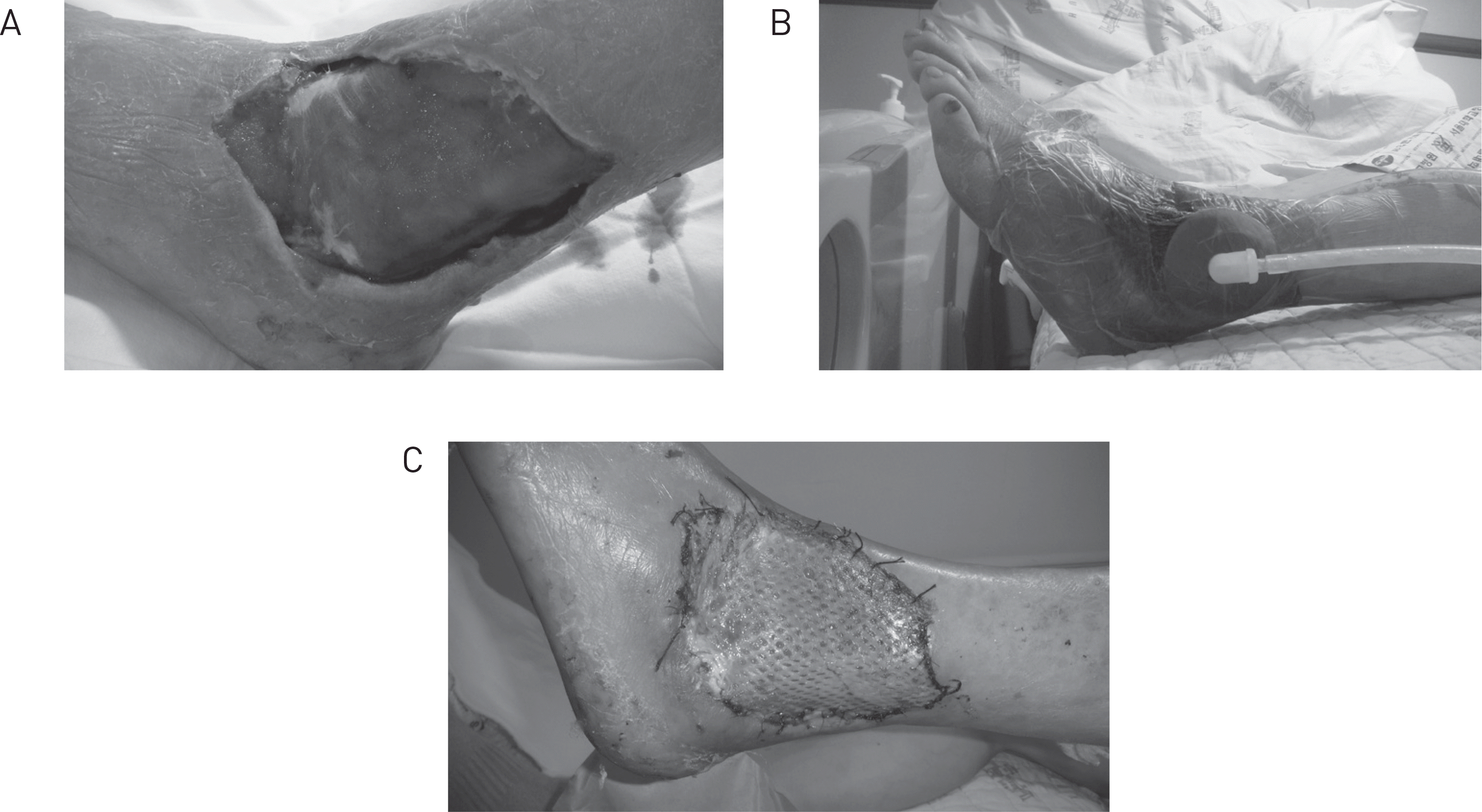
Fig. 4.
Case of diabetic arthropathic patient. (A) Severe bone destruction and resorption due to diabetic neuropathic arthropathy. Usually patients complain lesser degree of pain than nonneuropathic arthritic patients because of neuropathy. (B) We achieved successful ankle fusion by stable fixation of ankle joint and addition of autogenous bone graft.
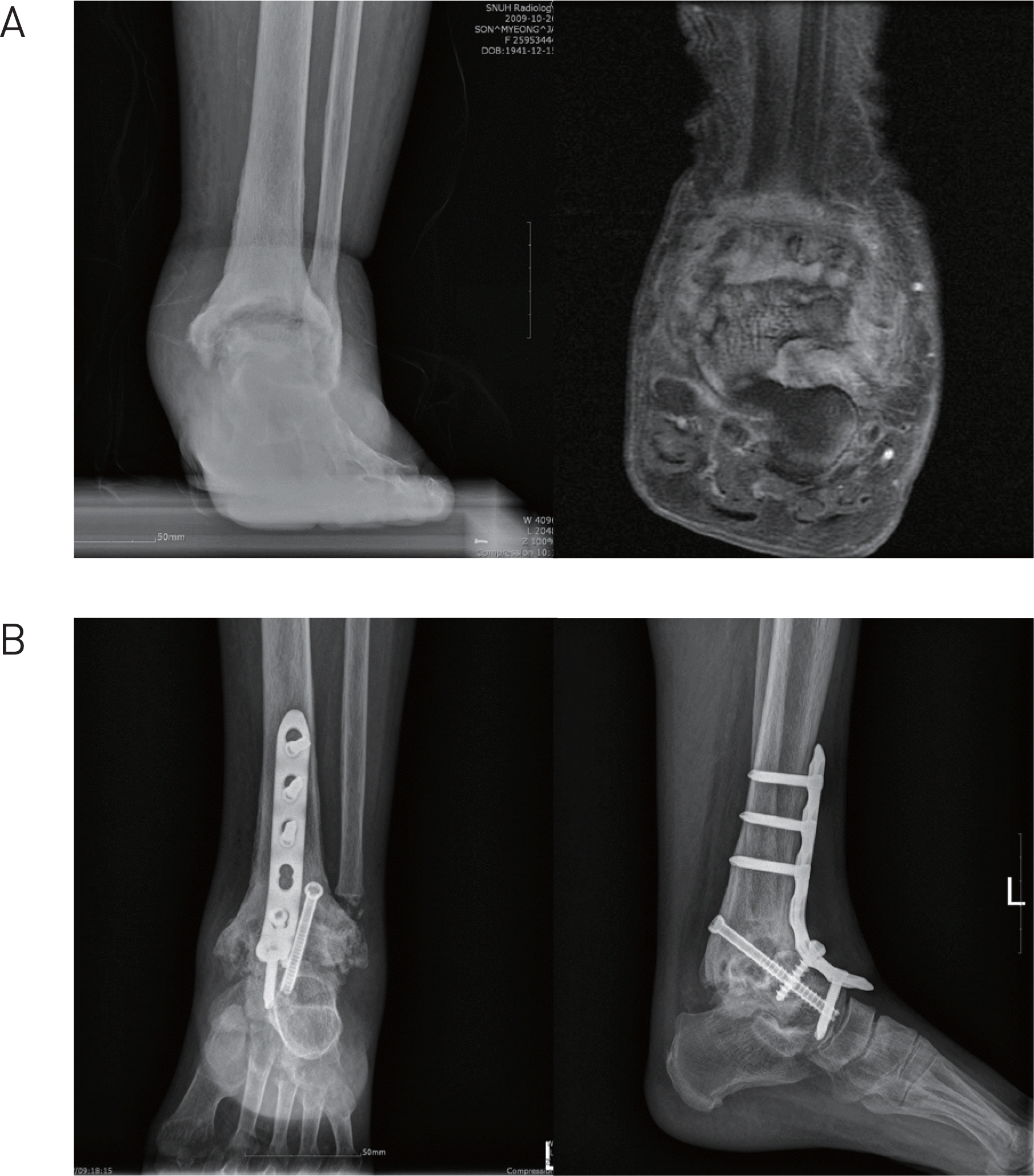
Table 1.
Wagner-Meggitt classification
Table 2.
The depth-ischemia classification of diabetic foot lesions
 XML Download