Journal List > Brain Neurorehabil > v.9(1) > 1054770

Jeong, Kim, and Lee: Relationship between Comorbid Cognitive Impairment and Functional Outcomes in Stroke Patients with Spatial Neglect

Abstract

Objective

The aim of this study was to investigate relationships between cognitive impairment and functional outcomes in stroke patients with spatial neglect.

Method

Twenty seven right hemisphere ischemic stroke patients who admitted to an inpatient rehabilitation unit were included. Patients were divided into two group based on KF-NAP scores: Neglect group (n=18) and non-neglect group (n=9). Neglect group (n=18) were subdivided into two groups by discharge FIM scores as poor outcome group (n=8, <70) and good outcome group (n=10, ≥70). Cognitive function assessments including attention (Attention & Calculation, Digit span test-Forward), memory (Digit span test-Backward, SVLT-Immediate recalls/Delayed recalls) and visual perception (LOTCA-Visual perception/Visuomotor organization), and stroke severity assessment including NIHSS, FMA, K-MMSE, and FIM were performed. Cognitive impairments and functional outcomes were compared between two groups.

Results

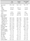
There were significant differences in NIHSS, FMA (affected U/E, affected L/E), discharge FIM, FIM gain and LOTCA-visuomotor organization between Neglect group and non- neglect group (p<0.05). Among the patients with spatial neglect, there were significant differences in KF-NAP, K-MMSE and assessment scores of memory and visual perception between poor outcome group and good outcome group. Attention, memory and visual perception were significantly correlated with KF-NAP scores (p<0.05).

Conclusion

Stroke patients with spatial neglect showed more severe impairment in cognitive, motor and general functional status, which resulted in delayed functional recovery. Spatial neglect patients with combined memory and visual perception impairment may show poor functional outcome.

Figures and Tables

Fig. 1

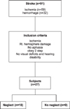
Inclusion criteria in this study.

bn-9-37-g001
Fig. 2

Anatomical analysis methods. Specific lesions related to spatial neglect. 1. Superior frontal gyrus, 2. Middle frontal gyrus, 3. Inferior frontal gyrus, 4. Superior parietal lobule, 5. Caudate nucleus, 6. Thalamus, 7. Inferior parietal lobule, 8. Internal capsule-anterior limb, 9. Basal ganglia, 10. Insular cortex, 11. Internal capsule- posterior limb, 12. Hippocampus, 13. Superior temporal gyrus 14. Middle temporal gyrus.

bn-9-37-g002
Table 1

Comparison of Characteristics and Cognitive Function in Neglect Group and Non-neglect Group

bn-9-37-i001
Total
N=27
Neglect
N=18
Non-neglect
N=9
Age (years) 67.6 ± 10.6 66.3 ± 11.8 70.1 ± 7.7
SEX
 Male 14 (51.9) 11 (61.1) 3 (33.3)
 Female 13 (48.1) 7 (38.9) 6 (66.7)
Lesion location
 ACA 2 (7.4) 2 (11.1) 0 (0)
 MCA 25 (92.6) 16 (88.9) 9 (100)
Initial NIHSS* 9.2 ±4.2 10.8 ± 3.8 5.4 ± 2.9
Initial FMA
 Affected total* 42.8 ± 33.2 32.6 ± 25.9 63.3 ± 38.1
 Affected U/E* 27.6 ± 25.9 20.8 ± 19.8 40.9 ± 28.3
 Unaffected U/E 61.0 ± 13.2 58.8 ± 15.9 65.4 ± 1.1
 Affected L/E* 15.3 ± 11.1 11.8 ± 9.8 22.4 ± 10.5
 Unaffected L/E 30.6 ± 10.3 29.2 ± 12.4 33.4 ± 1.1
Initial FIM* 68.9 ± 21.9 60.0 ± 18.1 86.7 ± 18.2
Discharge FIM* 80.7 ± 26.6 68.1 ± 21.5 106.0 ± 15.7
FIM gain* 11.8 ± 10.6 8.1 ± 7.3 19.2 ± 12.5
Initial K-MMSE 21.1 ± 6.1 19.7 ± 6.3 24.0 ± 4.5
A&C (K-MMSE) 2.0 ± 1.8 1.6 ± 1.6 2.7 ± 2.1
Digit span-F 5.7 ± 1.9 5.7 ± 1.8 5.7 ± 2.1
Digit span-B 2.7 ± 1.4 2.7 ± 1.6 2.7 ± 0.9
SVLT-IR 14.1 ± 6.3 13.9 ± 7.4 14.3 ± 3.4
SVLT-DR 4.2 ± 3.0 4.2 ± 3.6 4.1 ± 1.4
LOTCA-VP 13.1 ± 3.7 12.2 ± 4.3 15.0 ± 0.7
LOTCA-VMO* 14.3 ± 6.4 11.9 ± 5.1 19.1 ± 6.2

Values are mean ± standard deviation or N (%). A&C: Attention & Calculation, Digit span-F: Digit span test-Forward, Digit span-B: Digit span test-Backward, SVLT-IR: SVLT-Immediate recalls, SVLT-DR: SVLT-Delayed recalls, LOTCA-VP: LOTCA-Visual perception, LOTCA-VMO: LOTCA- Visuomotor organization. Mann-Whitney test by *p<0.05.

Table 2

Comparison of Cognitive Function and Functional Outcomes in Neglect Group and Non-neglect Group by ANCOVA Analysis

bn-9-37-i002
Neglect group Non-neglect group p
Mean ± SD 95% CI Mean ± SD 95% CI
A&C (K-MMSE) 1.8 ± 0.4 0.855~2.776 2.2 ± 0.7 0.790~3.727 .638
Digit span-F 5.9 ± 0.4 4.956~6.989 5.1 ± 0.7 3.613~6.721 .422
Digit span-B 2.9 ± 0.3 2.202~3.665 2.2 ± 0.5 1.127~3.362 .340
SVLT-IR* 16.1 ± 1.2 13.464~18.802 9.9 ± 51.9 5.876~14.037 .025
SVLT-DR 5.0 ± 0.6 3.664~6.453 2.4 ± 1.0 0.306~4.571 .065
LOTCA-VP 13.5 ± 0.6 12.123~14.898 12.3 ± 1.0 10.191~14.433 .381
LOTCA-VMO 12.8 ± 1.3 10.101~15.691 17.2 ± 2.0 12.986~21.482 .125
D/C FIM* 77.0 ± 2.4 71.937~82.069 88.1 ± 3.7 80.360~95.849 .033
FIM gain* 8.0 ± 2.4 3.011~13.143 19.1 ± 3.7 11.434~26.923 .033

Values are mean ± standard deviation. A&C: Attention & Calculation, Digit span-F: Digit span test-Forward, Digit span-B: Digit span test-Backward, SVLT-IR: SVLT-Immediate recalls, SVLT-DR: SVLT-Delayed recalls, LOTCA-VP: LOTCA-Visual perception, LOTCA-VMO: LOTCA- Visuomotor organization, D/C FIM: Discharge FIM. ANCOVA by *p<0.05.

Table 3

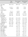
Comparison of Characteristics and Cognitive Function in Neglect group by Functional Outcomes

bn-9-37-i003
Total
N=18
Poor outcome
N=8
Good outcome
N=10
Age (years) 66.3 ± 11.8 69.9 ± 9.4 63.5 ± 13.2
SEX
 Male 11 (61.1) 4 (50) 7 (70)
 Female 7 (38.9) 4 (50) 3 (30)
Lesion location
 ACA 2 (11.1) 1 (12.5) 1 (10)
 MCA 16 (88.9) 7 (87.5) 9 (90)
KF-NAP* 14.4 ± 6.6 18.3 ± 8.0 11.3 ± 2.8
Initial NIHSS 10.8 ± 3.8 12.5 ± 4.0 9.4 ± 3.2
Initial FMA
 Affected U/E* 20.8 ± 22.6 27.6 ± 25.0 15.4 ± 20.0
 Unaffected U/E 58.8 ± 15.9 54.1 ± 21.0 62.5 ± 10.0
 Affected L/E* 11.8 ± 9.8 9.0 ± 7.2 14.0 ± 11.3
 Unaffected L/E 29.1 ± 12.2 24.5 ± 8.4 33.0 ± 14.2
Initial FIM* 60.0 ± 18.1 46.3 ± 17.5 70.9 ± 9.2
FIM gain* 8.1 ± 7.3 3.5 ± 2.9 11.7 ± 7.8
Initial K-MMSE* 19.7 ± 6.3 16.0 ± 7.4 22.6 ± 3.3
A&C (K-MMSE) 1.6 ± 1.6 2.1 ± 1.4
Digit span-F 5.7 ± 1.8 6.3 ± 2.0
Digit span-B 2.7 ± 1.6 3.1 ± 1.5
SVLT-IR** 14.0 ± 7.4 18.5 ± 6.2
SVLT-DR** 4.2 ± 3.6 6.5 ± 3.1
LOTCA-VP** 12.2 ± 4.3 15.1 ± 1.1
LOTCA-VMO** 12.0 ± 5.1 14.2 ± 4.9

Values are mean ± standard deviation or N (%). A&C: Attention & Calculation, Digit span-F: Digit span test-Forward, Digit span-B: Digit span test-Backward, SVLT-IR: SVLT-Immediate recalls, SVLT-DR: SVLT-Delayed recalls, LOTCA-VP: LOTCA-Visual perception, LOTCA-VMO: LOTCA- Visuomotor organization. Mann-Whitney test by *p<0.05, **p<0.01.

Table 4

Correlation between Neglect Severity and Cognitive Impairment

bn-9-37-i004
KF-NAP
Coefficient p
Attention & Calculation -.383 0.117
Digit span test-Forward* -.561 0.016
Digit span test-Backward -.445 0.064
SVLT-Immediate recalls* -.515 0.029
SVLT-Delayed recalls* -.581 0.011
LOTCA
 -Visual perception*** -.738 0.000
LOTCA
 -Visuomotor organization** -.687 0.002

Spearman Correlation by *p<0.05, **p<0.01, ***p<0.001.

Table 5

Comparison of Lesion Location between Neglect Group and Noneglect Group

bn-9-37-i005
Lesion Neglect (N=18) No neglect (N=9)
SFG 1 (6) 0 (0)
MFG* 12 (67) 1 (11)
IFG 9 (50) 1 (11)
SPL 4 (22) 0 (0)
IPL* 9 (50) 0 (0)
STG* 8 (44) 0 (0)
MTG* 9 (50) 0 (0)
P-O-J 4 (22) 0 (0)
Hippo 1 (6) 0 (0)
I* 10 (56) 1 (11)
Cn 4 (22) 1 (11)
BG 7 (39) 5 (56)
Th 0 (0) 2 (22)
IC-Al 1 (6) 0 (0)
IC-Pl 0 (0) 1 (11)
PVWM-A 4 (22) 3 (33)
PVWM-P 6 (33) 0 (0)

Values are N (%), Kai-square test by *p<0.05. Abbreviation: SFG: Superior frontal gyrus, MFG: Middle frontal gyrus, IFG: Inferior frontal gyrus, SPL: Superior Parietal lobule, IPL: Inferior Parietal lobule, STG: Superior Temporal gyrus, MTG: Middle Temporal gyrus, P-O-J: Parieto-occipital junction, Hippo: Hippocampus, I: Insular, Cn: Caudate nucleus, BG: basal ganglia, Th: Thalamus, IC-Al: Internal capsule-anterior limb, IC-Pl: Internal capsule-posterior limb, PVWM-A: Periventricular white matter-anterior part, PVWM-P: Periventricular white matter-posterior part.

References

1. Bowen A, Lincoln N. Cognitive rehabilitation for spatial neglect following stroke. Cochrane Lib. 2007; 2:1–52.
crossref
2. Urbanski MA, De Schotten MT, Rodrigo S, Oppenheim C, Touzé E, Méder JF, Bartolomeo PA. DTI-MR tractography of white matter damage in stroke patients with neglect. Exp Brain Res. 2011; 208(4):491–505.
crossref
3. Paolucci S, Antonucci G, Grasso MG, Pizzamiglio L. The role of unilateral spatial neglect in rehabilitation of right brain–damaged ischemic stroke patients: a matched comparison. Arch Phys Med Rehabil. 2001; 82:743–749.
crossref
4. Halligan PW, Fink GR, Marshall JC, Vallar G. Spatial cognition: evidence from visual neglect. Trends Cognit Sci. 2003; 7:125–133.
crossref
5. Vallar G. Extrapersonal visual unilateral spatial neglect and its neuroanatomy. Neuroimage. 2001; 14:S52–S58.
crossref
6. Mesulam MM. Spatial attention and neglect: parietal, frontal andcingulate contributions to the mental representation and attentional targeting of salient extrapersonal events. Phil Trans R Soc Lond B. 1999; 354:1325–1346.
crossref
7. Chen P, Chen CC, Hreha K, Goedert KM, Barrett AM. Kessler Foundation Neglect Assessment Process Uniquely Measures Spatial Neglect During Activities of Daily Living. Arch Phys Med Rehabil. 2015; 96(5):869–876.
crossref
8. Yoo EY, Jung MY, Park SY, Choi EH. Current trends of Occupational Therapy Assessment Tool by Korean Occupational Therapist. J Korean Soc Occup Ther. 2006; 14(3):27–37.
9. Plummer P, Morris ME, Dunai J. Assessment of unilateral neglect. Phys Ther. 2003; 83:732–740.
crossref
10. Chen P, Hreha K, Pitteri M. Kessler Foundation Neglect Assessment Process: KF-NAP 2014 manual. West Orange: Kessler Foundation;2014.
11. Gillen R, Tennen H, McKee T. Unilateral spatial neglect: relation to rehabilitation outcomes in patients with right hemisphere stroke. Arch Phys Med Rehabil. 2005; 86:763–767.
crossref
12. Pedersen PM, Jorgensen HS, Nakayama H, Raaschou HO, Olsen TS. Hemineglect in acute stroke—incidence and prognostic implications. The Copenhagen Stroke Study. Am J Phys Med Rehabil. 1997; 76:122–122.
13. Verdon V, Schwartz S, Lovblad KO, et al. Neuroanatomy of hemispatial neglect and its functional components: a study using voxel-based lesion-symptom mapping. Brain. 2010; 133(Pt 3):880–894.
crossref
14. Husain M, Rorden C. Non-spatially lateralized mechanisms in hemispatial neglect. Nat Rev Neurosci. 2003; 4:26–36.
crossref
15. Chen P, Hreha K. Kessler Foundation Neglect Assessment Process: KF-NAP 2015 manual. West Orange: Kessler Foundation;2015.
16. Wojciulik E, Husain M, Clarke K, Driver J. Spatial working memory deficit in unilateral neglect. Neuropsychologia. 2001; 39(4):390–396.
crossref
17. Gainotti G, Tiacci C. The relationships between disorders of visual perception and unilateral spatial neglect. Neuropsychologia. 1971; 9(4):451–158.
crossref
18. Kang Y, Na DL. Seoul Neuropsychological Screening Battery (SNSB). 1st ed. Incheon: Human Brain Research & Consulting Co;2003.
19. Katherine Salter BA, Mark Hartley BA, Norine Foley B. Impact of early vs delayed admission to rehabilitation on functional outcomes in persons with stroke. J Rehabil Med. 2006; 38:113–117.
crossref
20. Katz N, Itzkovich M, Averbuch S, Elazar B. Loewenstein Occupational Therapy Cognitive Assessment (LOTCA) battery for brain-injured patients: Reliability and validity. Am J Occup Ther. 1989; 43:184–192.
crossref
21. Azouvi P, Olivier S, de Montety G, Samuel C, Louis-Dreyfus A, Tesio L. Behavioral Assessment of unilateral neglect: Study of the psychometric properties of the Catherine Bergego Scale. Arch Phys Med Rehabil. 2003; 84(1):51–57.
crossref
22. Chen P, Hreha K, Fortis P, Goedert KM, Barrett AM. Functional assessment of spatial neglect: A review of the Catherine Bergego Scale and an introduction of the Kessler Foundation Neglect Assessment Process. Top Stroke Rehabil. 2012; 19:423–435.
crossref
23. Ling FM. Stroke Rehabilitation: Predicting Inpatient Length of Stay and Discharge Placement. Hong Kong J Occup Ther. 2004; 14(1):3–11.
crossref
24. Moeller T, Rief E. Pocket atlas of sectional anatomy [vol 1 head and neck]. New York: thieme;2007.
25. Karnath HO, Berger MF, Küker W, Rorden C. The anatomy of spatial neglect based on voxelwise statistical analysis: a study of 140 patients. Cerebral Cortex. 2004; 14(10):1164–1172.
crossref
26. Karnath HO, Rennig J, Johannsen L, Rorden C. The anatomy underlying acute versus chronic spatial neglect: a longitudinal study. Brain. 2011; 134:903–912.
crossref
27. Karnath HO, Himmelbach M, Rorden C. The subcortical anatomy of human spatial neglect: putamen, caudate nucleus and pulvinar. Brain. 2002; 125(2):350–360.
crossref
28. Karnath HO, Rorden C, Ticini LF. Damage to white matter fiber tracts in acute spatial neglect. Cerebral Cortex. 2009; 19(10):2331–2337.
crossref
29. Wade D, Skilbeck C, David R, Langton-Hewer R. Stroke: a critical approach to diagnosis, treatment and management. London: Chapman and Hall;1985.
30. Driver J, Mattingley JB. Parietal neglect and visual awareness. Nat Neurosci. 1998; 1:17–12.
crossref
31. Jehkonen M, Ahonen JP, Dastidar P, et al. Visual neglect as a predictor of functional outcome one year after stroke. Acta Neurol Scand. 2000; 101:195–201.
crossref
32. Denes G, Semenza C, Stoppa E, Lis A. Unilateral spatial neglect and recovery from hemiplegia. Brain. 1982; 105(3):543–552.
crossref
33. Brain Mort DJ, Malhotra P, Mannan SK, et al. The anatomy of visual neglect. Brain. 2003; 126(Pt 9):1986–1997.
crossref
34. Paolucci S, Antonucci G, Grasso MG, Pizzamiglio L. The role of unilateral spatial neglect in rehabilitation of right brain–damaged ischemic stroke patients: a matched comparison. Arch Phys Med Rehabil. 2001; 82(6):743–749.
crossref
35. Corbetta M, Kincade MJ, Lewis C, et al. Neural basis and recovery of spatial attention deficits in spatial neglect. Nat Neurosci. 2005; 8:1603–1610.
crossref
36. Robertson IH, Mattingley JB, Rorden C, Driver J. Phasic alerting of neglect patients overcomes their spatial deficit in visual awareness. Nature. 1998; 395(6698):169–172.
crossref
37. Stone SP, et al. Unilateral neglect: a common but heterogeneous syndrome. Neurology. 1998; 50:1902–1905.
crossref
38. Baddeley AD, Hitch GJ. Working memory. In : Bower GA, editor. Recent advances in learning and motivation, Vol. 8. New York: Academic Press;1974. p. 47–90.
39. Katz N, Hartman-Maeir A, Ring H, Soroker N. Relationships of cognitive performance and daily function of clients following right hemisphere stroke: predictive and ecological validity of the LOTCA battery. Occup Participation Healt. 2000; 20(1):3–17.
crossref
40. LaBar KS, Gitelman DR, Parrish TB, Mesulam MM. Neuroanatomic overlap of working memory and spatial attention networks: a functional MRI comparison within subjects. Neuroimage. 1999; 10(6):695–704.
crossref
41. Hampshire A, Chamberlain SR, Monti MM, Duncan J, Owen AM. The role of the right inferior frontal gyrus: inhibition and attentional control. Neuroimage. 2010; 50(3):1313–1319.
crossref
42. Aron AR, Robbins TW, Poldrack RA. Inhibition and the right inferior frontal cortex. Trends Cogn Sci. 2004; 8:170–177.
crossref
43. Clower DM, West RA, Lynch JC, Strick PL. The inferior parietal lobule is the target of output from the superior colliculus, hippocampus, and cerebellum. J Neurosci. 2001; 21(16):6283–6291.
crossref
44. Corbetta M, Shulman GL. Spatial neglect and attention networks. Annu Rev Neurosci. 2011; 34:569–599.
crossref
TOOLS
Similar articles