

 PDF
PDF ePub
ePub Citation
 Print
Print


Introduction
Glucose is a major energy source for maintenance of brain function. Persistent hypoglycemia can cause irreversible brain damage and lead to various neurological manifestations including cognitive impairment, abnormal behavior, dysarthria, aphasia, tremor, hemiplegia, seizure and coma.123456 Previous studies have focused on the global outcomes such as Glasgow Outcome Scale rather than specific focal symptoms.45 Dysphagia and voiding difficulty have been rarely reported in the patients with hypoglycemic encephalopathy (HE).
The authors report two cases of patients with HE who had dysphagia and voiding difficulty along with impaired ambulation and cognitive dysfunction. The patients showed improvement of neurological symptoms after approximately 3~4 weeks of inpatient rehabilitation.
Case Reports
1) Case 1
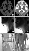
A 76-year-old man with diabetes mellitus had been controlled with oral hypoglycemic drugs (metformin, voglibose, and glimepiride) for 20 years and was admitted to our hospital because of an altered mental state. The day before admission, the patient skipped lunch and dinner and kept alert until 10 PM. The patient's son found him drowsy at 8 AM in the morning. On arrival to the hospital, his initial blood glucose level was 30 mg/mL. Brain magnetic resonance imaging (MRI) performed on admission showed no acute lesions (Fig. 1A, B). After admission, his hypoglycemic state was controlled. Even after correcting the hypoglycemic state, disorientation persisted for a day. Follow-up MRI was not taken during the hospital stay.
He was transferred to the department of rehabilitation at 17 days after onset. In ambulatory activities, his standing balance was poor and minimal assistance was required to walk with a walker (Fig. 1E). He scored 19 on Mini-Mental State Examination-Korean (MMSE-K) indicating cognitive dysfunction. His MMSE-K score was 24 in the one year ago. He had repeated cough during meals after the onset of hypoglycemic encephalopathy and aspiration was noted by fluid test in videofluoroscopic swallowing study (VFSS) (Fig. 1C). Diet modification and thickener were recommended to prevent aspiration. He used intermittent catheterization for voiding and the voiding volume was 400 to 500 ml (Table 1).
Comprehensive rehabilitation was conducted and the patient received strengthening exercise, functional electrical stimulation, balance training, gait training and dysphagia therapy including chin tuck education, oro-motor exercise and pharyngeal constrictor stimulation. Approximately three weeks later, he showed remarkable improvements in gait, swallowing, and voiding. Aspiration was not noted for all kinds of test in a follow-up VFSS (Fig. 1D). He could walk for approximately 200 m without assistance (Fig. 1F) and void urine without catheterization with a residual urine volume less than 50 cc. The electroencephalogram (EEG) was taken one month after onset and did reveal abnormal findings. However, his cognitive function improved little (Table 1). Nine months later, the MMSE score was 23, which was almost same with the level before hypoglycemic encephalopathy.
2) Case 2
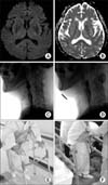
A 75-year-old woman with a history of diabetes had been controlled with oral hypoglycemic drugs (metformin and glimepiride) and was admitted to our hospital because of an altered mental state. She had no problems in eating, voiding and cognition before the hypoglycemic event. She was found by a neighbor after she was not present at regular meetings for two days. On arrival to the hospital, blood glucose testing revealed hypoglycemia. Brain MRI was performed one day after admission and did not show acute lesion (Fig. 2A, B). Follow-up MRI was taken two weeks later and there was no interval change.
She was transferred to the department of rehabilitation at one month after onset. She was unable to stand without support because of poor balance (Fig. 2E). She used nasogastric tube for deeding, and aspiration was noted during swallowing by fluid test in VFSS (Fig. 2C). She had voiding difficulty and required Foley catheter. Her cognitive function was severely impaired (Table 1).
After 1 month of comprehensive rehabilitation, she showed remarkable improvements in gait and swallowing. Penetration or aspiration was not noted for all kinds of tests in a follow-up VFSS (Fig. 2D). She was able to walk with minimal assistance (Fig. 2F). She experienced voiding difficulty after removal of the Foley catheter; therefore, the catheter was reinserted. Her cognitive function changed a little (Table 1). EEG was conducted 6 months after onset and indicated diffuse cerebral dysfunction.
Discussion
According to the International Diabetes Federation, there were 366 million people with diabetes in 2011, and this number is expected to rise to 552 million by 2030.7 Hypoglycemic episodes are not rare in patients with diabetes. The incidence of hypoglycemia with blood glucose levels <50 mg/dL was 7% among 274 patients with type 1 or type 2 diabetes aged ≥18 years, who were admitted as inpatients.8 Most of patients with hypoglycemic events recover without neurological sequalae.1 Severe hypoglycemia can cause neurological deterioration and, sometimes, it is life threatening.4 Outcomes of neurological symptom regarding weakness or dysarthria were variable from full recovery to persistent sequelae.23
The swallowing difficulty after HE has not been discussed widely. Sohn et al. reported a case with swallowing difficulty after HE which was responsive to rehabilitative treatment. The swallowing difficulty was improved and feeding method was progressed from nasogastric tube to oral feeding of thickened diet.9 The voiding difficulty also has not been discussed widely in patients with HE. The voiding difficulty may be affected by autonomic neuropathy in diabetic patient and benign prostate hypertrophy in elderly male. Transrectal ultrasound revealed benign prostatic hyperplasia in case 1. However, two patients in both cases had no problems in voiding before the hypoglycemic event and there was no other pathologic cause to explain the voiding difficulty. We assume that the voiding difficulty was derived from HE mainly and, particularly the symptom was aggravated by benign prostatic hyperplasia in case 1. Cognitive deficits after HE have been presented in many reports. Kim et al. described a patient who presented with persistent cognitive dysfunction, in which the MMSE-K score was 2 at two weeks after onset. The cognitive dysfunction persisted for more than one month and the patient scored 6 at follow-up MMSE-K.6 In our case report, the patient of case 1 already had suffered cognitive dysfunction (MMSE-K: 24) before the hypoglycemic event. The MMSE-K score got worsen to 19 after the hypoglycemia. The MMSE-K score was improved to the premorbid level after nine months. These finding suggested cognitive dysfunction by HE.
The brain is highly susceptible to hypoglycemia. Brain lesions was detected on MRI in patients with severe HE.10 High signal intensity was detected in the cerebral cortex, basal ganglia, and hippocampus on diffusion-weighted MRI (DWI).61011 Recently, the value of MRI was discussed as a predictive value of outcome.10 The patients with focal lesions on DWI showed better prognoses than the patients with diffuse lesions.11 This is consistent with our finding. Our patients showed no acute lesion in brain MRI and the outcome was good.
Reports about the progress of rehabilitation after HE are rare. Our two patients in the present report participated in rehabilitation programs from the subacute stage for more than three weeks. Although this report presented only two patients and did not have controls, the authors thought that functional status can be improved after comprehensive rehabilitation in patients with HE. Further studies with a large sample size and controls are needed to verify the effect of rehabilitation on the long-term outcomes of HE.
Various neurological symptoms have been reported after severe hypoglycemia,123456 but dysphagia and voiding difficulty have been rarely reported. Here we reported two patients with HE who showed impaired ambulation, cognitive dysfunction, dysphagia, and voiding difficulty. After rehabilitation, the recovery in swallowing and locomotor function was rapid and satisfactory; however, cognitive function showed minimal improvement. MRI findings may predict outcomes and rehabilitation in the subacute stage may improve outcomes in patients with HE.

 XML Download