

 PDF
PDF ePub
ePub Citation
 Print
Print


Introduction
Valproate is commonly prescribed for the treatment of seizure disorders, psychiatric conditions, including bipolar disorder, and chronic pain syndromes. Most of its side effects are innocuous, such as transient sedation, postural tremor, and reversible thrombocytopenia. But, it can also cause a serious complication like VHE which is an unusual idiosyncratic reaction characterized by a decreasing level of consciousness, cognitive slowing, focal neurologic deficit, vomiting, drowsiness, and lethargy, sometimes progressing to coma and even death.
For the diagnosis of VHE, patients should be on valproate therapy, presence of elevated level of serum ammonia, normal liver function test, and improvement in clinical feature on withdrawal of valproate. But, it may be overlooked as the range of serum valproate and liver function tests are mostly within normal ranges. The management of VHE is discontinuation of valproate and generally supportive, such as careful monitoring, precaution of fall down and serial follow-up of laboratory tests.1 By reporting a case of VHE, we are willing to inform that physicians in all areas of practice must be aware of this potentially serious adverse effects and suspect it in any patient on valproate therapy who demonstrates altered mentality or behavior.
Case Report
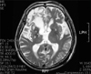
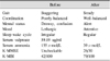
A 65-year-old man was admitted to the Department of Rehabilitation, he developed lethargy, confusion and staggering gait since several weeks. History taking showed that he had been prescribed zonisamide (400 mg/day) as a prophylactic anticonvulsant medication for prevention of post-stroke seizure since he received parieto-temporal craniotomy due to epidural hemorrhage. About 4 weeks before switched it over to valproate (1,000 mg/day), he was drowsy but responsive to pain. On physical examination, the motor and sensory functions were intact as well as the deep tendon reflexes without showing pathologic reflexes such as Babinski's sign and ankle clonus. No generalized spasticity was presented. He showed defective coordination in both upper and lower limbs with severe degree of gait ataxia. Muscle strength in both upper and lower extremities measured by the Medical Research Council Scale was roughly grade 4 and 3 respectively, and balance tested by Berg Balance Scale was total score of 18, which indicate a state of high fall risk. The Korean version of the mini-mental state examination could not be performed initially due to drowsy mentality. The Korean-modified Barthel Index was 42 out of total 100, identifying decreased activity of individual daily living. On second day of symptom onset, laboratory tests including a complete blood count, liver function tests, renal function parameters, serum electrolytes, blood sugar, thyroid function tests and nutritional parameters like vitamin B12 showed to be within normal limits. The level of serum valproate was 59.1 ug/ml, which was within therapeutic range (50~100 ug/ml). On day 4, the level of blood ethanol was also normal. But, he showed still decreased responsiveness and ataxia without Romberg's sign. Brain magnetic resonance imaging was performed on fifth day of symptom onset without any remarkable findings except old lesion (Fig. 1). Also, there was no abnormality of visceral organs in abdominopelvic computed tomography. He continued taking valproate, even though worsening in consciousness and balance. Finally, we ended up checking serum ammonia level on day 7, which showed extraordinary high level up to 155 umol/L (normal range: 12~47 umol/L). On the impression of VHE, we promptly stopped valproate and started lactulose therapy which was given orally at a speed of 30 ml per hour and received 50 ml lactulose enema every 4 hours in a few days of discontinuation of valproate. On day 10, ammonia level came down to 39µmol/L and the cognitive function got better as follows: the Korean version of the mini-mental state examination and Korean-modified Barthel Index was improved up to 26 and 79 respectively (Table 1). On day 11, we gradually reduced administration of lactulose. On day 12, he was able to stand independently with stable gait pattern and balance. Also, disturbed sleep-wake cycle returned to normal and the patient had become more alert and attentive. After 2 months of physical therapy such as lower extremity strengthening, he was discharged with phenytoin to replace valproate. The patient recovered rapidly that ended up walking without support. So far, he is getting along with nothing in particular happening.
Discussion
Valproate is a fatty acid that has antiseizure activity. Asymptomatic hyperammonemia is presented half of valproate treated patients compared to treated with other mood-stabilizing anticonvulsant. Valproate is metabolized through hepatic and renal metabolic pathways. Hepatic metabolism of valproate involves three major pathways: glucuronidation (30~40%), β-oxidation in mitochondria (40%), and ω-oxidation in cytoplasm (10%). Normally, β-oxidation predominates and produces the nontoxic metabolites. However, during long-term or high-dose valproate therapy or as a result of acute overdose, metabolism of valproate shifts toward ω-oxidation producing level of 2-propyl-4-pentanoic acid (4-en-valproate) and propionic acid causing hepatotoxicity and elevated ammonia level. Stimulation of renal glutaminase by 4-envalproate leads to increases renal glutamine uptake and increased ammonia release. Additionally, valproate produces carnitine deficiency by several known mechanisms. Mitochondrial disorders, some organic acidemia and fatty acid oxidation disorders can also cause this syndromes.2
The idiosyncratic toxicity of valproate is largely limited to hepatotoxicity with transient elevation of serum transaminase, amylase and ammomia. One study reported VHE from the administration of multiple drugs with valproate but, in spite of short-term medication and therapeutic dose of valproate only, this patient was drowsy and showed gait disturbance.3
Asymptomatic hyperammonemia has been reported 51.4% of valproate-treated patients compared to 21.7% of the patients treated with other mood-stabilizing anticonvulsant.2 VHE can also occur in the presence of normal serum ammonia levels, if extravascular brain ammonia levels are elevated.
VHE can be acute or subacute with progression of ataxia and lethargy to coma and death. The ability to stand and to walk in a well-coordinated effortless fashion requires the integrity of the entire nervous system. Relatively subtle deficits localized to one part of central or peripheral nervous system will produce characteristic abnormalities. Specific gait disturbances are as follows: cerebellar ataxia (absent Romberg's sign), sensory ataxia (present Romberg's sign), parkinsonian gait, spastic gait, hemiparetic gait, steppage gait, antalgic gait, hysterical gait, gait disorder of elderly. This patient presented absent Romberg's sign after getting well and showed normal liver function test with high ammonia and normal renal function test.
VHE may present with unilateral or bilateral neurological signs or seizures. Ictal or postictal behavioral changes misdiagnosed as worsening of psychiatric symptoms or worsening of nonconvulsive seizures can result in the unfortunate decision to increase rather than decrease valproate dosage. Reduced motor activity, lethargy, or confusion may be misattributed to worsening of psychosis or mood disorder and also lead to inappropriate increase of valproate dose.2 Hypoactive encephalopathy may be misdiagnosed as depression and may lead to inappropriate dose increase. Raised serum ammonia levels may be the only abnormal finding. In most individuals with VHE, serum valproate levels are within the therapeutic range and there is no correlation between serum valproate levels and hyperammonemia. Raised serum and CSF glutamine were elevated in 80% of the VHE patients in a study (n = 7); hence, this may be useful adjunctive laboratory tests for the diagnosis of VHE.4 EEG findings in patients with VHE are characterized by signs of severe encephalopathy. Progressive clinical improvement correlates with normalization of EEG and serum ammonia levels. The results of MRI are bilateral T2 hyperintensities in the cerebellar white matter and globus pallidus.5 It is possible that the cortical changes was not shown as differing serum ammonia levels result in a different pattern of brain injury.6 Duration of valproate therapy, age and gender are not risk factors.7 There does not seem to be any relationship between the daily doses of valproate and the appearance and severity of VHE. Long duration of valproate treatment has not been associated with the onset of VHE.8
VHE is rare especially when valproate is used as monotherapy.9 In a retrospective study, no case of hyperammonemia was reported in patients on chronic valproate monotherapy (n = 16) as compared to 16 cases of hyperammonemia in patients on valproate and anticonvulsant polytherapy (n = 47).7 Significant differences in mean basal level of ammonia between valproate monotherapy and anticonvulsant polytherapy (valproate, phenytoin, topiramate and phenobarbital) (p<0.01) is noted.9
Co-administration of certain drugs appears to predispose to VHE. But, this patient does not have any abnormality of liver functions and metabolic disorders and not take any toxic drugs or neuroleptics developing hyperammonemia except only valproate. He restored shortly without sequelae after discontinuing valproate.
In patients with severe VHE, the condition can be assessed and monitored with the Glasgow Coma scale which measures eye opening, verbal behavior and general motor responsiveness. Detection of minimal or subclinical VHE is difficult, so use of neuropsychological testing has been shown to be more sensitive compared to observational methods. Neuropsychological tests of motor speed, visual perception and construction, concentration and attention (e.g. Trails A and B, line tracing, serial dotting, and the digit symbol tests) may be useful when VHE is suspected but not clinically obvious.
Discontinuation of valproate is recommended for symptomatic patients and L-carnitine supplementation can be effective. Lactulose, neomycin and rifaximin may be necessary. Hemodialysis is the most effective treatment for rapidly reducing serum ammonia and improving consciousness.10
The diagnosis of VHE may be overlooked when the serum valproate concentration and liver function tests are within expected normal ranges. So physicians should be alert to this potential complication of valproate and measure serum ammonia in patient with alterations in mental status or unexplained behavioral change.
 XML Download