

 PDF
PDF ePub
ePub Citation
 Print
Print


Abstract
Objective
To investigate the effect of regular exercise program on cognitive function in chronic cerebral hypoperfused rat.
Method
Forty-eight male Sprague-Dawley rats were used. Chronic cerebral hypoperfusion was induced by bilateral common carotid arteries occlusion (BCCAO). All rats were randomly divided into 4 groups: normal rats (group A); normal rats with regular exercise program (group B); BCCAO rats (group C); BCCAO rats with regular exercise program (group D). Regular exercise program was composed of daily 30-minute treadmill exercise for 4 weeks. Cognitive function was evaluated by Morris water maze (MWM) test. The activities of superoxide dismutase (SOD) and the level of malondialdehyde (MDA) were checked. The neurons were microscopically analyzed on Hematoxylin-Eosin and Cresyl violet stains.
Results
After regular exercise program, there was significant difference in the escape latency among 4 groups in hidden platform trial of MWM test (p<0.05). There was significant difference in the number of crossings among 4 groups in probe trial of MWM test (p<0.05). The activities of SOD of group A and group D were significantly higher than those of group C, respectively (p<0.05). Histopathological study displayed the formation of apoptotic cell bodies and pyknotic cells in group C and group D. There were more normal neurons in group D than group C.
Figures and Tables
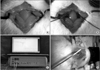 | Fig. 1These figures show sham operation (A), bilateral common carotid arteries occlusion (B), LASER Doppler system (C), and the setting of its probe (D).
|
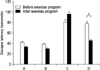 | Fig. 2The results of Morris water maze test: the changes of escape latency before and after the exercise program in hidden platform trial. Escape latency of group D was significantly shortened after regular exercise program (p<0.05). Group A: normal rats, Group B: normal rats with exercise, Group C: BCCAO rats, Group D: BCCAO rats with exercise. *p<0.05 by Wilcoxon Signed rank test.
|
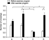 | Fig. 3The results of Morris water maze test: the number of crossings before and after the exercise program in probe trial. After regular exercise program, the number of crossings was significantly increased in group B and group D, respectively (p<0.05). After regular exercise program, the number of crossings of group C was less than those of the other groups (p<0.05). Group A: normal rats, Group B: normal rats with exercise, Group C: BCCAO rats, Group D: BCCAO rats with exercise. *p<0.05 by Kruskal-Wallis test. †p<0.05 by Wilcoxon Signed rank test.
|
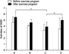 | Fig. 4The activities of superoxide dismutase. After regular exercise program, the activities of SOD were increased in all groups. Especially, the activities of SOD of group A and group D were more increased than those of group C (p<0.05). SOD: superoxide dismutase, Group A: normal rats, Group B: normal rats with exercise, Group C: BCCAO rats, Group D: BCCAO rats with exercise. *p<0.05 by Kruskal-Wallis test.
|
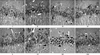 | Fig. 5Histopathological findings of the hippocampus of rats: upper panel - before the exercise program, lower panel - after the exercise program. The formation of apoptotic bodies (arrow) and pyknotic cells (arrow head) were displayed in group C and group D. There were more normal neurons in group D than group C. Hematoxylin and Eosin stain (×400). I, V: group A (normal rats), II, VI: group B (normal rats with exercise), III, VII: group C (BCCAO rats), IV, VIII: group D (BCCAO rats with exercise).
|
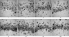 | Fig. 6Histopathological findings of the hippocampus of rats: upper panel - before the exercise program, lower panel - after the exercise program. The formation of apoptotic bodies (arrow) and pyknotic cells (arrow head) were displayed in group C and group D. There were more normal neurons in group D than group C. Cresyl violet stain (×400). I, V: group A (normal rats), II, VII: group B (normal rats with exercise), III, VII: group C (BCCAO rats), IV, VIII: group D (BCCAO rats with exercise).
|
References
1. Jhoo JH, Kim KW, Huh Y, Lee SB, Park JH, Lee JJ, Choi EA, Han C, Choo IH, Youn JC, Lee DY, Woo JI. Prevalence of dementia and its subtype in an elderly urban Korean population: results from the Korean Longitudinal Study on Health and Aging (KLoSHA). Dement Geriatr Cogn Disord. 2008. 26:270–276.
2. Rockwood K, Wentzel C, Hachinski V, Hogan DB, MacKnight C, McDowell I. Prevalence and outcomes of vascular cognitive impairment. Neurology. 2000. 54:447–451.
3. Román GC, Erkinjuntti T, Wallin A, Pantoni L, Chui HC. Subcortical ischaemic vascular dementia. Lancet Neurol. 2002. 1:426–436.
4. Jellinger KA. The enigma of vascular cognitive disorder and vascular dementia. Acta Neuropathol. 2007. 113:349–388.
5. Sarti C, Pantoni L, Bartolini L, Inzitari D. Cognitive impairment and chronic cerebral hypoperfusion: what can be learned from experimental models. J Neurol Sci. 2002. 203:263–266.
6. Wakita H, Tomimoto H, Akiguchi I, Kimura J. Glial activation and white matter changes in the rat brain induced by chronic cerebral hypoperfusion: an immunohistochemical study. Acta Neuropathol. 1994. 87:484–492.
7. Booth FW, Laye MJ. The future: genes, physical activity and health. Acta Physiol (Oxf). 2010. 199:549–556.
8. Kramer AF, Hahn S, Cohen NJ, Banich MT, McAuley E, Harrison CR, Chason J, Vakil E, Bardell L, Boileau RA, Colcombe A. Ageing, fitness and neurocognitive function. Nature. 1999. 400:418–419.
9. Ang ET, Dawe GS, Wong PTH, Moochhala S, Ng YK. Alteration in spatial learning and memory after forced exercise. Brain Research. 2006. 1113:186–193.
10. Cotman CW, Berchtold NC. Exercise: a behavioral intervention to enhance brain health and plasticity. Trends Neurosci. 2002. 25:295–301.
11. Neeper SA, Gomez-finilla F, Choi J, Cotman C. Exercise and brain neurotrophins. Nature. 1995. 373:109.
12. Lee MH, Kim H, Kim SS, Lee TH, Lim BV, Chang HK, Jang MH, Shin MC, Shin MS, Kim CJ. Treadmill exercise suppresses ischemia-induced increment in apoptosis and cell proliferation in hippocampal dentate gyrus of gerbils. Life Sci. 2003. 73:2455–2465.
13. Laurin D, Verreault R, Lindsay J, MacPherson K, Rockwood K. Physical activity and risk of cognitive impairment and dementia in elderly persons. Arch Neurol. 2001. 58:498–504.
14. van Praag H, Kempermann G, Gage FH. Running increases cell proliferation and neurogenesis in the adult mouse dentate gyrus. Nat Neurosci. 1999. 2:266–270.
15. Jee YS, Ko IG, Sung YH, Lee JW, Kim YS, Kim SE, Kim BK, Seo JH, Shin MS, Lee HH, Cho HJ, Kim CJ. Effects of treadmill exercise on memory and c-Fos expression in the hippocampus of the rats with intracerebroventricular injection of streptozotocin. Neurosci Lett. 2008. 443:188–192.
16. Chae CH, Jung SL, An SH, Park BY, Wang SW, Cho IH, Cho JY, Kim HT. Treadmill exercise improves cognitive function and facilitates nerve growth factor signaling by activating mitogen-activated protein kinase/extracellular signal-regulated kinase1/2 in the streptozotocin-induced diabetic rat hippocampus. Neuroscience. 2009. 164:1665–1673.
17. Morris RG, Garrud P, Rawlins JN, O'Keefe J. Place navigation impaired in rats with hippocampal lesions. Nature. 1982. 297:681–683.
18. Payan HM, Levine S, Strebel R. Effects of cerebral ischemia in various strains of rats. Proc Soc Exp Biol Med. 1965. 120:208–209.
19. Hicks L, Birren JE. Aging, brain damage and psychomotor slowing. Psychol Bull. 1970. 74:377–396.
20. Chodzko-Zajko WJ, Moore KA. Physical fitness and cognitive functioning in aging. Exerc Sport Sci Rev. 1994. 22:195–220.
21. Teri L, Logsdon RG, McCurry SM. Exercise intervention for dementia and cognitive impairment: the Seattle protocols. J Nutr Health Aging. 2008. 12:391–394.
22. Farkas E, Luiten PCM, Bari F. Permanent, bilateral common carotid artery occlusion in the rat: a model for chronic cerebral hypoperfusion-related neurodegenerative diseases. Brain Res Rev. 2007. 54:162–180.
23. Milner B, Squire LR, Kandel ER. Cognitive neuroscience and the study of memory. Neuron. 1998. 20:445–468.
24. Alaei H, Moloudi R, Sarkaki AR. Effects of treadmill running on mid-term memory and swim speed in the rat with Morris water maze test. J Bodyw Mov Ther. 2008. 12:72–75.
25. Adlard PA, Perreau VM, Cotman CW. The exercise-induced of BDNF within hippocampus varies across life-span. Neurobiol Aging. 2005. 26:511–520.
26. Kuhn HG, Dickson-Anson H, Gage FH. Neurogenesis in the dentate gyrus of the adult rat: age related decrease of neuronal progenitor proliferation. J Neurosci. 1996. 16:2027–2033.
27. Radák Z, Kaneko T, Tahara S, Nakamoto H, Pucsok J, Sasvári M, Csaba Nyakas C, Goto S. Regular exercise improves cognitive function and decreases oxidative damage in rat brain. Neurochem Int. 2001. 38:17–23.
28. Muralikrishna Adibhatla R, Hatcher JF. Phospholipase A2, reactive oxygen species, and lipid peroxidation in cerebral ischemia. Free Radic Biol Med. 2006. 40:376–387.
29. Halliwell B. Reactive oxygen species and the central nervous system. J Neurochem. 1992. 59:1609–1623.
30. Smith MA, Perry G. Alzheimer disease protein-protein interaction and oxidative stress. Bol Estud Med Biol. 1996. 44:5–10.
31. Nita DA, Nita V, Spulber S, Moldovan M, Popa DP, Zagrean AM, Zagrean L. Oxidative damage following cerebral ischemia depends on reperfusion-a biochemical study in rat. J Cell Mol Med. 2001. 5:163–170.
32. Aytac E, Oktay Seymen H, Uzun H, Dikmen G, Altug T. Effects of iloprost on visual evoked potentials and brain tissue oxidative stress after bilateral common carotid artery occlusion. Prostaglandins Leukot Essent Fatty Acids. 2006. 74:373–378.
33. Samorajski T, Delaney C, Dunlap WP. Effect of exercise on longevity, body weight, locomotor performance, and passive--avoidance memory of C57B1 mice. Neurobiol Aging. 1985. 6:17–24.
34. Somani SM, Ravi R, Rybak LP. Effect of exercise training on antioxidant system in brain regions of rat. Pharmacol Biochem Behav. 1995. 50:635–639.
 XML Download