

 PDF
PDF ePub
ePub Citation
 Print
Print


Abstract
Purpose
This study was designed to assess the effects of a rehabilitation program on clinical symptoms, subacromial space parameters and the supraspinatus vascularity in individuals with subacromial impingement syndrome (SIS).
Methods
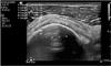
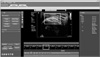
Thirty-five participants (exercise group with SIS [EG]=11, non-exercise group with SIS [NEG]=10, control group [CG]=14) took part in this study. Only EG participated in 6-week rehabilitation program. Outcomes were evaluated at baseline, 6 weeks, and 10 weeks. Changes in symptoms and functional limitations were assessed using Shoulder Pain and Disability Index (SPADI) questionnaire. Changes in acromiohumeral distance (AHD) and supraspinatus tendon thickness (STT)/AHD were assessed using ultrasonographic measures. Quantitative analysis of tendon blood flow was performed by determining four regions of interest with power Doppler quantification and analysis software to normalize data for interpretation of the mean ratio of colored pixel to the region of interest (vascularization index [VI]) and the intensity per pixel (flow index [FI]).
Figures and Tables
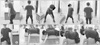 | Fig. 4Rehabilitation program. (A) Robbery exercise for lower trapezius. (B) Lawn mower exercise for lower trapezius. (C) Low exercise for lower trapezius. (D) Inferior glide exercise for lower trapezius. (E) Prone shoulder flexion at 135° abduction for lower trapezius. (F) Push-ups with a plus for serratus anterior.
|
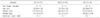
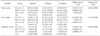
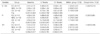
Table 2
Comparison of Shoulder Pain and Disability Index score between groups at baseline and after rehabilitation program
![]()
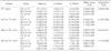
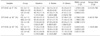
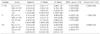
Table 5
The QLAB analysis of power Doppler ultrasound in bursal lateral region of supraspinatus tendon
![]()
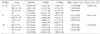
Table 6
The QLAB analysis of power Doppler ultrasound in articular lateral region of supraspinatus tendon
![]()
References
1. Seitz AL, McClure PW, Finucane S, Boardman ND 3rd, Michener LA. Mechanisms of rotator cuff tendinopathy: intrinsic, extrinsic, or both? Clin Biomech (Bristol, Avon). 2011; 26:1–12.

2. Hegedus EJ, Cook C, Brennan M, Wyland D, Garrison JC, Driesner D. Vascularity and tendon pathology in the rotator cuff: a review of literature and implications for rehabilitation and surgery. Br J Sports Med. 2010; 44:838–847.
3. Lewis JS. Rotator cuff tendinopathy: a model for the continuum of pathology and related management. Br J Sports Med. 2010; 44:918–923.
4. Codman EA. The shoulder: rupture of the supraspinatus tendon and other lesions in or about the subacromial bursa. Brevard County: Krieger;1934.
5. Roy JS, Moffet H, McFadyen BJ. Upper limb motor strategies in persons with and without shoulder impingement syndrome across different speeds of movement. Clin Biomech (Bristol, Avon). 2008; 23:1227–1236.
6. Ludewig PM, Reynolds JF. The association of scapular kinematics and glenohumeral joint pathologies. J Orthop Sports Phys Ther. 2009; 39:90–104.
7. Roy JS, Moffet H, Hebert LJ, Lirette R. Effect of motor control and strengthening exercises on shoulder function in persons with impingement syndrome: a single-subject study design. Man Ther. 2009; 14:180–188.
8. Worsley P, Warner M, Mottram S, et al. Motor control retraining exercises for shoulder impingement: effects on function, muscle activation, and biomechanics in young adults. J Shoulder Elbow Surg. 2013; 22:e11–e19.
9. Desmeules F, Minville L, Riederer B, Cote CH, Fremont P. Acromio-humeral distance variation measured by ultrasonography and its association with the outcome of rehabilitation for shoulder impingement syndrome. Clin J Sport Med. 2004; 14:197–205.
10. Savoie A, Mercier C, Desmeules F, Fremont P, Roy JS. Effects of a movement training oriented rehabilitation program on symptoms, functional limitations and acromiohumeral distance in individuals with subacromial pain syndrome. Man Ther. 2015; 20:703–708.
11. Goodmurphy CW, Osborn J, Akesson EJ, Johnson S, Stanescu V, Regan WD. An immunocytochemical analysis of torn rotator cuff tendon taken at the time of repair. J Shoulder Elbow Surg. 2003; 12:368–374.
12. Rudzki JR, Adler RS, Warren RF, et al. Contrast-enhanced ultrasound characterization of the vascularity of the rotator cuff tendon: age- and activity-related changes in the intact asymptomatic rotator cuff. J Shoulder Elbow Surg. 2008; 17:1 Suppl. 96S–100S.
13. Levy O, Relwani J, Zaman T, Even T, Venkateswaran B, Copeland S. Measurement of blood flow in the rotator cuff using laser Doppler flowmetry. J Bone Joint Surg Br. 2008; 90:893–898.
14. Longo UG, Franceschi F, Ruzzini L, Rabitti C, Morini S, Maffulli N, et al. Histopathology of the supraspinatus tendon in rotator cuff tears. Am J Sports Med. 2008; 36:533–538.
15. Adler RS, Fealy S, Rudzki JR, et al. Rotator cuff in asymptomatic volunteers: contrast-enhanced US depiction of intratendinous and peritendinous vascularity. Radiology. 2008; 248:954–961.
16. Pappas GP, Blemker SS, Beaulieu CF, McAdams TR, Whalen ST, Gold GE. In vivo anatomy of the Neer and Hawkins sign positions for shoulder impingement. J Shoulder Elbow Surg. 2006; 15:40–49.
17. Michener LA, Subasi Yesilyaprak SS, Seitz AL, Timmons MK, Walsworth MK. Supraspinatus tendon and subacromial space parameters measured on ultrasonographic imaging in subacromial impingement syndrome. Knee Surg Sports Traumatol Arthrosc. 2015; 23:363–369.
18. McClure P, Michener L. Measures of adult shoulder function: the American shoulder and elbow surgeons standardized shoulder form patient self report section (ASES), disabilities of the arm, shoulder, and hand (DASH), shoulder disability questionnaire, shoulder pain and disability index (SPADI), and simple shoulder test. Arthritis Care Res (Hoboken). 2003; 49:S50–S58.
19. Bigliani LU, Levine WN. Subacromial impingement syndrome. J Bone Joint Surg Am. 1997; 79:1854–1868.
20. Chen SK, Simonian PT, Wickiewicz TL, Otis JC, Warren RF. Radiographic evaluation of glenohumeral kinematics: a muscle fatigue model. J Shoulder Elbow Surg. 1999; 8:49–52.
21. Green S, Buchbinder R, Hetrick S. Physiotherapy interventions for shoulder pain. Cochrane Database Syst Rev. 2003; (2):CD004258.
22. Girometti R, De Candia A, Sbuelz M, Toso F, Zuiani C, Bazzocchi M. Supraspinatus tendon US morphology in basketball players: correlation with main pathologic models of secondary impingement syndrome in young overhead athletes: preliminary report. Radiol Med. 2006; 111:42–52.
23. Cholewinski JJ, Kusz DJ, Wojciechowski P, Cielinski LS, Zoladz MP. Ultrasound measurement of rotator cuff thickness and acromio-humeral distance in the diagnosis of subacromial impingement syndrome of the shoulder. Knee Surg Sports Traumatol Arthrosc. 2008; 16:408–414.
24. Azzoni R, Cabitza P, Parrini M. Sonographic evaluation of subacromial space. Ultrasonics. 2004; 42:683–687.
25. Graichen H, Bonel H, Stammberger T, et al. Three-dimensional analysis of the width of the subacromial space in healthy subjects and patients with impingement syndrome. AJR Am J Roentgenol. 1999; 172:1081–1086.
26. Hebert LJ, Moffet H, Dufour M, Moisan C. Acromiohumeral distance in a seated position in persons with impingement syndrome. J Magn Reson Imaging. 2003; 18:72–79.
27. Maenhout A, Van Eessel V, Van Dyck L, Vanraes A, Cools A. Quantifying acromiohumeral distance in overhead athletes with glenohumeral internal rotation loss and the influence of a stretching program. Am J Sports Med. 2012; 40:2105–2112.
28. Soslowsky LJ, Thomopoulos S, Tun S, et al. Neer Award 1999: overuse activity injures the supraspinatus tendon in an animal model: a histologic and biomechanical study. J Shoulder Elbow Surg. 2000; 9:79–84.
29. Carpenter JE, Flanagan CL, Thomopoulos S, Yian EH, Soslowsky LJ. The effects of overuse combined with intrinsic or extrinsic alterations in an animal model of rotator cuff tendinosis. Am J Sports Med. 1998; 26:801–807.
30. McCreesh KM, Riley SJ, Crotty JM. Neovascularity in patellar tendinopathy and the response to eccentric training: a case report using Power Doppler ultrasound. Man Ther. 2013; 18:602–605.
 XML Download