Abstract
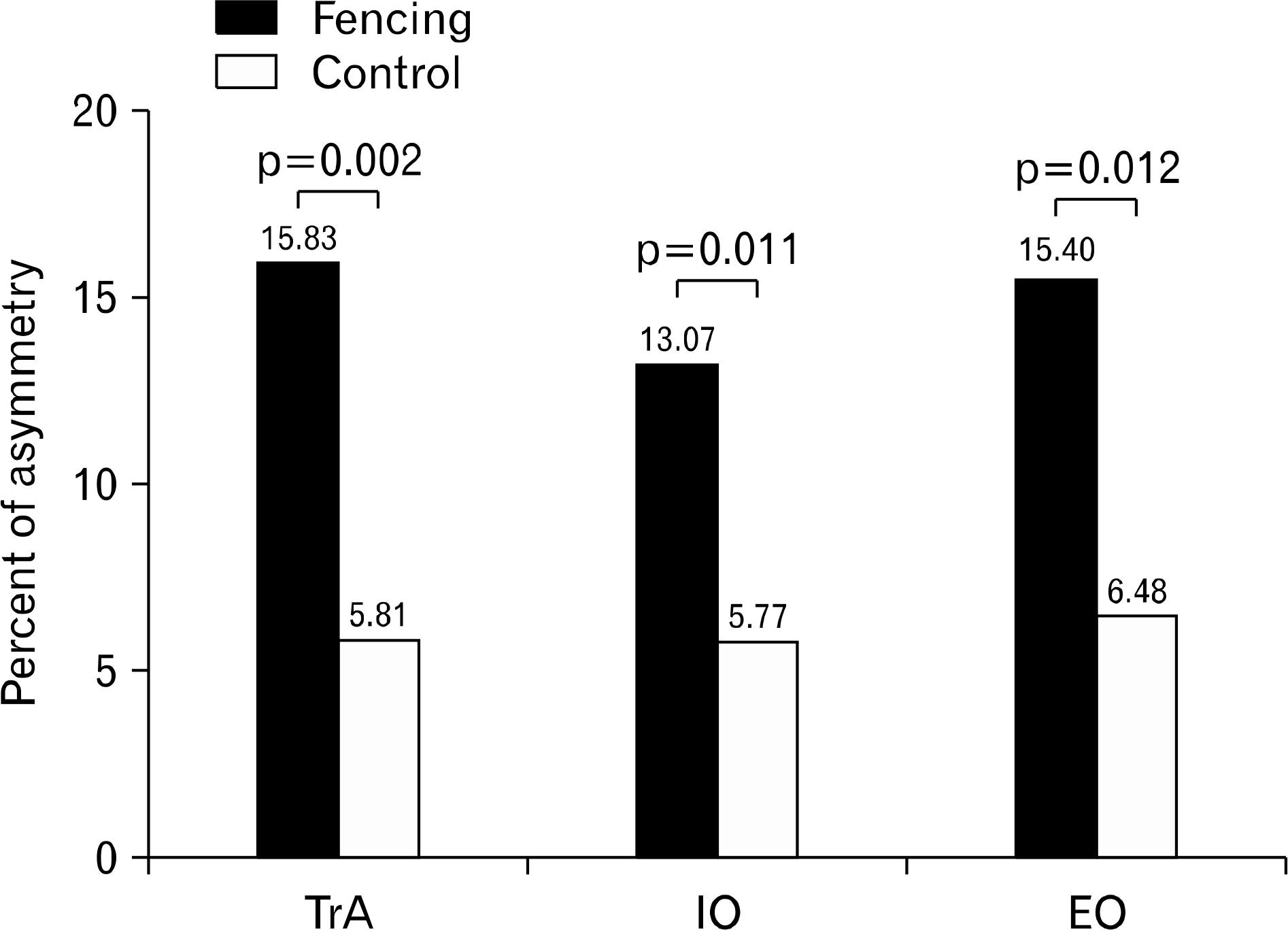
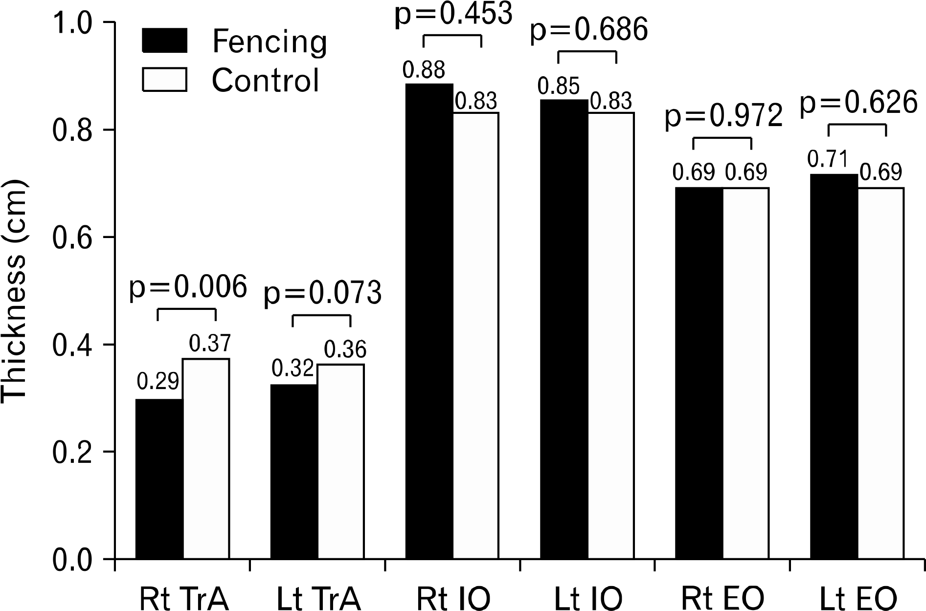
The purpose of this study is to compare the side-to-side thickness and asymmetry in the lateral abdominal (LAM) wall muscle group between fencing players and matched controls. Twenty fencing players (10 males and 10 females) and 20 matched controls participated in this study. The resting thicknesses of the transversus abdominis (TrA), internal oblique (IO), and external oblique (EO) of the LAM on both sides of the abdominal wall were measured in each group using 7.5 MHz linear array ultrasonography. Statistical analysis showed that the asymmetry of the fencers was 15% TrA, 13% IO, and 15% EO, whereas the control group showed 5% TrA, 5% IO, and 6% EO. The LAM was more asymmetric in the fencers than in the controls (p<0.05). The thickness of the right TrA was 0.37 cm in the controls, which was significantly greater than the 0.29 cm thickness in the fencers (p<0.05). The thicknesses of the left TrA and both IO and EO did not differ significantly between fencers and controls (p>0.05). The thickness of the TrA, IO, and EO of the side-to-side LAM wall was more asymmetric in the fencers than in the controls. This suggests that clinicians may find benefits in providing scientific baseline data on muscle asymmetry when treating and managing fencing athletes.
Go to : 
References
1. Richardson C, Jull G, Hodges P, Hides J. Therapeutic exercises for spinal segmental stabilization in low back pain: scientific basis and clinical approach. Edinburgh: Churchill Livingstone;1999.
2. Hodges PW, Gurfinkel VS, Brumagne S, Smith TC, Cordo PC. Coexistence of stability and mobility in postural control: evidence from postural compensation for respiration. Exp Brain Res. 2002; 144:293–302.

3. Geiger RA, Allen JB, O'Keefe J, Hicks RR. Balance and mobility following stroke: effects of physical therapy interventions with and without biofeedback/forceplate training. Phys Ther. 2001; 81:995–1005.

4. Dickstein R, Sheffi S, Ben Haim Z, Shabtai E, Markovici E. Activation of flexor and extensor trunk muscles in hemiparesis. Am J Phys Med Rehabil. 2000; 79:228–34.

5. Richardson CA, Snijders CJ, Hides JA, Damen L, Pas MS, Storm J. The relation between the transversus abdominis muscles, sacroiliac joint mechanics, and low back pain. Spine (Phila Pa 1976). 2002; 27:399–405.

6. Masse-Alarie H, Flamand VH, Moffet H, Schneider C. Corticomotor control of deep abdominal muscles in chronic low back pain and anticipatory postural adjustments. Exp Brain Res. 2012; 218:99–109.

7. Joseph LH, Hussain RI, Naicker AS, Htwe O, Pirunsan U, Paungmali A. Pattern of changes in local and global muscle thickness among individuals with sacroiliac joint dysfunction. Hong Kong Physiother J. 2015; 33:28–33.

8. Rankin G, Stokes M, Newham DJ. Abdominal muscle size and symmetry in normal subjects. Muscle Nerve. 2006; 34:320–6.

9. Tahan N, Khademi-Kalantari K, Mohseni-Bandpei MA, Mikaili S, Baghban AA, Jaberzadeh S. Measurement of superficial and deep abdominal muscle thickness: an ultrasonography study. J Physiol Anthropol. 2016; 35:17.

10. Sitilertpisan P, Pirunsan U, Puangmali A, et al. Comparison of lateral abdominal muscle thickness between weightlifters and matched controls. Phys Ther Sport. 2011; 12:171–4.

11. Adamczyk W, Bialy M, Stranc T. Asymmetry of lateral abdominal wall muscles in static and dynamic conditions: a preliminary study of professional basketball players. Sports Sci. 2016; 31:e15–18.

12. Balius R, Pedret C, Galilea P, Idoate F, Ruiz-Cotorro A. Ultrasound assessment of asymmetric hypertrophy of the rectus abdominis muscle and prevalence of associated injury in professional tennis players. Skeletal Radiol. 2012; 41:1575–81.

13. Sanchis-Moysi J, Idoate F, Dorado C, Alayon S, Calbet JA. Large asymmetric hypertrophy of rectus abdominis muscle in professional tennis players. PLoS One. 2010; 5:e15858.

14. Hides J, Stanton W, Freke M, Wilson S, McMahon S, Richardson C. MRI study of the size, symmetry and function of the trunk muscles among elite cricketers with and without low back pain. Br J Sports Med. 2008; 42:809–13.

15. Guilhem G, Giroux C, Couturier A, Chollet D, Rabita G. Mechanical and muscular coordination patterns during a high-level fencing assault. Med Sci Sports Exerc. 2014; 46:341–50.

16. Frere J, Gopfert B, Nuesch C, et al. Kinematical and EMG-classifications of a fencing attack. Int J Sports Med. 2011; 32:28–34.

17. Roi GS, Bianchedi D. The science of fencing: implications for performance and injury prevention. Sports Med. 2008; 38:465–81.
18. Turner A, James N, Dimitriou L, et al. Determinants of Olympic fencing performance and implications for strength and conditioning training. J Strength Cond Res. 2014; 28:3001–11.

19. Stetts DM, Freund JE, Allison SC, Carpenter G. A rehabilitative ultrasound imaging investigation of lateral abdominal muscle thickness in healthy aging adults. J Geriatr Phys Ther. 2009; 32:60–6.

20. Mannion AF, Pulkovski N, Gubler D, et al. Muscle thickness changes during abdominal hollowing: an assessment of between-day measurement error in controls and patients with chronic low back pain. Eur Spine J. 2008; 17:494–501.

21. Davarian S, Maroufi N, Ebrahimi E, Parnianpour M, Fara-hmand F. Normal postural responses preceding shoulder flexion: co-activation or asymmetric activation of transverse abdominis? J Back Musculoskelet Rehabil. 2014; 27:545–51.

22. Morris SL, Lay B, Allison GT. Transversus abdominis is part of a global not local muscle synergy during arm movement. Hum Mov Sci. 2013; 32:1176–85.

24. Chow JW, Park SA, Tillman MD. Lower trunk kinematics.
25. Tsolaks C, Bogdanis GC, Vagenas G. Anthropometric profile and limb asymmetries in young male and female fencers. J Hum Mov Stud. 2006; 50:201–15.
26. Margonato V, Roi GS, Cerizza C, Galdabino GL. Maximal isometric force and muscle cross-sectional area of the forearm in fencers. J Sports Sci. 1994; 12:567–72.

27. Roi GS, Mognoni P. Lo spadista modello. Rivista di cultura sportiva. 1987; 6:50–7.
28. Nystrom J, Lindwall O, Ceci R, Harmenberg J, Svedenhag J, Ekblom B. Physiological and morphological characteristics of world class fencers. Int J Sports Med. 1990; 11:136–9.

Go to : 
 | Fig. 1.Ultrasound imaging of the lateral abdominal wall muscles taken during the resting state. (A) Fencing group, (B) control group. EO: external oblique, IO: internal oblique, TrA: transversus abdominis. |
 | Fig. 2.Differences between the fencing and control groups in the percent of asymmetry in muscle thickness of the lateral abdominal wall. TrA: transversus abdominis, IO: internal oblique, EO: external oblique. |
 | Fig. 3.Lateral abdominal wall thickness in fencing and control group. Rt: right, TrA: transversus abdominis, Lt: left, IO: internal oblique, EO: external oblique. |
Table 1.
Demographic characteristics of subjects
Table 2.
Comparison of the asymmetry of the lateral abdominal muscle between the fencing and matched control groups
| Asymmetric | Fencing group (n=20) | Control group (n=20) | t-value | p-value |
|---|---|---|---|---|
| TrA (%) | 15.80±12.15 | 5.81±2.36 | 3.61 | 0.002∗ |
| IO (%) | 13.07±11.24 | 5.77±3.90 | 2.74 | 0.011∗ |
| EO (%) | 15.40±14.21 | 6.48±3.27 | 2.73 | 0.012∗ |
Table 3.
Comparison of the lateral abdominal muscle thickness between the fencing and matched control groups
| Thickness | Fencing group (n=20) | Control group (n=20) | p-value |
|---|---|---|---|
| Right TrA (cm) | 0.29±0.85 | 0.37±0.84 | 0.006∗ |
| Left TrA (cm) | 0.32±0.07 | 0.36±0.08 | 0.073 |
| Right IO (cm) | 0.88±0.20 | 0.83±0.19 | 0.453 |
| Left IO (cm) | 0.85±0.17 | 0.83±0.18 | 0.686 |
| Right EO (cm) | 0.69±0.12 | 0.69±0.22 | 0.972 |
| Left EO (cm) | 0.71±0.14 | 0.69±0.22 | 0.626 |


 PDF
PDF ePub
ePub Citation
 Print
Print


 XML Download