Abstract
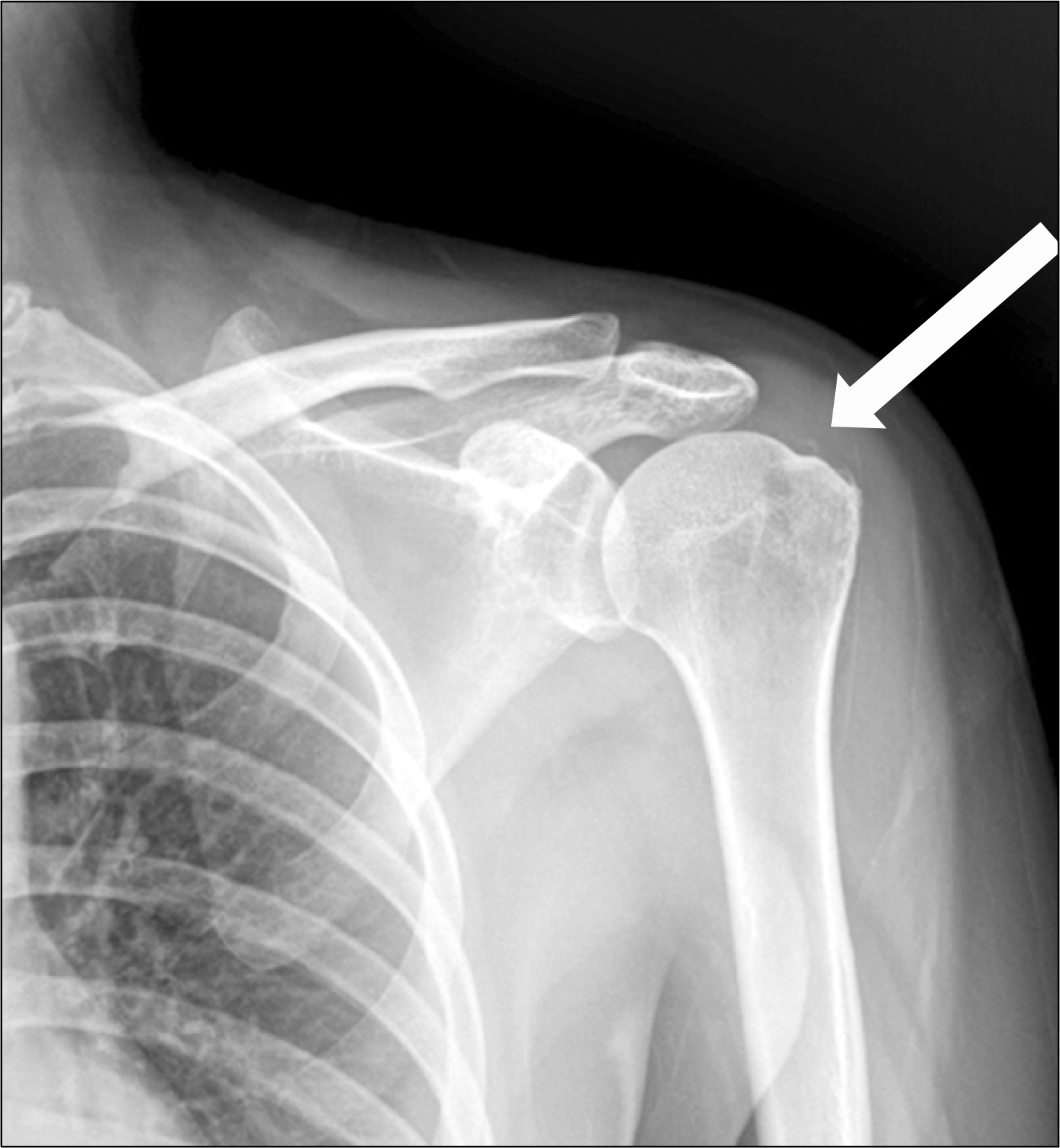
Isolated rupture of infraspinatus after barbotage for calcific tendinitis has not been reported in the literature. We report on a case of isolated infraspinatus rupture and suprascapular nerve neuropathy after steroid injection and barbotage of calcific tendinitis in rotator cuff. At 6-month follow-up after surgery, satisfactory clinical and radiological outcomes were observed with daily activity level. The author reports this case and review the literature.
References
1. de Witte PB, Selten JW, Navas A, et al. Calcific tendinitis of the rotator cuff: a randomized controlled trial of ultrasound-guided needling and lavage versus subacromial corticosteroids. Am J Sports Med. 2013; 41:1665–73.
2. Suzuki K, Potts A, Anakwenze O, Singh A. Calcific tendinitis of the rotator cuff: management options. J Am Acad Orthop Surg. 2014; 22:707–17.
3. Bhatia M, Singh B, Nicolaou N, Ravikumar KJ. Correlation between rotator cuff tears and repeated subacromial steroid injections: a case-controlled study. Ann R Coll Surg Engl. 2009; 91:414–6.

4. Tillander B, Franzen LE, Karlsson MH, Norlin R. Effect of steroid injections on the rotator cuff: an experimental study in rats. J Shoulder Elbow Surg. 1999; 8:271–4.

5. Gatt DL, Charalambous CP. Ultrasound-guided barbotage for calcific tendonitis of the shoulder: a systematic review including 908 patients. Arthroscopy. 2014; 30:1166–72.

6. Boykin RE, Friedman DJ, Higgins LD, Warner JJ. Suprascapular neuropathy. J Bone Joint Surg Am. 2010; 92:2348–64.

7. Sergides NN, Nikolopoulos DD, Boukoros E, Papagianno-poulos G. Arthroscopic decompression of an entrapped suprascapular nerve due to an ossified superior transverse scapular ligament: a case report. Cases J. 2009; 2:8200.

8. Kim SH, Koh YG, Sung CH, Moon HK, Park YS. Iatrogenic suprascapular nerve injury after repair of type II SLAP lesion. Arthroscopy. 2010; 26:1005–8.

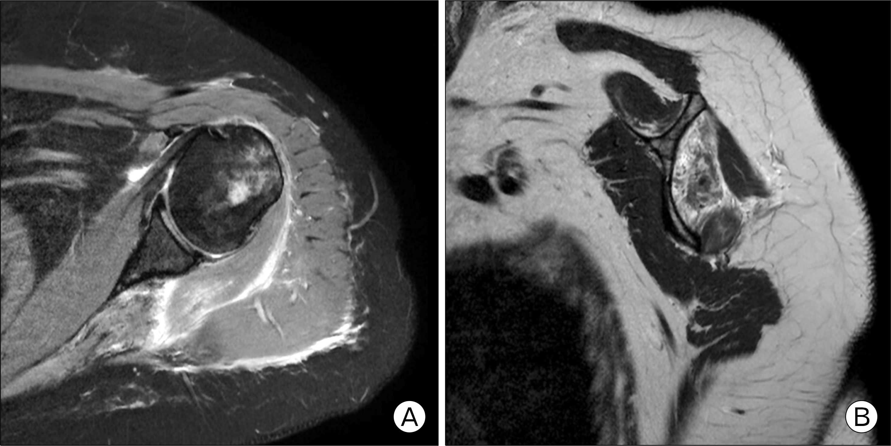
Fig. 2.
(A) Magnetic resonance imaging showed that the distal region of the infraspinatus tendon had a full-thickness tear, while the proximal region was retracted medially past the glenoid cavity with fatty atrophy. (B) An incomplete full-thickness tear of the teres minor muscle was also present.
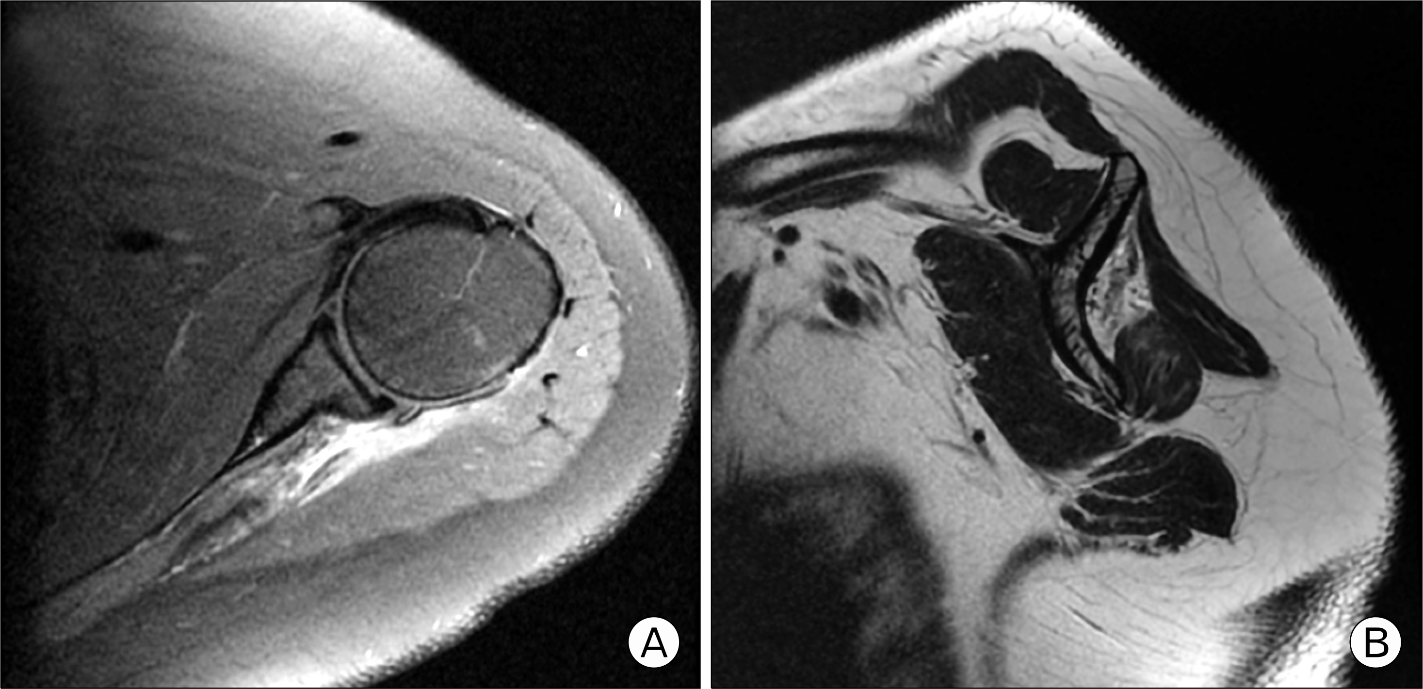
Fig. 3.
(A) An infraspinatus tear accompanied by calcification was observed. (B) Arthroscopic infraspinatus tendon repair was performed using knotless anchor suture.
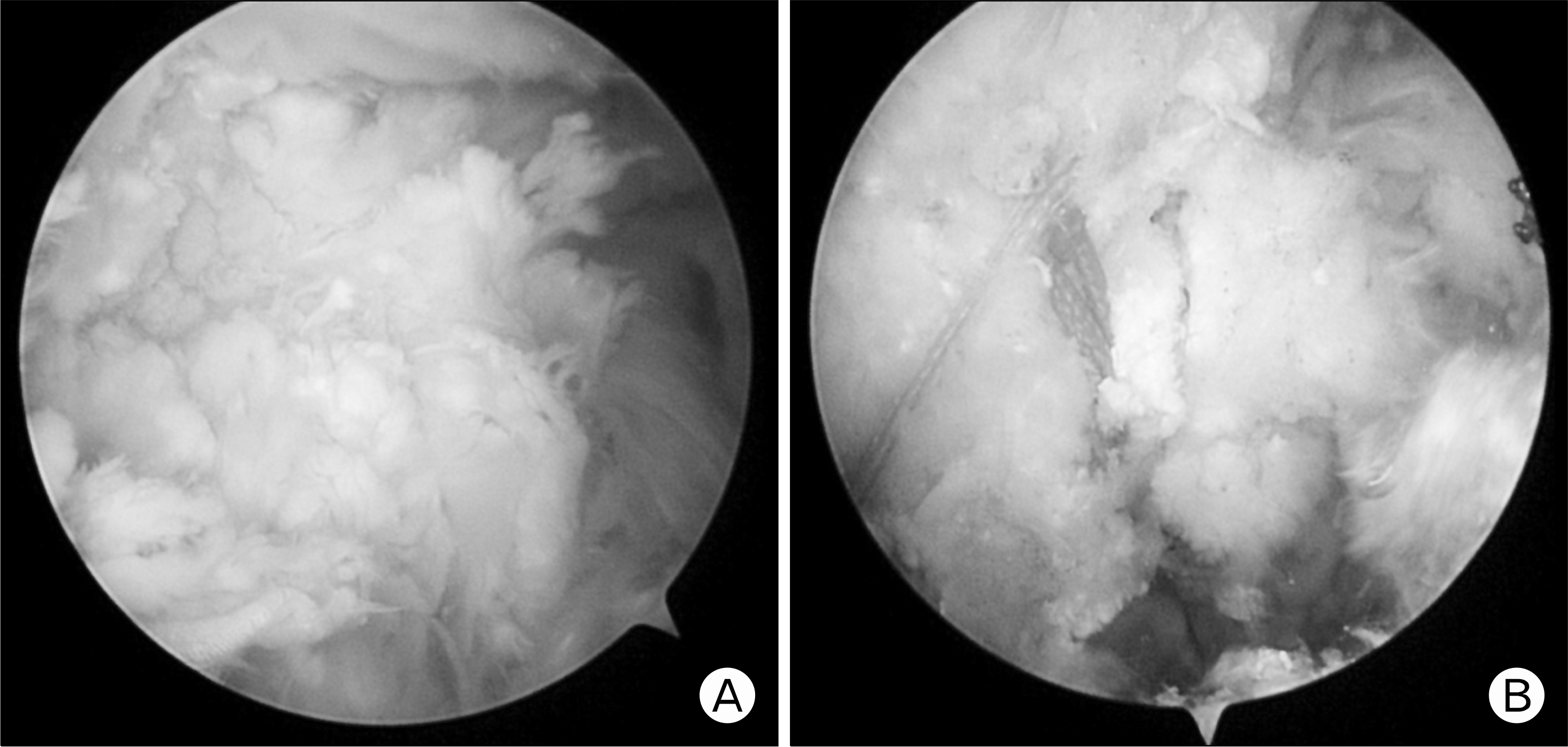
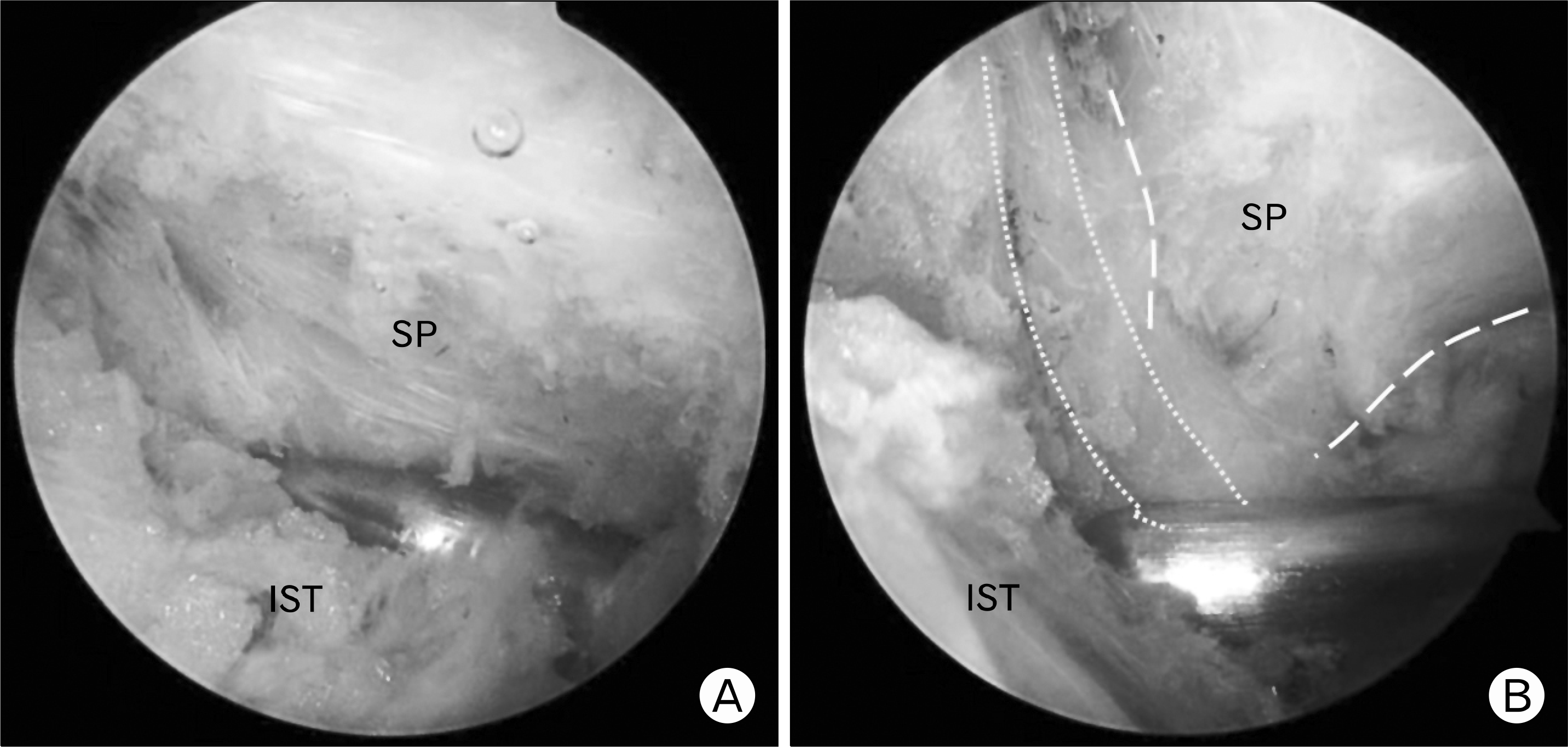
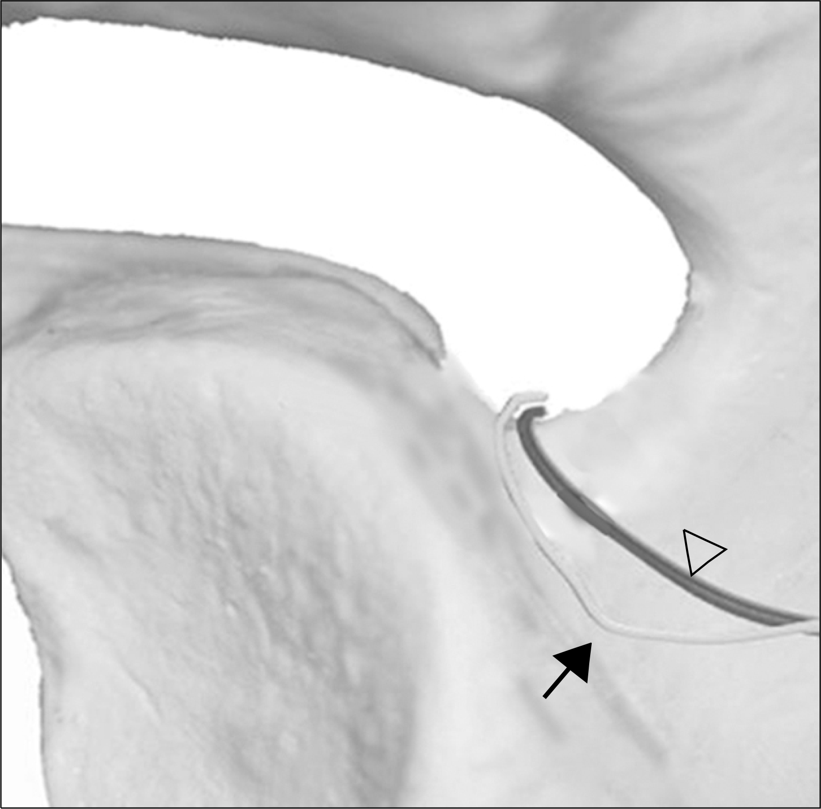
Fig. 4.
(A) Arthroscopic suprascapular nerve release was performed using metal trocar. (B) The dotted line shows suprascapular nerve compressed by scapular spine. SP: scapular spine, IST: infraspinatus.


 PDF
PDF ePub
ePub Citation
 Print
Print


 XML Download