

 PDF
PDF ePub
ePub Citation
 Print
Print


Abstract
The purpose of this study was to evaluate the humeral tunnel characters and clinical relevance according to entry point of the humeral tunnel in the baseball players. It was hypothesized that the medial collateral ligament (MCL) reconstruction with nonanatomical starting location of the humeral tunnel (inferior edge of the medial epicondyle: group NA) provided less favorable radiological and clinical outcomes compared to that with anatomical starting location (original footprint of the MCL: group A). The retrospective case review yielded 19 consecutive athletes who underwent isolated MCL reconstruction using the docking technique. Three dimensional-computed tomography scan was performed at 3 months, and the iso-surfacing by marching cubes algorithm were applied to evaluate the length and angle of humeral tunnel. Three outcome measures were used in this study: the visual analog scale for pain, range of motion and the Conway scale. The angle of the humeral tunnel was measured 12.2o (range, 7.9o−25.2o) in the group NA and 15.5o (range, 9.8o−30.4o) in the group A (p<0.05). The mean length of humeral tunnel is measured 16.3 mm (range, 11.7−20.1 mm) in the group NA and 15.2 mm (range, 10.3−19.1 mm) in the group A (p<0.05). MCL reconstruction brought substantial improvement in pain and function. However, between-group comparison revealed no statistical differences in all outcome measurements. The MCL reconstruction using the docking technique provided favorable clinical outcomes in baseball players. Although the humeral tunnel angle and length were different depending on the humeral entry points, clinical differences between the two entry points were not found.
References
1. Jones KJ, Osbahr DC, Schrumpf MA, Dines JS, Altchek DW. Ulnar collateral ligament reconstruction in throwing athletes: a review of current concepts. AAOS exhibit selection. J Bone Joint Surg Am. 2012; 94:e49.
2. Argo D, Trenhaile SW, Savoie FH 3rd, Field LD. Operative treatment of ulnar collateral ligament insufficiency of the elbow in female athletes. Am J Sports Med. 2006; 34:431–7.

3. Bruce JR, Andrews JR. Ulnar collateral ligament injuries in the throwing athlete. J Am Acad Orthop Surg. 2014; 22:315–25.
4. Freehill MT, Safran MR. Diagnosis and management of ulnar collateral ligament injuries in throwers. Curr Sports Med Rep. 2011; 10:271–8.
5. Ford GM, Genuario J, Kinkartz J, Githens T, Noonan T. Return-to-play outcomes in professional baseball players after medial ulnar collateral ligament injuries: comparison of operative versus nonoperative treatment based on magnetic resonance imaging findings. Am J Sports Med. 2016; 44:723–8.
6. Jobe FW, Stark H, Lombardo SJ. Reconstruction of the ulnar collateral ligament in athletes. J Bone Joint Surg Am. 1986; 68:1158–63.
7. Rohrbough JT, Altchek DW, Hyman J, Williams RJ 3rd, Botts JD. Medial collateral ligament reconstruction of the elbow using the docking technique. Am J Sports Med. 2002; 30:541–8.
8. Dodson CC, Thomas A, Dines JS, Nho SJ, Williams RJ 3rd, Altchek DW. Medial ulnar collateral ligament reconstruction of the elbow in throwing athletes. Am J Sports Med. 2006; 34:1926–32.
9. Paletta GA Jr, Wright RW. The modified docking procedure for elbow ulnar collateral ligament reconstruction: 2-year follow-up in elite throwers. Am J Sports Med. 2006; 34:1594–8.
10. Koh JL, Schafer MF, Keuter G, Hsu JE. Ulnar collateral ligament reconstruction in elite throwing athletes. Arthroscopy. 2006; 22:1187–91.
11. Bowers AL, Dines JS, Dines DM, Altchek DW. Elbow medial ulnar collateral ligament reconstruction: clinical relevance and the docking technique. J Shoulder Elbow Surg. 2010; 19(2 Suppl):110–7.
12. Byram IR, Khanna K, Gardner TR, Ahmad CS. Characterizing bone tunnel placement in medial ulnar collateral ligament reconstruction using patient-specific 3-dimensional computed tomography modeling. Am J Sports Med. 2013; 41:894–902.
13. Newman TS, Yi H. A survey of the marching cubes algorithm. Comput Graph. 2006; 30:854–79.
14. Keller RA, Steffes MJ, Zhuo D, Bey MJ, Moutzouros V. The effects of medial ulnar collateral ligament reconstruction on Major League pitching performance. J Shoulder Elbow Surg. 2014; 23:1591–8.
15. Merolla G, Del Sordo S, Paladini P, Porcellini G. Elbow ulnar collateral ligament reconstruction: clinical, radiographic, and ultrasound outcomes at a mean 3-year follow-up. Musculoskelet Surg. 2014; 98(Suppl 1):87–93.
16. Savoie FH 3rd, Morgan C, Yaste J, Hurt J, Field L. Medial ulnar collateral ligament reconstruction using hamstring allograft in overhead throwing athletes. J Bone Joint Surg Am. 2013; 95:1062–6.
17. Hechtman KS, Zvijac JE, Wells ME, Botto-van Bemden A. Long-term results of ulnar collateral ligament reconstruction in throwing athletes based on a hybrid technique. Am J Sports Med. 2011; 39:342–7.
Fig. 1.
Images of three dimensional reconstructed computed tomography showing the starting point of the humeral tunnel: (A) inferior point, (B) anteroinferior point.
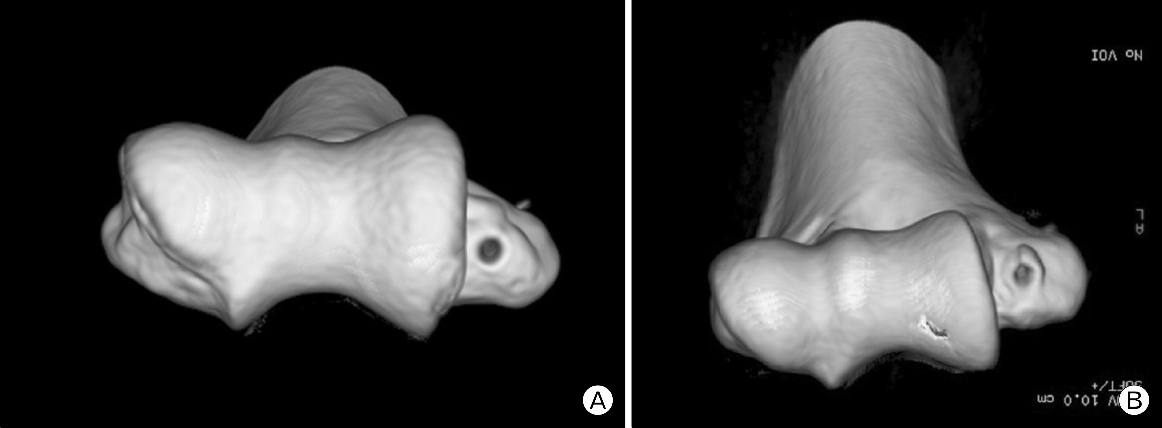
Fig. 2.
A photograph of screenshot showing measuring process of the length and angle of the humeral tunnel using the iso-surfacing by marching cubes algorithm (A) and formation of angle with humeral axis (B).
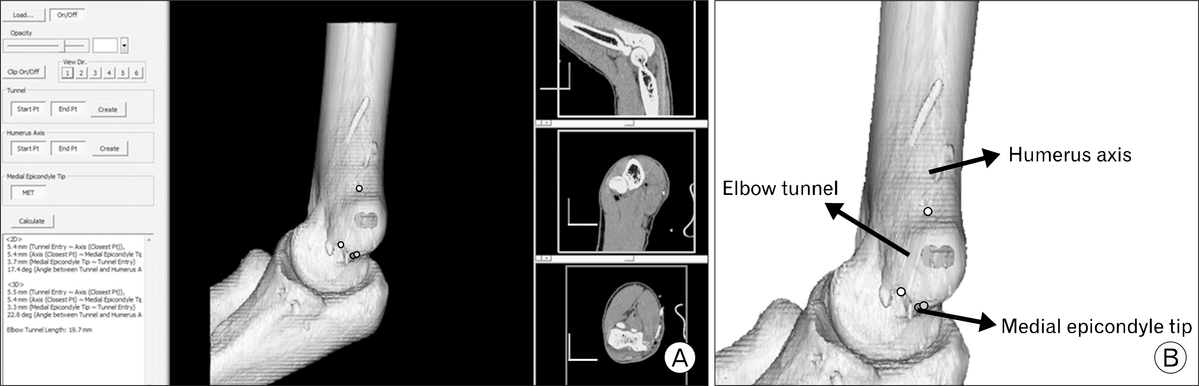
Table 1.
Demographics
| Variable | Group NA∗ (n=9) | Group A† (n=10) |
|---|---|---|
| Age (yr) | 21.9 (17−28) | 20.8 (18−29) |
| Side (right:left) | 7:2 | 9:1 |
| Follow-up (mo) | 25.1 (24−36) | 24.6 (24−32) |
| Grade‡ | 5, 2, 2 | 3, 4, 3 |
| Position§ | 7, 1, 1 | 8, 2, 0 |
| Arthroscopic spur | 5 | 4 |
| resection |
Table 2.
Clinical outcomes
| Variable | Group NA∗ (n=9) | Group A† (n=10) | p-value |
|---|---|---|---|
| Preoperative VAS for pain, mean (range) | 6.0 (3−8) | 6.3 (3−8) | NS |
| Postoperative VAS for pain, mean (range) | 0.5 (0−4) | 0.4 (0−3) | NS |
| Preoperative range of motion‡ | 3.3/132.1/77.8/64.4 | 1.0/133.0/80.0/72.5 | NS |
| Postoperative range of motion | 0/137.2/80.0/73.3 | 0.5/135.5/79.0/76.0 | NS |
| Conway scale§ | 7, 1, 1, 0 | 9, 0, 0, 1 | NS |
 XML Download