Abstract
The purpose of this study is to evaluate the ultrasonographic findings of little leaguer's shoulder among adolescent baseball players. Forty-two little leaguer's shoulder patients (age, 11–16 years; mean, 13.8 years; right, 39; left, 3), based on plain X-ray, were examined by bilateral shoulder ultrasonography. All patients were divided into groups on the basis of sonographic abnormalities and bilateral differences of physeal gap were measured in the cases of significant physeal widening. Sonographic abnormalities of dominant shoulder were physeal irregularity (45%), physeal fragmentation (21%), periosteal thickening (36%) and physeal widening (83%) that was the most common abnormalities. Seven of 42 patients (group A) had only physeal irregularity with minimal physeal widening, 26 patients (group B) had more than 1-mm physeal widening compared with nondominant shoulder. Nine patients (group C) had both physeal widening and fragmentation. Mean physeal gaps of the dominant and nondominant shoulders in 35 patients (group B and C) were 3.4±0.8 mm and 1.4±0.1 mm, respectively (p=0.013) and increased average physeal gap of dominant shoulder was 2.0±0.8 mm. Among three groups of patients, the duration of symptom was significant longer in group C (p=0.011). Physeal widening and fragmentation were associated with progression of the disease, but physeal irregularity was relatively early sonographic finding. Ultrasonography is a useful tool to evaluate the status of proximal humeral epiphysis and can aid early diagnosis of little leaguer's shoulder in the field.
Go to : 
References
1. Adams JE. Little league shoulder: osteochondrosis of the proximal humeral epiphysis in boy baseball pitchers. Calif Med. 1966; 105:22–5.
3. Barnett LS. Little League shoulder syndrome: proximal humeral epiphyseolysis in adolescent baseball pitchers. A case report. J Bone Joint Surg Am. 1985; 67:495–6.
4. Ocguder DA, Tosun O, Bektaser B, Cicek N, Ipek A, Bozkurt M. Ultrasonographic evaluation of the shoulder in asymptomatic overhead athletes. Acta Orthop Belg. 2010; 76:456–61.
5. Wang HK, Lin JJ, Pan SL, Wang TG. Sonographic evaluations in elite college baseball athletes. Scand J Med Sci Sports. 2005; 15:29–35.

6. Brasseur JL, Lucidarme O, Tardieu M, et al. Ultrasonographic rotator-cuff changes in veteran tennis players: the effect of hand dominance and comparison with clinical findings. Eur Radiol. 2004; 14:857–64.

7. Gainor BJ, Piotrowski G, Puhl J, Allen WC, Hagen R. The throw: biomechanics and acute injury. Am J Sports Med. 1980; 8:114–8.

8. Olsen SJ 2nd, Fleisig GS, Dun S, Loftice J, Andrews JR. Risk factors for shoulder and elbow injuries in adolescent baseball pitchers. Am J Sports Med. 2006; 34:905–12.

9. Lyman S, Fleisig GS, Andrews JR, Osinski ED. Effect of pitch type, pitch count, and pitching mechanics on risk of elbow and shoulder pain in youth baseball pitchers. Am J Sports Med. 2002; 30:463–8.

10. Pappas AM, Zawacki RM, McCarthy CF. Rehabilitation of the pitching shoulder. Am J Sports Med. 1985; 13:223–35.

11. Burkhart SS, Morgan CD, Kibler WB. The disabled throwing shoulder: spectrum of pathology. Part I. pathoanatomy and biomechanics. Arthroscopy. 2003; 19:404–20.

12. Dameron TB Jr, Reibel DB. Fractures involving the proximal humeral epiphyseal plate. J Bone Joint Surg Am. 1969; 51:289–97.

13. Mariscalco MW, Saluan P. Upper extremity injuries in the adolescent athlete. Sports Med Arthrosc. 2011; 19:17–26.

14. Obembe OO, Gaskin CM, Taffoni MJ, Anderson MW. Little Leaguer's shoulder (proximal humeral epiphysiolysis): MRI findings in four boys. Pediatr Radiol. 2007; 37:885–9.

15. Song JC, Lazarus ML, Song AP. MRI findings in Little Leaguer's shoulder. Skeletal Radiol. 2006; 35:107–9.

16. Lyman S, Fleisig GS, Waterbor JW, et al. Longitudinal study of elbow and shoulder pain in youth baseball pitchers. Med Sci Sports Exerc. 2001; 33:1803–10.

17. Zaremski JL, Krabak BJ. Shoulder injuries in the skeletally immature baseball pitcher and recommendations for the prevention of injury. PM R. 2012; 4:509–16.

Go to : 
 | Fig. 1.Scanning technique and corresponding sonographic images of both shoulder in a little leaguer shoulder player. (A) Three-dimensional computed tomography of shoulder shows anatomical relationship between anterolateral corner of acromion (arrow) and proximal humeral epiphysis (dotted box indicates the location of the transducer). (B) Transducer is placed on lateral aspect of shoulder behind vertical imaginary line from acromion to upper arm. (C) Longitudinal images of both shoulders show hyperechoic irregular growth plate with bony fragmentation and widening of proximal humeral growth plate (asterisks indicate periosteum). D: deltoid muscle, E: proximal humeral epiphysis, M: proximal humeral metaphysis. |
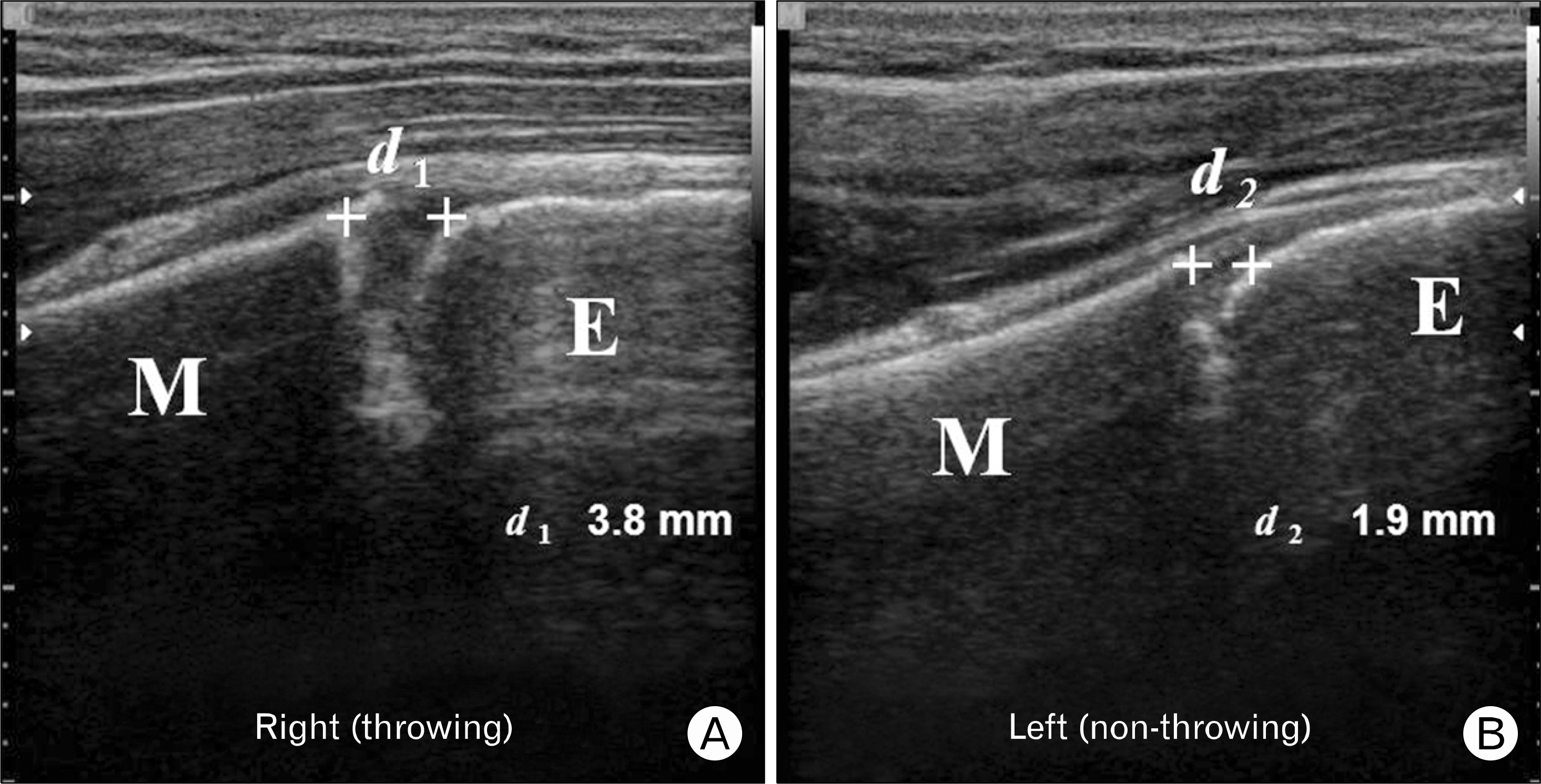
 | Fig. 2.Bilateral measurement of physeal gap in right little leaguer shoulder player. Increased physeal gap (d1–d2=1.9 mm) is noted as right throwing side and non-throwing side, 3.8 mm and 1.9 mm, respectively. M: proximal humeral metaphysis, E: proximal humeral epiphysis. |
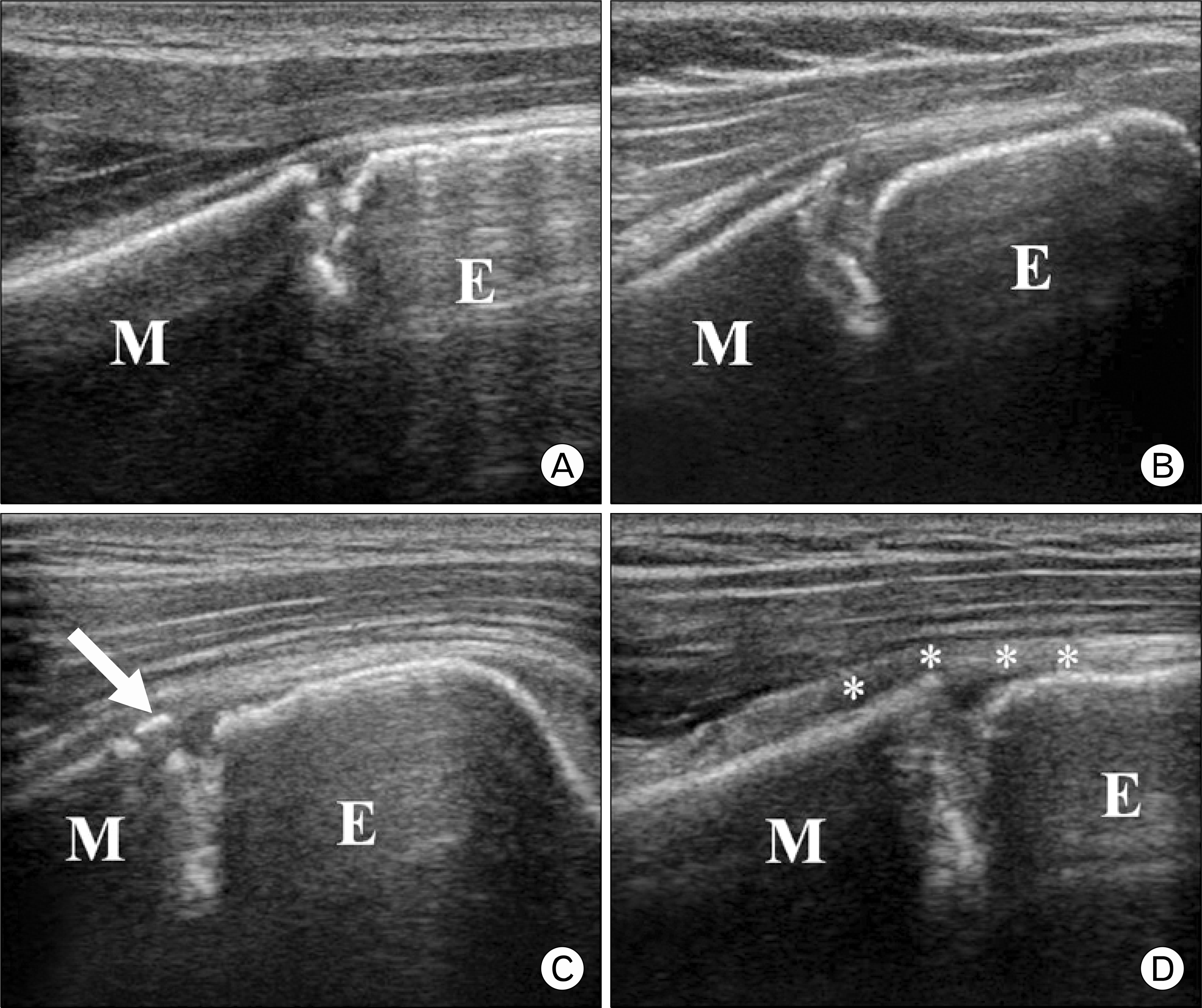
 | Fig. 3.Various sonographic abnormalities of proximal humeral physes in throwing side of little leaguer shoulder players. (A) Irregular physis and minimal increased physeal gap, (B) widening of physeal gap more than 1 mm and physeal irregularity, (C) fragmentation of physis (arrow) and (D) periosteal thickening (asterisks indicate periosteum). M: proximal humeral metaphysis, E: proximal humeral epiphysis. |
 | Fig. 4.Classification of group of little leaguer shoulder players according to sonographic images. (A) Only physeal irregularity with minimal increased gap (<1 mm). (B) Physeal widening with or without physeal irregularity and (C) physeal widening and fragmentation of physis. M: proximal humeral metaphysis, E: proximal humeral epiphysis. |
Table 1.
Demographic characteristics of the patients (n=42)
| Characteristics | Value |
|---|---|
| Age (yr) | 13.8±2.5 |
| Height (cm) | 161.8±10.9 |
| Weight (kg) | 57.2±11.3 |
| Body mass index (kg/m2) | 21.7±2.8 |
| Training history (yr) | 3.4±2.9 |
| Symptom duration (mo) | 2.3±2.2 |
| Dominant (right:left) | 39:3 |
| Position | |
| Pitcher | |
| Pitcher only | 8 (19) |
| Pitcher or fielder | 12 (29) |
| Fielder | |
| Catcher | 6 (14) |
| In-fielder | 10 (24) |
| Out-fielder | 6 (14) |
| Pitch count/wk∗ | 550±80.5 |
Table 2.
Abnormal sonographic findings (n=42)
| Variable | No. (%) |
|---|---|
| Physeal irregularity | 19 (45) |
| Physeal widening (>1 mm) | 35 (83) |
| Physeal fragmentation | 9 (21) |
| Periosteal thickening | 15 (36) |
Table 3.
Sonographic classification of little league shoulder players (n=42)
Table 4.
Bilateral proximal humeral physeal gap in players with group B and C
| Variable | Group B & C (n=35) | Group B (n=26) | Group C (n=9) |
|---|---|---|---|
| Injured shoulder (mm) | 3.4±0.8 | 3.1±0.3 | 4.5±0.6 |
| Uninjured shoulder (mm) | 1.4±0.1 | 1.4±0.2 | 1.4±0.9 |
| Difference (mm) | 2.0±0.8 | 1.7±0.6 | 3.0±0.4 |
| p-value∗ | 0.013 | 0.002 | 0.016 |
Table 5.
Demographic comparison among groups
| Variable | Group A (n=7) | Group B (n=26) | Group C (n=9) | p-value |
|---|---|---|---|---|
| Height (cm) | 160.7±8.9 | 161.7±9.7 | 161.8±7.5 | 0.224 |
| Weight (kg) | 57.6±9.3 | 57.2±10.2 | 58.6±11.7 | 0.364 |
| Body mass index (kg/m2) | 22.3±1.5 | 21.8±1.7 | 22.3±1.8 | 0.473 |
| Training history (yr) | 3.2±1.6 | 3.4±1.5 | 3.2±1.7 | 0.238 |
| Symptom duration (mo) | 1.1±2.3 | 2.2±3.3 | 4.2±2.1 | 0.011∗ |
| Position (pitcher†:fielder‡) | 4:3 (57) | 12:14 (46) | 4:5 (44) | 0.066 |
| Pitch count/wk§ | 560±70.8 | 550±57.5 | 570±76.4 | 0.243 |


 PDF
PDF ePub
ePub Citation
 Print
Print


 XML Download