Abstract
Rupture of the pectoralis major muscle may occur in youngers or athletes associated with extreme sports, especially during the weight training. It is uncommon, but the incidence is increased by the recent growth of athletic population. In young active individuals, ruptures of the pectoralis major muscle have the best results after surgical repair. However, if diagnosis of the pectoralis major muscle rupture is missed or delayed, the patient will be limited to return to sport activity. The object of this paper is to report our experience of pectoralis major muscle rupture in 3 cases.
REFERENCES
1.ElMaraghy AW., Devereaux MW. A systematic review and comprehensive classification of pectoralis major tears. J Shoulder Elbow Surg. 2012. 21:412–22.

2.Haley CA., Zacchilli MA. Pectoralis major injuries: evaluation and treatment. Clin Sports Med. 2014. 33:739–56.
3.Lee BI., Kwon SW., Lee HU, et al. Rupture of the pectoralis major muscle during bench-pressing: a case report. J Korean Orthop Soc Sports Med. 2007. 6:115–8.
4.Fung L., Wong B., Ravichandiran K., Agur A., Rindlisbacher T., Elmaraghy A. Three-dimensional study of pectoralis major muscle and tendon architecture. Clin Anat. 2009. 22:500–8.

5.Beloosesky Y., Grinblat J., Katz M., Hendel D., Sommer R. Pectoralis major rupture in the elderly: clinical and sonographic findings. Clin Imaging. 2003. 27:261–4.
6.Schepsis AA., Grafe MW., Jones HP., Lemos MJ. Rupture of the pectoralis major muscle. Outcome after repair of acute and chronic injuries. Am J Sports Med. 2000. 28:9–15.
7.Bak K., Cameron EA., Henderson IJ. Rupture of the pectoralis major: a meta-analysis of 112 cases. Knee Surg Sports Trauma-tol Arthrosc. 2000. 8:113–9.

8.de Castro Pochini A., Andreoli CV., Belangero PS, et al. Clinical considerations for the surgical treatment of pectoralis major muscle ruptures based on 60 cases: a prospective study and literature review. Am J Sports Med. 2014. 42:95–102.
9.Sherman SL., Lin EC., Verma NN, et al. Biomechanical analysis of the pectoralis major tendon and comparison of techniques for tendo-osseous repair. Am J Sports Med. 2012. 40:1887–94.

10.Zafra M., Munoz F., Carpintero P. Chronic rupture of the pectoralis major muscle: report of two cases. Acta Orthop Belg. 2005. 71:107–10.
Fig. 1.
(A) Clinical photograph of the patient before the operation. Note the asymmetric absent anterior axillary fold and swelling and ecchymosis on the affected (right) side. (B) Axial T2-weighted fat suppression magnetic resonance image at the level of the proximal humerus. The pectoralis major tendon is detached medially from the normal insertion site and attenuated (arrow). Adjacent area of white arrow reflect hematoma.
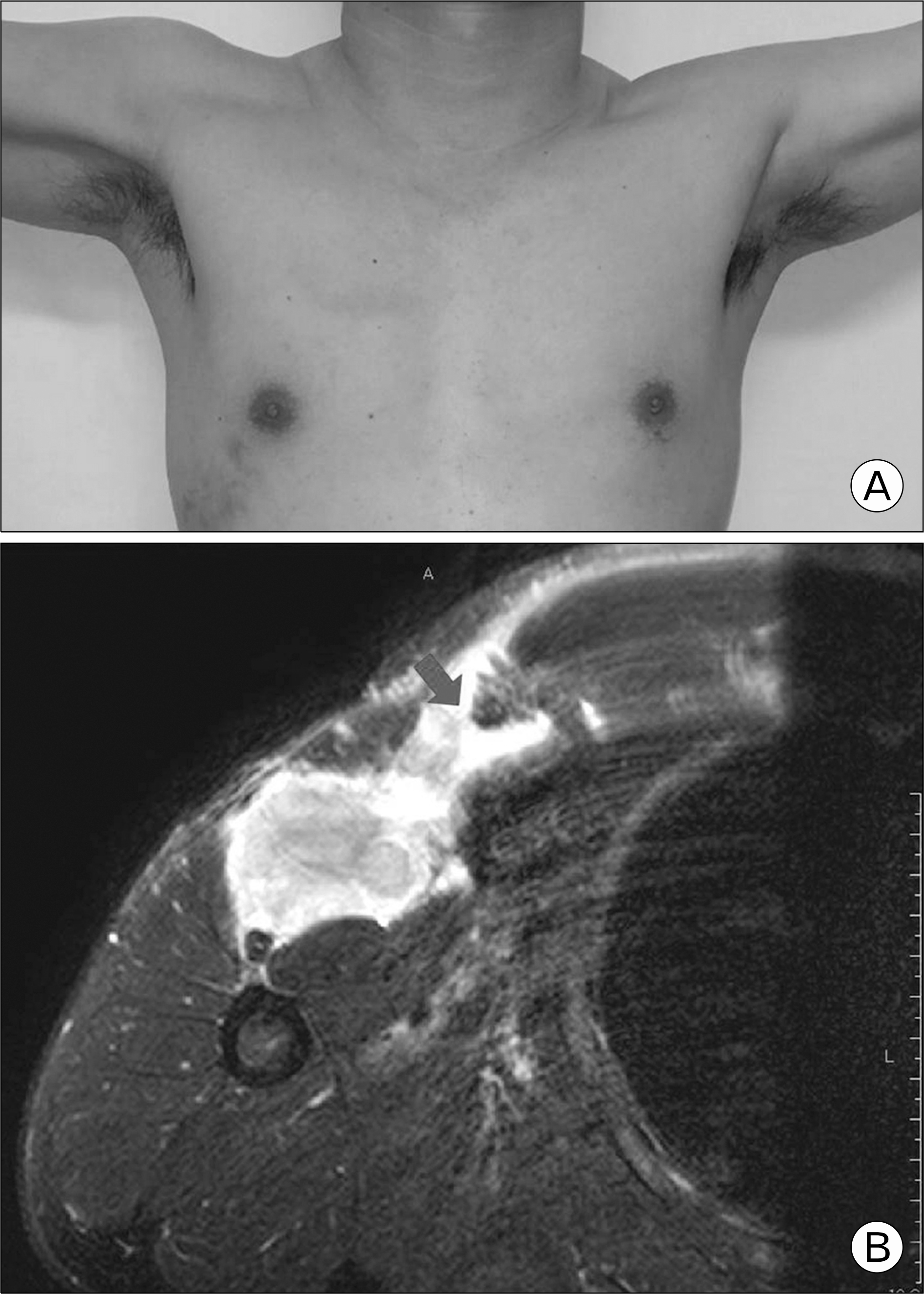
Fig. 2.
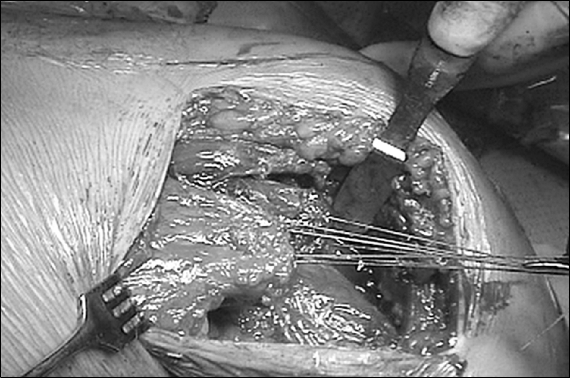
Operative photograph. The pectoralis major tendon is completely ruptured from lateral lip of bicipital groove. The stumps of pectoralis major tendons are delaminated. Posterior laminar is retracted medially more than anterior laminar.
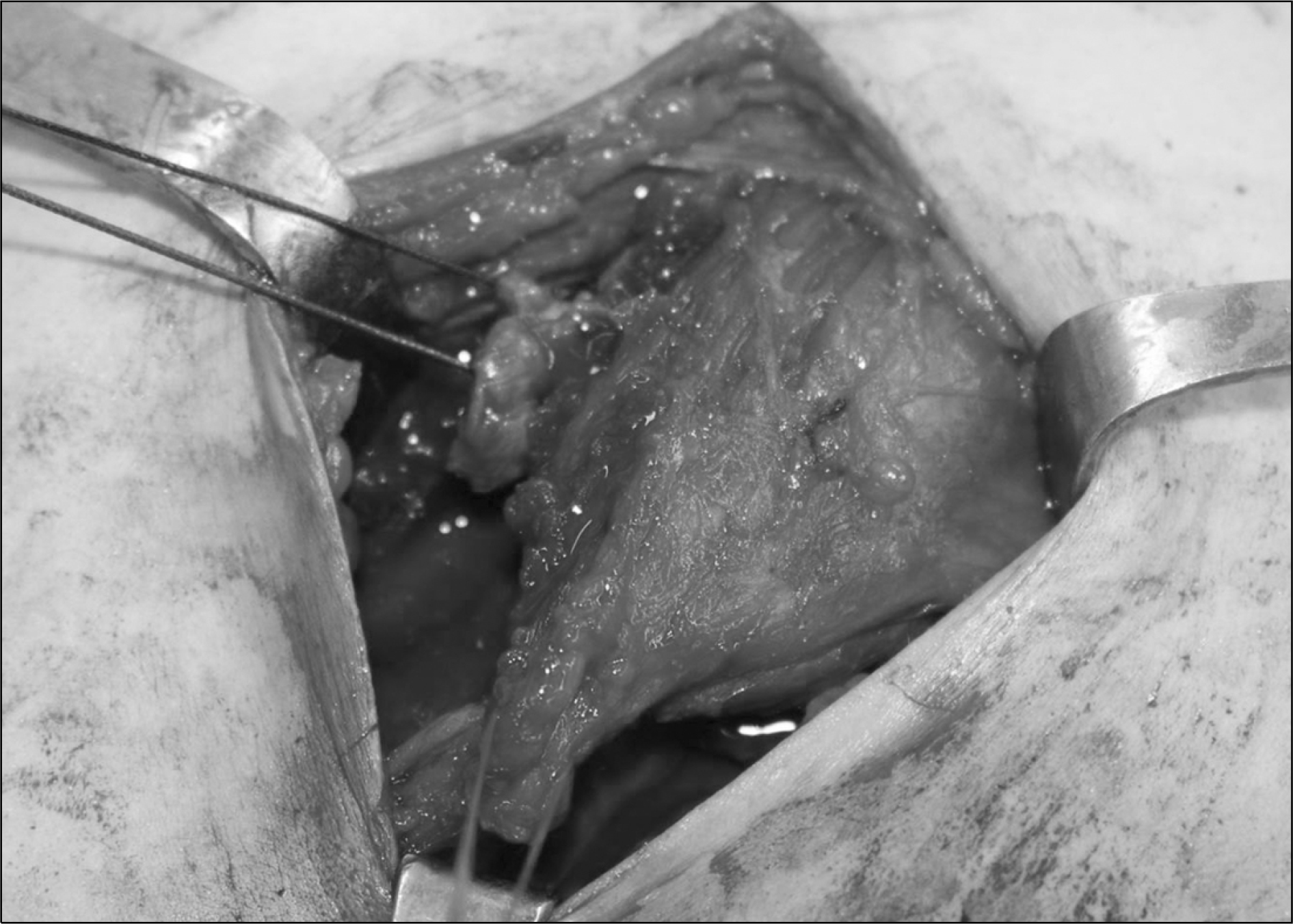
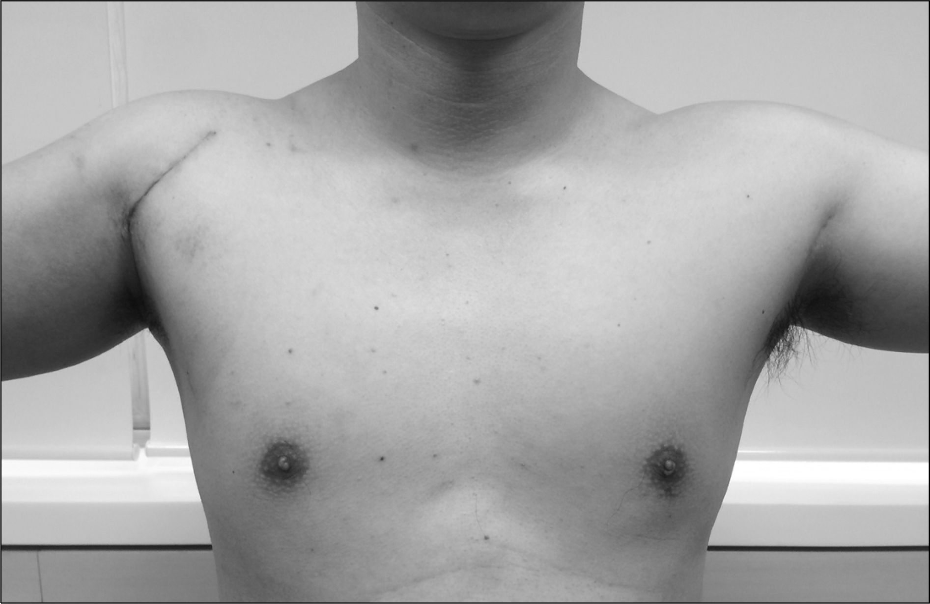
Fig. 3.
Clinical image. At 1 month after surgery, the axillary fold was restored and the tendon of pectoralis major muscle was palpated directly.
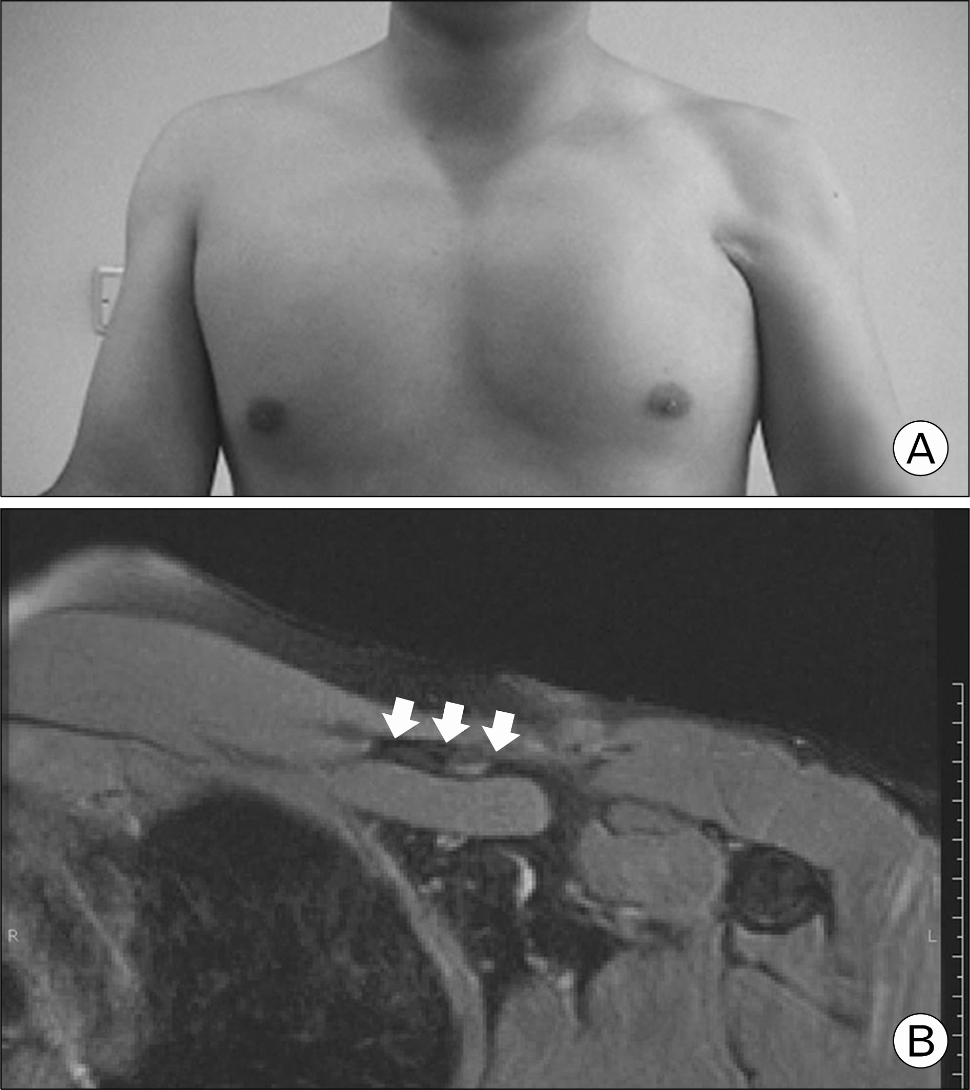
Fig. 4.
(A) Clinical photograph of the patient before the operation. Note the asymmetric absent anterior axillary fold on the affected (left) side. (B) Axial T2-weighted fat suppression magnetic resonance image at the level of the proximal humerus. The pectoralis major tendon is detached medially from the normal insertion site and attenuated (white arrows). Adjacent area of white arrow reflect fatty infiltration.


 PDF
PDF ePub
ePub Citation
 Print
Print


 XML Download