Abstract
The aim of this study was to evaluate the postoperative outcomes of anterior cruciate ligament (ACL) reconstructionuction using 2 additional fixation technique on tibial side. Between October 2008 and February 2012, sixty consecutive patients who underwent ACL reconstruction with allograft for ACL injuries were retrospectively enrolled. All patients were reconstructed with fresh frozen achilles tendon or posterior tibialis tendon allograft. Fixation on tibial side with bioabsorbable suture anchor (BSA) was in 30 patients (group A) and metal screw fixation was in 30 patients (group B). The data was collected at preoperatively and at least 1 years postoperatively, which included KT-2000 arthrometer objectively, and Tegner score, Lysholm score, and International Knee Documentation Committee (IKDC) scores subjectively. At the final follow up, the KT-2000 arthrometer improved significantly with an average of 3.28 mm anterior translation in the group A, 3.56 mm in group B. The preoperative mean Lysholm, Tegner and IKDC score was 46.14, 4.86, 63.17 in the group A, and 45.30, 4.40, 54.07 in the group B. The postoperative mean Lysholm, Tegner and IKDC score was 83.80, 8.14, 75.57 in the group A, and 88.75, 7.62, 65.10 in the group B. All functional outcomes were improved significantly (p=0.004) in both groups, but no differences were noted between the 2 groups (p>0.05). Both additional fixation techniques using BSA or metal screw fixation on tibial side in ACL reconstruction improved functional outcomes significantly. BSA technique seems to provide adequate strength suitable for early rehabilitation after ACL reconstruction.
Go to : 
REFERENCES
1. Dragoo JL, Braun HJ, Durham JL, Chen MR, Harris AH. Incidence and risk factors for injuries to the anterior cruciate ligament in National Collegiate Athletic Association football: data from the 2004-2005 through 2008-2009 National Collegiate Athletic Association Injury Surveillance System. Am J Sports Med. 2012; 40:990–5.
2. Almqvist KF, Willaert P, De Brabandere S, Criel K, Verdonk R. A longterm study of anterior cruciate ligament allograft reconstruction. Knee Surg Sports Traumatol Arthrosc. 2009; 17:818–22.

3. Brand JC Jr, Nyland J, Caborn DN, Johnson DL. Soft-tissue interference fixation: bioabsorbable screw versus metal screw. Arthroscopy. 2005; 21:911–6.

4. Giurea M, Zorilla P, Amis AA, Aichroth P. Comparative pullout and cyclic-loading strength tests of anchorage of hamstring tendon grafts in anterior cruciate ligament reconstruction. Am J Sports Med. 1999; 27:621–5.
5. Magen HE, Howell SM, Hull ML. Structural properties of six tibial fixation methods for anterior cruciate ligament soft tissue grafts. Am J Sports Med. 1999; 27:35–43.

6. Tetsumura S, Fujita A, Nakajima M, Abe M. Biomechanical comparison of different fixation methods on the tibial side in anterior cruciate ligament reconstruction: a biomechanical study in porcine tibial bone. J Orthop Sci. 2006; 11:278–82.

7. Yoo JC, Ahn JH, Kim JH, et al. Biomechanical testing of hybrid hamstring graft tibial fixation in anterior cruciate ligament reconstruction. Knee. 2006; 13:455–9.

8. Hill PF, Russell VJ, Salmon LJ, Pinczewski LA. The influence of supplementary tibial fixation on laxity measurements after anterior cruciate ligament reconstruction with hamstring tendons in female patients. Am J Sports Med. 2005; 33:94–101.

9. Jansson KA, Linko E, Sandelin J, Harilainen A. A prospective randomized study of patellar versus hamstring tendon autografts for anterior cruciate ligament reconstruction. Am J Sports Med. 2003; 31:12–8.

10. Kim MK, Na SI, Lee JM, Park JY. Comparison of bioabsorbable suture anchor fixation on the tibial side for anterior cruciate ligament reconstruction using free soft tissue graft: experimental laboratory study on porcine bone. Yonsei Med J. 2014; 55:760–5.

11. Beynnon BD, Johnson RJ, Abate JA, Fleming BC, Nichols CE. Treatment of anterior cruciate ligament injuries, part 2. Am J Sports Med. 2005; 33:1751–67.

12. Clark R, Olsen RE, Larson BJ, Goble EM, Farrer RP. Cross-pin femoral fixation: a new technique for hamstring anterior cruciate ligament reconstruction of the knee. Arthroscopy. 1998; 14:258–67.

13. Howell SM, Deutsch ML. Comparison of endoscopic and two-incision techniques for reconstructing a torn anterior cruciate ligament using hamstring tendons. Arthroscopy. 1999; 15:594–606.

14. Scheffler SU, Sudkamp NP, Gockenjan A, Hoffmann RF, Weiler A. Biomechanical comparison of hamstring and patellar tendon graft anterior cruciate ligament reconstruction techniques: The impact of fixation level and fixation method under cyclic loading. Arthroscopy. 2002; 18:304–15.
Go to : 
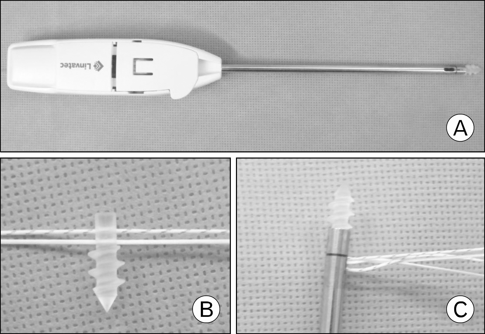
 | Fig. 2.(A) Bioabsorbable suture anchor (Duet Suture Anchor, Linvatec Corp., Largo, FL, USA). (B) Separated suture anchor tip and FiberWire (Arthrex, Naples, FL, USA) connecting the tendon. (C) Reassembled bioabsorbable suture anchor |
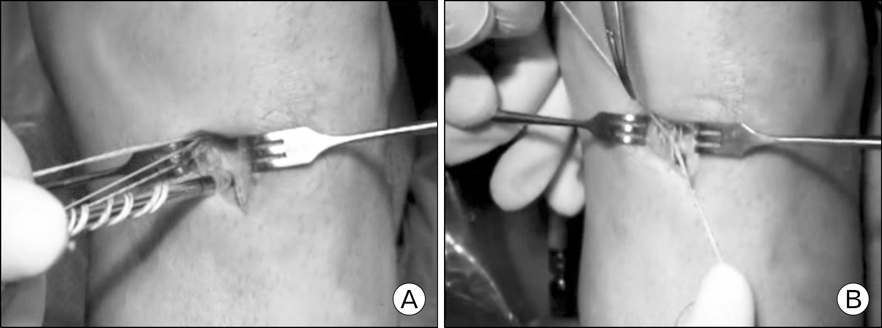
 | Fig. 3.(A) Bioabsorbable suture anchor was inserted on tibial drilling site. (B) FiberWire on both sides was tied. |
Table 1.
Comparison of clinical outcome of both group


 PDF
PDF ePub
ePub Citation
 Print
Print


 XML Download