

 PDF
PDF ePub
ePub Citation
 Print
Print


Abstract
Intramedullary screw fixation and bicortical screw fixation are widely used operation methods in the surgical treatment of Jones fractures. The purpose of this study is to evaluate of mechnical stability in two kind of Jones fracture. Using Mimics, three-dimensional models of the fifth metatarsal were reconstructed form computed tomography images of a 23-year-old Korean healthy male. Normal and osteoporotic bone models were made by changing bone density or thickness of cortical and cancellous bone. Two kinds of fixation techniques, i.e., intramedullary and bicortical screw fixation models, were simulated and muscles forces related to the fifth metatarsal base were applied. Maximum contact pressure difference were measured as 20,818 MPa, 12,155 MPa in normal bone, 23,371 MPa, 13,765 MPa in 85% cancellous osteoporotic bone, 24,310 MPa and 14,264 MPa in 75% cancellos osteoporotic model, 21,337 MPa, 20,971 MPa in −0.5 mm cortical osteoporotic bone, 26,322 MPa and 36,153 MPa in −1 mm cortical osteoporotic model, respectively for intramedullary screw fixation and bicortical screw fixation. Displacements on fracture interface were 0.208 mm, 0.126 mm in normal bone while 0.229 mm, 0.127 mm in 85% cancellos osteoporotic model, 0.241 mm, 0.127 mm in 75% cancellos osteoporotic model, 0.223 mm, 0.271 mm in −0.5 mm cortical osteoporotic model, 0.292 mm, 0.480 mm in −1 mm cortical osteoporotic model, respectively for intramedullary screw fixation and bicortical screw fixation. Bicortical screw fixation is superior in mechanical stability than intramedullary screw fixation for normal bone quality Jones fractures. For cortical osteoporotic bone Jones fractures, however, intramedullary screw fixation can give a better mechanical stability than bicortical screw fixation.
Go to : 
REFERENCES
1. Jones R. Fracture of the base of the fifth metatarsal bone by indirect violence. Ann Surg. 1902; 35:697–700.
2. Clapper MF, O'Brien TJ, Lyons PM. Fractures of the fifth metatarsal. Analysis of a fracture registry. Clin Orthop Relat Res. 1995; 315:238–41.
3. Pietropaoli MP, Wnorowski DC, Werner FW, Fortino MD. Intramedullary screw fixation of Jones fractures: a biomechanical study. Foot Ankle Int. 1999; 20:560–3.

4. Dameron TB Jr. Fractures and anatomical variations of the proximal portion of the fifth metatarsal. J Bone Joint Surg Am. 1975; 57:788–92.
5. Kavanaugh JH, Brower TD, Mann RV. The Jones fracture revisited. J Bone Joint Surg Am. 1978; 60:776–82.
6. Mahajan V, Chung HW, Suh JS. Fractures of the proximal fifth metatarsal: percutaneous bicortical fixation. Clin Orthop Surg. 2011; 3:140–6.
7. Portland G, Kelikian A, Kodros S. Acute surgical management of Jones' fractures. Foot Ankle Int. 2003; 24:829–33.
8. Husain ZS, DeFronzo DJ. A comparison of bicortical and intramedullary screw fixations of Jones' fractures. J Foot Ankle Surg. 2002; 41:146–53.
9. Bucholz RW, Heckman JD, Court-Brown CM. Rockwood & Green's fractures in adults. 6th ed.Philadelphia, USA: Lippincott Williams & Wilkins;2006.
10. Johnson JT, Labib SA, Fowler R. Intramedullary screw fixation of the fifth metatarsal: an anatomic study and improved technique. Foot Ankle Int. 2004; 25:274–7.
11. Ritzel H, Amling M, Posl M, Hahn M, Delling G. The thickness of human vertebral cortical bone and its changes in aging and osteoporosis: a histomorphometric analysis of the complete spinal column from thirty-seven autopsy specimens. J Bone Miner Res. 1997; 12:89–95.
12. Baroud G, Nemes J, Ferguson SJ, Steffen T. Material changes in osteoporotic human cancellous bone following infiltration with acrylic bone cement for a vertebral cement augmentation. Comput Methods Biomech Biomed Engin. 2003; 6:133–9.
13. Rohr ES, Johnson JE, Zhao L, Harris GF. Three-dimensional finite element analysis of the fifth metatarsal jones fracture. Harris GF, Smith PA, Marks RM, editors. editors.Foot and ankle motion analysis: clinical treatment and technology. Florida: Talyer & Francis;2007. p. 347–62.
14. Lawrence SJ, Botte MJ. Jones' fractures and related fractures of the proximal fifth metatarsal. Foot Ankle. 1993; 14:358–65.
15. Josefsson PO, Karlsson M, Redlund-Johnell I, Wendeberg B. Jones fracture. Surgical versus nonsurgical treatment. Clin Orthop Relat Res. 1994; 299:252–5.
16. Quill GE Jr. Fractures of the proximal fifth metatarsal. Orthop Clin North Am. 1995; 26:353–61.
17. Hatch RL, Alsobrook JA, Clugston JR. Diagnosis and management of metatarsal fractures. Am Fam Physician. 2007; 76:817–26.
18. Vertullo CJ, Glisson RR, Nunley JA. Torsional strains in the proximal fifth metatarsal: implications for Jones and stress fracture management. Foot Ankle Int. 2004; 25:650–6.
19. Stewart IM. Jones's fracture: fracture of base of fifth metatarsal. Clin Orthop. 1960; 16:190–8.
20. Chao EY, Inoue N, Koo TK, Kim YH. Biomechanical considerations of fracture treatment and bone quality maintenance in elderly patients and patients with osteoporosis. Clin Orthop Relat Res. 2004; 425:12–25.
Go to :
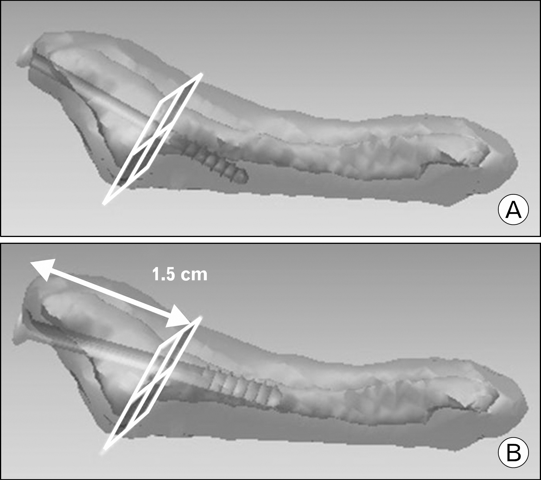 | Fig. 1.Bicortical screw fixation (A), Intramedullary screw fixation (B) model. Rectangular grid is fracture plane. |
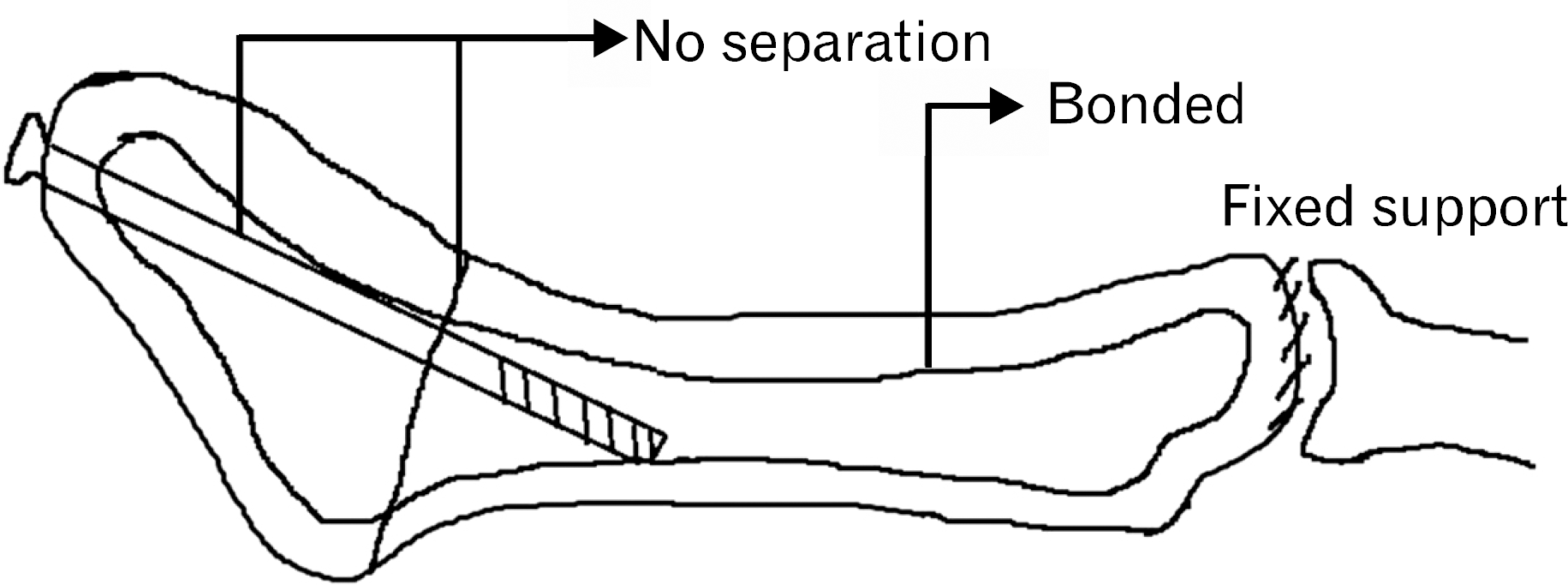 | Fig. 2.Boundary condition. Interface between cortical and cancellous bone is bonded. Bonded: no movement, complete union state, Fracture site: interface between screw and bone are no separation, No separation: movement can occur but contact is maintained, Fixed support: fifth meta-tarsophalangeal joint is fixed state. |
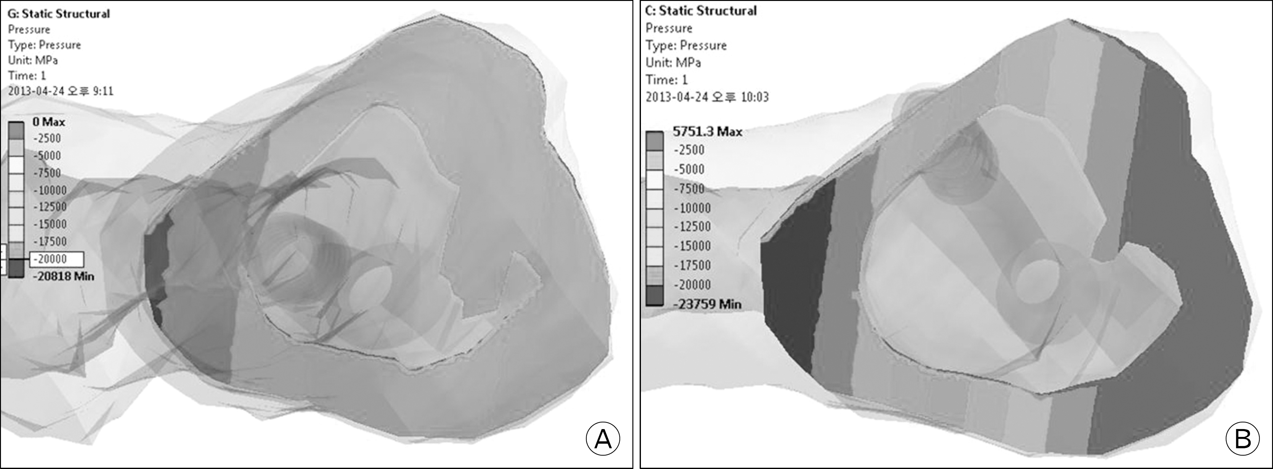 | Fig. 3.Contact pressure on fracture plane. Contact pressure distribution is displayed by gradation scale. (A) is displayed by similar color. But, (B) is displayed by various color (sharp contrast). In other words, (A) is more evenly distributed contact pressure and (B) is too concentrated or too little contact pressure. It means mechanical stable or unstable, respectively. |
Table 1.
Magnitudes of the muscle forces related to the fifth metatarsal base
| Category | Peroneus brevis | Peroneus tertius | Dorsal interosseous | Plantar interosseous | Flexor digiti minimi brevis |
|---|---|---|---|---|---|
| PSCA (cm2) | 11.50 | 3.10 | 2.72 | 1.38 | 2.00 |
| Force (N) | 287.5 | 77.5 | 68.0 | 34.5 | 50.0 |
Table 2.
Contact pressure and displacement on fracture plane
 XML Download