Abstract
This study was conducted to evaluate the diagnostic utility of magnetic resonance imaging (MRI) for the patients having problems confined to cross-legged posture. The study subjects were 128 cases (male 87.5%) and 120 patients from October 2008 to June 2013. Average age of male patients was 50 years old (range, 21−72 years old), and female 45 years old (range, 18−76 years old). The rate of positive MRI findings was compared according to abnormal physical findings. The average duration of symptoms was 11.7 months. The most frequent complains was on the back (41.9% at rest, 57% when taking the posture). Patrick test was positive for 33.6% of cases, simple radiography was abnormal only for 20% of cases. Bone scan was normal for all 98 cases. Only 21.9% of 128 cases showed abnormal MRI findings which were managed with conservative treatment. Limitation in the range of hip joint motion was not statistically associated with abnormal findings of MRI (p=0.148). Normal Patrick test was associated with normal MRI finding (p=0.001). Among normal cases on both physical and simple bone X-ray film, 88.6% were normal at MRI. In conclusion, for patients with physical complaints from the cross-legged posture, diagnostic utility of MRI is relatively low when they show normal on both physical examination and simple radiography.
Go to : 
REFERENCES
1. Cotten A, Boutry N, Demondion X, et al. Acetabular labrum: MRI in asymptomatic volunteers. J Comput Assist Tomogr. 1998; 22:1–7.

2. Leunig M, Podeszwa D, Beck M, Werlen S, Ganz R. Magnetic resonance arthrography of labral disorders in hips with dysplasia and impingement. Clin Orthop Relat Res. 2004; 418:74–80.

3. Kim KW, Lee TH, Nam WD, Rhyu KH. Normal adult hip range of motion focusingon hip flexion. J Korean Orthop Assoc. 2006; 41:361–7.

4. Suk SI, Lee CK, Baek GH, et al. Orthopaedics. 7th ed.Seoul: Choisin Medical;2013.
5. Kenna C, Murtagh J. Patrick or fabere test to test hip and sacroiliac joint disorders. Aust Fam Physician. 1989; 18:375.
6. Kleinfield SL, Daniel D, Ndetan H. Faculty perception of clinical value of five commonly used orthopedic tests. J Chiropr Educ. 2011; 25:164–8.

7. Magee DJ. Orthopedic physical assessment. Philadelphia: W.B. Saunders;1992.
8. Tannast M, Siebenrock KA, Anderson SE. Femoroacetabular impingement: radiographic diagnosis–what the radiologist should know. AJR Am J Roentgenol. 2007; 188:1540–52.

9. Ito K, Leunig M, Ganz R. Histopathologic features of the acetabular labrum in femoroacetabular impingement. Clin Orthop Relat Res. 2004; 429:262–71.

10. Bredella MA, Stoller DW. MR imaging of femoroacetabular impingement. Magn Reson Imaging Clin N Am. 2005; 13:653–64.

11. Espinosa N, Rothenfluh DA, Beck M, Ganz R, Leunig M. Treatment of femoroacetabular impingement: preliminary results of labral refixation. J Bone Joint Surg Am. 2006; 88:925–35.
12. Mitchell B, McCrory P, Brukner P, O'Donnell J, Colson E, Howells R. Hip joint pathology: clinical presentation and correlation between magnetic resonance arthrography, ultrasound, and arthroscopic findings in 25 consecutive cases. Clin J Sport Med. 2003; 13:152–6.

13. Haims A, Katz LD, Busconi B. MR arthrography of the hip. Radiol Clin North Am. 1998; 36:691–702.

14. Kellgren JH, Lawrence JS. Radiological assessment of osteo- arthrosis. Ann Rheum Dis. 1957; 16:494–502.
15. Czerny C, Hofmann S, Urban M, et al. MR arthrography of the adult acetabular capsular-labral complex: correlation with surgery and anatomy. AJR Am J Roentgenol. 1999; 173:345–9.

16. Ferguson SJ, Bryant JT, Ito K. The material properties of the bovine acetabular labrum. J Orthop Res. 2001; 19:887–96.

17. Edwards DJ, Lomas D, Villar RN. Diagnosis of the painful hip by magnetic resonance imaging and arthroscopy. J Bone Joint Surg Br. 1995; 77:374–6.

18. Petersilge CA. Current concepts of MR arthrography of the hip. Semin Ultrasound CT MR. 1997; 18:291–301.
19. Lee HS. Basic Korean dictionary. 3rd ed.Paju: Minjoong seorim;2014.
20. Park KS, Yoon TR, Haq RU. Posttraumatic osteonecrosis of the femoral head after nine years of posterior femoral head fracture dislocation. J Korean Orthop Assoc. 2014; 49:153–8.

21. Morimoto T, Sonohata M, Mawatari M. Validity of the patrick test for osteoarthritis of the hip and sciatica. Spine J Meet Abstr. 2011. GP138.
Go to : 
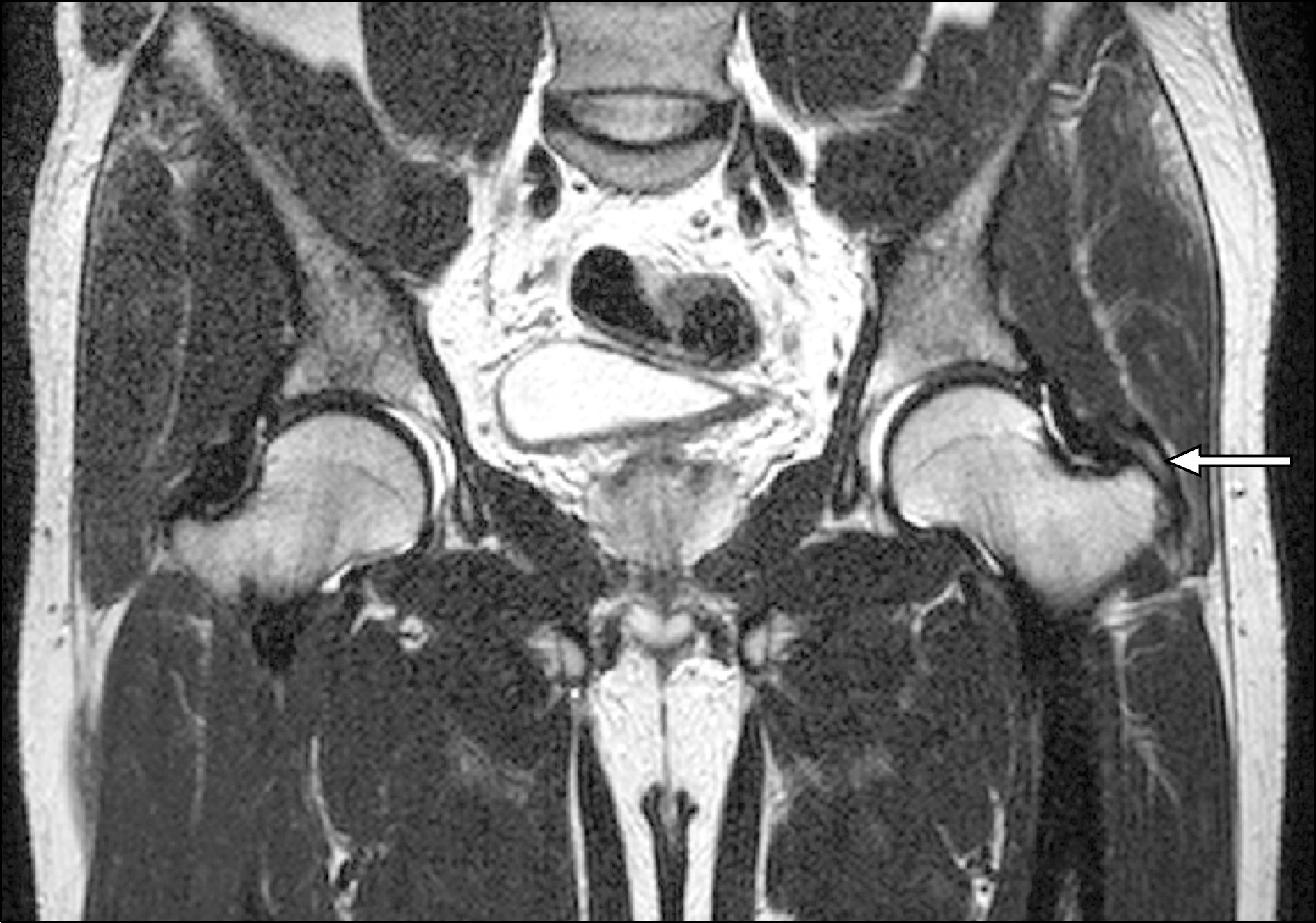
 | Fig. 1.A 55-year-old male has left buttock pain in cross legged position. Increased signal intensity of distal gluteus minimus tendon around posterior greater trochanter area is observed in the left side on the magnetic resonance imaging (arrow) and this is consistent with tendinopathy. |
Table 1.
Patients' characteristics
Table 2.
Abnormal findings of magnetic resonance imaging
| Abnormal finding | Cases (%) (n=28) |
|---|---|
| Tendinosis | 11 (39.3) |
| Ostepenia | 5 (17.9)∗ |
| Synovitis | 4 (14.3) |
| Strain | 4 (14.3) |
| Calcific tendinitis | 2 (7.1) |
| Labral lesion | 1 (3.6) |
| Others | 1 (3.6) |
Table 3.
The relationship between abnormal physical findings and magnetic resonance imaging results among patients with normal simple X-ray film
| Characteristic | Magnetic resonance imaging | p-value | ||
|---|---|---|---|---|
| Normal | Abnormal | All | ||
| Hip ROM | 0.148∗ | |||
| Normal | 78 (81.2) | 18 (18.8) | 96 (100.0) | |
| Limited | 4 (57.1) | 3 (42.9) | 7 (100.0) | |
| All | 82 (79.6) | 21 (20.4) | 103 (100.0) | |
| Patrick test | 0.001 | |||
| Negative | 63 (88.7) | 8 (11.3) | 71 (100.0) | |
| Positive | 19 (59.4) | 13 (40.6) | 32 (100.0) | |
| All | 82 (79.6) | 21 (20.4) | 103 (100.0) | |
| LOM & Patrick test | 0.003∗ | |||
| Normal | 61 (88.4) | 8 (11.6) | 69 (100.0) | |
| Abnormal | 21 (61.8) | 13 (38.2) | 34 (100.0) | |
| All | 82 (79.6) | 21 (20.4) | 103 (100.0) | |


 PDF
PDF ePub
ePub Citation
 Print
Print


 XML Download