

 PDF
PDF ePub
ePub Citation
 Print
Print


Abstract
We evaluated abnormalities in medial portion of elbow in high-school weightlifter compared with the non weightlifter using a stress radiography and ultrsonography. The experimental group(G1) was 26 high school weightlifters with an average age of 17 years old (range, 16—18 years). The control group (G2) were comprised of 25 age matched general students. Both groups received physical examination, simple and valgus stress radiography and ultrasonography on both side of elbow. Physical examination showed 26.9% (14/52 elbows) tenderness and 19.2% (10/52 elbows) valgus laxity in G1, no tenderness and laxity in G2. There were no differences in medial joint gaps on simple radiography (G1, 3.3 mm, G2, 2.7 mm; p>0.05), but the valgus stress view showed 5.6±0.8 mm medial joint gap in G1 and 3.8±0.8 mm in G2 (p<0.001). Ultrasonography in G1, angular deformity was found in 67.3% (36/52) and G2 all in normal (p<0.01). The horizontal distance was an average 4.9±1.23 mm for the G1 and 3.1±0.78 mm for the G2 (p<0.001). Vertical distance of the proximal portion of the ulna was average 0.58±0.94 mm for the G1 and 1.59±0.49 mm for the G2 (p<0.001). In G1, angular deformity of male was 50% (15/30 elbows) and female was 95% (21/22 elbows) (p<0.001). Change of horizontal and vertical distance were larger in female (p<0.05). In conclusion, there were increased incidence of medial elbow joint laxity in high school weightlifter, especially in female, regardless of career. Sustained valgus laxity could be prone to ulnar collateral ligament injury and should be evaluated with ultrasonography-assisted dynamic study.
Go to : 
References
1. Andrews JR, Timmerman LA. Outcome of elbow surgery in professional baseball players. Am J Sports Med. 1995; 23:407–13.

2. Elliott B, Fleisig G, Nicholls R, Escamilia R. Technique effects on upper limb loading in the tennis serve. J Sci Med Sport. 2003; 6:76–87.
3. Fleisig GS, Andrews JR, Dillman CJ, Escamilla RF. Kinetics of baseball pitching with implications about injury mechanisms. Am J Sports Med. 1995; 23:233–9.
4. Hamilton CD, Glousman RE, Jobe FW, Brault J, Pink M, Perry J. Dynamic stability of the elbow: electromyographic analysis of the flexor pronator group and the extensor group in pitchers with valgus instability. J Shoulder Elbow Surg. 1996; 5:347–54.
5. Jobe FW, Stark H, Lombardo SJ. Reconstruction of the ulnar collateral ligament in athletes. J Bone Joint Surg Am. 1986; 68:1158–63.
6. Loftice J, Fleisig GS, Zheng N, Andrews JR. Biomechanics of the elbow in sports. Clin Sports Med. 2004; 23:519–30.
7. Werner SL, Fleisig GS, Dillman CJ, Andrews JR. Biomechanics of the elbow during baseball pitching. J Orthop Sports Phys Ther. 1993; 17:274–8.
8. Bennett JB, Tullos HS. Ligamentous and articular injuries in the athlete. Morrey BF, editor. The elbow and its disorders. Philadelphia: WB Saunders;1985. p. 502–22.
9. Rijke AM, Goitz HT, McCue FC, Andrews JR, Berr SS. Stress radiography of the medial elbow ligaments. Radiology. 1994; 191:213–6.
10. Ellenbecker TS, Mattalino AJ, Elam EA, Caplinger RA. Medial elbow joint laxity in professional baseball pitchers. A bilateral comparison using stress radiography. Am J Sports Med. 1998; 26:420–4.
11. Lee GA, Katz SD, Lazarus MD. Elbow valgus stress radiography in an uninjured population. Am J Sports Med. 1998; 26:425–7.
12. Thompson WH, Jobe FW, Yocum LA, Pink MM. Ulnar collateral ligament reconstruction in athletes: muscle-splitting approach without transposition of the ulnar nerve. J Shoulder Elbow Surg. 2001; 10:152–7.
13. Azar FM, Andrews JR, Wilk KE, Groh D. Operative treatment of ulnar collateral ligament injuries of the elbow in athletes. Am J Sports Med. 2000; 28:16–23.
14. Timmerman LA, Andrews JR. Undersurface tear of the ulnar collateral ligament in baseball players: a newly recognized lesion. Am J Sports Med. 1994; 22:33–6.
15. Jobe FW, Kvitne RS. Elbow instability in the athlete. Instr Course Lect. 1991; 40:17–23.
16. Mirowitz SA, London SL. Ulnar collateral ligament injury in baseball pitchers: MR imaging evaluation. Radiology. 1992; 185:573–6.
17. Timmerman LA, Schwartz ML, Andrews JR. Preoperative evaluation of the ulnar collateral ligament by magnetic resonance imaging and computed tomography arthrography. Evaluation in 25 baseball players with surgical confirmation. Am J Sports Med. 1994; 22:26–31.
18. Sugimoto K, Matui N, Taneda Y, Oyabu N, Nakano Y, Goto H. Ultrasonographic evaluation of the ulnar collateral ligament of the elbow joint. J Jpn Soc Orthop Ultrason. 1994; 6:187–8.
19. Sugimoto K, Ohta S. The natural course of the MCL injury of the elbow. J Jpn Soc Orthop Ultrason. 1997; 9:15–9.
20. Miller TT, Adler RS, Friedman L. Sonography of injury of the ulnar collateral ligament of the elbow-initial experience. Skeletal Radiol. 2004; 33:386–91.
21. De Smet AA, Winter TC, Best TM, Bernhardt DT. Dynamic sonography with valgus stress to assess elbow ulnar collateral ligament injury in baseball pitchers. Skeletal Radiol. 2002; 31:671–6.
22. Sasaki J, Takahara M, Ogino T, Kashiwa H, Ishigaki D, Kanauchi Y. Ultrasonographic assessment of the ulnar collateral ligament and medial elbow laxity in college baseball players. J Bone Joint Surg Am. 2002; 84:525–31.
23. Nazarian LN, McShane JM, Ciccotti MG, O'Kane PL, Harwood MI. Dynamic US of the anterior band of the ulnar collateral ligament of the elbow in asymptomatic major league baseball pitchers. Radiology. 2003; 227:149–54.
24. Callaway GH, Field LD, Deng XH, et al. Biomechanical evaluation of the medial collateral ligament of the elbow. J Bone Joint Surg Am. 1997; 79:1223–31.
25. Floris S, Olsen BS, Dalstra M, Sojbjerg JO, Sneppen O. The medial collateral ligament of the elbow joint: anatomy and kinematics. J Shoulder Elbow Surg. 1998; 7:345–51.
26. Sojbjerg JO, Ovesen J, Nielsen S. Experimental elbow instability after transection of the medial collateral ligament. Clin Orthop Relat Res. 1987; 218:186–90.
27. Drinkwater EJ, Galna B, McKenna MJ, Hunt PH, Pyne DB. Validation of an optical encoder during free weight resistance movements and analysis of bench press sticking point power during fatigue. J Strength Cond Res. 2007; 21:510–7.
Go to :
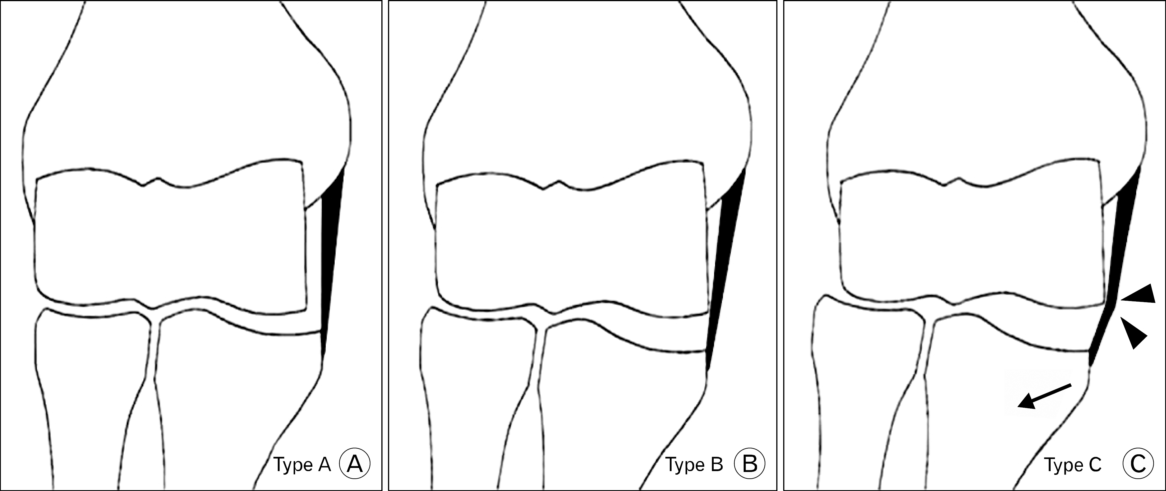 | Fig. 1.Three types of angular changes of the medial collateral ligament. (A) Type A: normal stable elbow joint. (B) Type B: increased medial elbow laxity, as manifested by widening of the medial joint space and lateral shift of the proximal part of the ulna. (C) Type C: increased lateral shift of the proximal part of the ulna (arrow) causing impingement of the ulnar collateral ligament on the trochlea (arrowheads). |
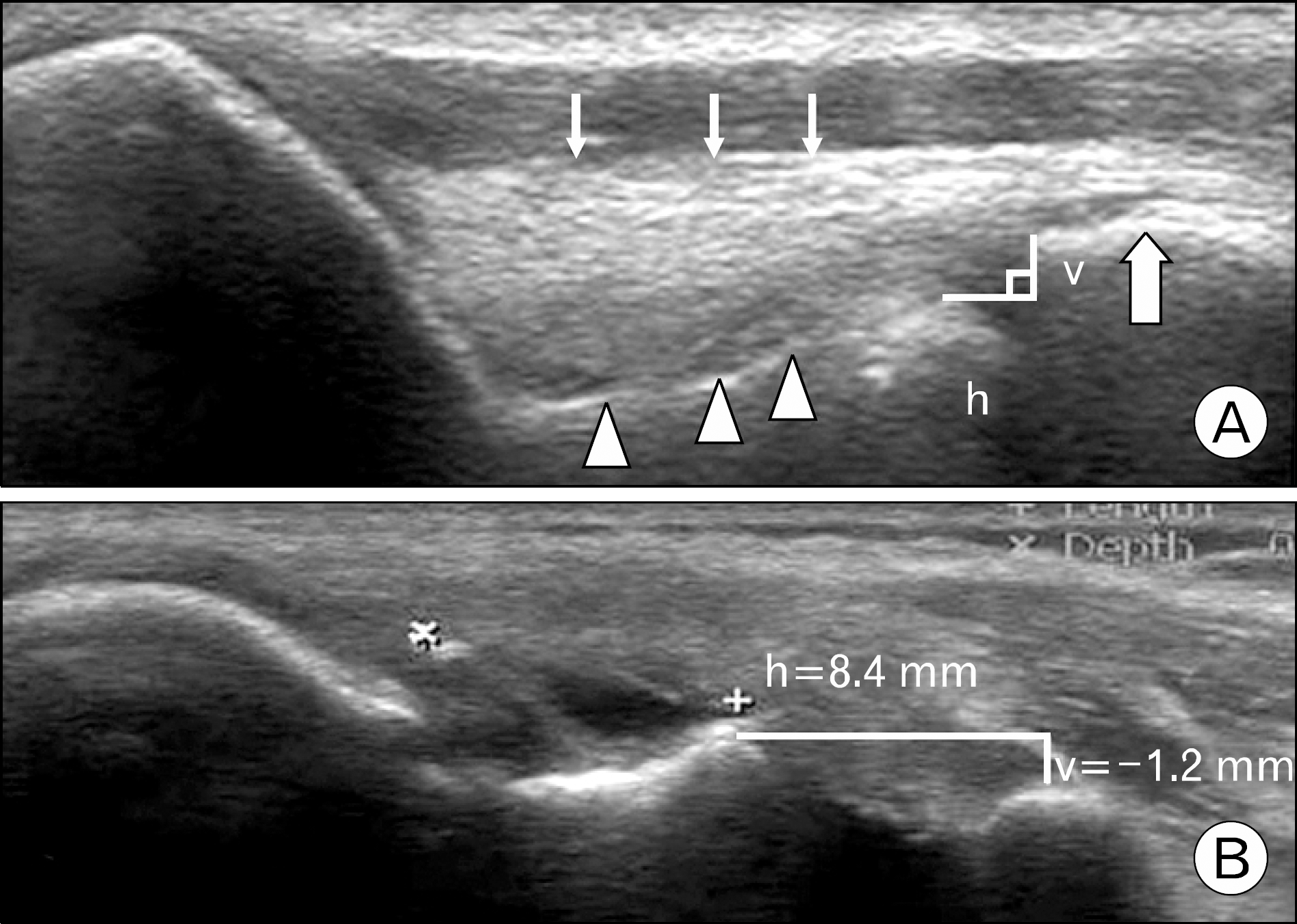 | Fig. 2.Ultrasonographic image of the elbow joint in normal and medial laxity. (A) Ultrasonography of normal elbow joint. The medial joint space is shown as a nonechoic space between the subchondral bone of the trochlea (arrowheads) and that of the coronoid process (large arrow). The ulnar collateral ligament (UCL) is identified as a band-like structure that attaches to the medial epicondyle and the tubercular portion of the coronoid process. The superficial surface of the ligament is seen outlined by a hyperechoic straight line (small arrows). (B) Ultrasonographic findings in ligament injury. The image showing type C angular changes with increased medial elbow laxity, as manifested by widening of the medial joint space (h=8.4 mm) and the lateral shift of the proximal part of the ulna (v=-1.2 mm). UCL tear is shown as a nonechoic gap between torn margin of the ligament (between × and +). h=horizontal distance of the medial joint space (with the assumption that the outline of the ulnar collateral ligament is a horizontal line), and v=vertical distance of the medial joint space. |
Table 1.
Valgus laxity on stress radiograph and angular deformity between weightlifter and control group
| Group | No. of elbow | Valgus stress view (mm) | Angular deformity | ||
|---|---|---|---|---|---|
| A | B | C | |||
| G1* | 52 | 5.60±0.80‡ | 16§ | 33 | 3 |
| Dominant | 26 | 5.54±0.66 | 5 | 20 | 1 |
| Non | 26 | 5.67±0.92 | 11 | 13 | 2 |
| dominant | |||||
| G2† | 50 | 3.81±0.74‡ | 38§ | 0 | 0 |
Table 2.
Horizontal and vertical distances of medial elbow joint on ultrasonographic image between weightlifter and control group
| Group | No. of elbow | Horizontal distance (mm) | Vertical distance (mm) |
|---|---|---|---|
| G1* | 52 | 4.92±1.23‡ | 0.58±0.94§ |
| Dominant | 26 | 5.06±1.12 | 0.48±0.86 |
| Non | 26 | 4.75±1.28 | 0.67±1.01 |
| dominant | |||
| G2† | 50 | 3.11±0.78‡ | 1.59±0.49§ |
Table 3.
Valgus laxity on stress radiograph and angular deformity between male and female in weightlifter group
| Gender N | No. of elbow | Angular deformity | ||
|---|---|---|---|---|
| A* | B† | C | ||
| Male | 30 | 15 | 15 | 0 |
| Female | 22 | 1 | 18 | 3 |
Table 4.
Horizontal and vertical distances of medial elbow joint on ultrasonographic image between male and female in weightlifter group
| Gender | No. of elbow | Horizontal distance (mm)* | Vertical distance (mm)† |
|---|---|---|---|
| Male | 30 | 4.47±1.13 | 0.85±1.06 |
| Female | 22 | 5.40±1.22 | 0.41±0.82 |
Table 5.
Horizontal and vertical distances of medial elbow joint on ultrasonographic image according to career
| Career | No. of elbow | Horizontal distance (mm)* | Vertical distance (mm)† |
|---|---|---|---|
| Total | 52 | 4.92±1.23 | 0.58±0.94 |
| 1−2 y | 14 | 5.14 | 0.61 |
| 3−4 y | 20 | 4.72 | 0.49 |
| >5 y | 18 | 5.0 | 0.61 |
 XML Download