

 PDF
PDF ePub
ePub Citation
 Print
Print


Abstract
The purpose of this study was to investigate clinical and radiological outcomes of multiple drilling in case of failed conservative treatment of juvenile osteochondritis dissecans in athletes. We treated 37 lesions from 30 athletic patients who failed conservative treatment for juvenile osteochondritis dissecans. Multiple drillings were done for 32 lesions and multiple drilling and bioabsorbable pin fixations were done for 5 lesions. Lysholm score, Hughston clinical scale were used for clinical evaluation before and last follow up of treatment. For radiologic evaluation we used magnetic resonance imaging at 3 months and 12 months after operation. Of all 37 lesions, 11 lesions were located on medial femoral condyle, 2 lesions on lateral femoral condyle and 24 lesions on trochlear groove. There were clinical and radiological improvement from Hughston scale after operative treatment. Twenty-five patients among 30 returned to the sports activity. There were no specific complications after operation. Multiple drilling and bioabsorbable pin fixation of juvenile athletic osteochondritis dissecans patients after failure of conservative treatment showed good clinical and radiologic results. So it would be helpful for juvenile athletic patients to return to sports activities.
Go to : 
References
1. Konig F. Ueber freie Korper in den Gelenken. Dtsch Z Chir. 1888; 27:90–109.
2. Williams JS Jr, Bush-Joseph CA, Bach BR Jr. Osteochondritis dissecans of the knee. Am J Knee Surg. 1998; 11:221–32.
3. Clanton TO, DeLee JC. Osteochondritis dissecans. History, pathophysiology and current treatment concepts. Clin Orthop Relat Res. 1982; 167:50–64.

4. Yoshida S, Ikata T, Takai H, Kashiwaguchi S, Katoh S, Takeda Y. Osteochondritis dissecans of the femoral condyle in the growth stage. Clin Orthop Relat Res. 1998; (346):162–70.
5. Murray JR, Chitnavis J, Dixon P, et al. Osteochondritis dissecans of the knee; longterm clinical outcome following arthroscopic debridement. Knee. 2007; 14:94–8.
6. Kocher MS, Tucker R, Ganley TJ, Flynn JM. Management of osteochondritis dissecans of the knee: current concepts review. Am J Sports Med. 2006; 34:1181–91.
7. Hefti F, Beguiristain J, Krauspe R, et al. Osteochondritis dissecans: a multicenter study of the European Pediatric Orthopedic Society. J Pediatr Orthop B. 1999; 8:231–45.
8. Hayan R, Phillipe G, Ludovic S, Claude K, Jean-Michel C. Juvenile osteochondritis of femoral condyles: treatment with transchondral drilling: analysis of 40 cases. J Child Orthop. 2010; 4:39–44.
9. Hughston JC, Hergenroeder PT, Courtenay BG. Osteochondritis dissecans of the femoral condyles. J Bone Joint Surg Am. 1984; 66:1340–8.
10. Anderson AF, Richards DB, Pagnani MJ, Hovis WD. Antegrade drilling for osteochondritis dissecans of the knee. Arthroscopy. 1997; 13:319–24.
11. Cahill BR. Osteochondritis dissecans of the knee: treatment of juvenile and adult forms. J Am Acad Orthop Surg. 1995; 3:237–47.
12. Cahill BR, Phillips MR, Navarro R. The results of conservative management of juvenile osteochondritis dissecans using joint scintigraphy: a prospective study. Am J Sports Med. 1989; 17:601–5.
13. Guhl JF. Arthroscopic treatment of osteochondritis dissecans. Clin Orthop Relat Res. 1982; 167:65–74.
14. Cepero S, Ullot R, Sastre S. Osteochondritis of the femoral condyles in children and adolescents: our experience over the last 28 years. J Pediatr Orthop B. 2005; 14:24–9.
15. Aglietti P, Buzzi R, Bassi PB, Fioriti M. Arthroscopic drilling in juvenile osteochondritis dissecans of the medial femoral condyle. Arthroscopy. 1994; 10:286–91.
16. Kocher MS, Micheli LJ, Yaniv M, Zurakowski D, Ames A, Adrignolo AA. Functional and radiographic outcome of juvenile osteochondritis dissecans of the knee treated with transarticular arthroscopic drilling. Am J Sports Med. 2001; 29:562–6.
17. Kouzelis A, Plessas S, Papadopoulos AX, Gliatis I, Lambiris E. Herbert screw fixation and reverse guided drillings, for treatment of types III and IV osteochondritis dissecans. Knee Surg Sports Traumatol Arthrosc. 2006; 14:70–5.
18. Fonseca F, Balaco I. Fixation with autogenous osteochondral grafts for the treatment of osteochondritis dissecans (stages III and IV). Int Orthop. 2009; 33:139–44.
19. Kobayashi T, Fujikawa K, Oohashi M. Surgical fixation of massive osteochondritis dissecans lesion using cylindrical osteochondral plugs. Arthroscopy. 2004; 20:981–6.
20. Miura K, Ishibashi Y, Tsuda E, Sato H, Toh S. Results of arthroscopic fixation of osteochondritis dissecans lesion of the knee with cylindrical autogenous osteochondral plugs. Am J Sports Med. 2007; 35:216–22.
21. Gudas R, Simonaityte R, Cekanauskas E, Tamosiunas R. A prospective, randomized clinical study of osteochondral autologous transplantation versus microfracture for the treatment of osteochondritis dissecans in the knee joint in children. J Pediatr Orthop. 2009; 29:741–8.
22. Aurich M, Anders J, Trommer T, et al. Histological and cell biological characterization of dissected cartilage fragments in human osteochondritis dissecans of the femoral condyle. Arch Orthop Trauma Surg. 2006; 126:606–14.
23. Din R, Annear P, Scaddan J. Internal fixation of undisplaced lesions of osteochondritis dissecans in the knee. J Bone Joint Surg Br. 2006; 88:900–4.
24. Larsen MW, Pietrzak WS, DeLee JC. Fixation of osteochondritis dissecans lesions using poly(l-lactic acid)/poly (glycolic acid) copolymer bioabsorbable screws. Am J Sports Med. 2005; 33:68–76.
25. Tuompo P, Arvela V, Partio EK, Rokkanen P. Osteochondritis dissecans of the knee fixed with biodegradable self-reinforced polyglycolide and polylactide rods in 24 patients. Int Orthop. 1997; 21:355–60.
26. Millington KL, Shah JP, Dahm DL, Levy BA, Stuart MJ. Bioabsorbable fixation of unstable osteochondritis dissecans lesions. Am J Sports Med. 2010; 38:2065–70.
27. Friederichs MG, Greis PE, Burks RT. Pitfalls associated with fixation of osteochondritis dissecans fragments using bioabsorbable screws. Arthroscopy. 2001; 17:542–5.
28. Scioscia TN, Giffin JR, Allen CR, Harner CD. Potential complication of bioabsorbable screw fixation for osteochondritis dissecans of the knee. Arthroscopy. 2001; 17:E7.
29. Eriksson E. Long-term prognosis of osteochondritis dissecans of the knee. Knee Surg Sports Traumatol Arthrosc. 2008; 16:435.
30. Ramirez A, Abril JC, Chaparro M. Juvenile osteochondritis dissecans of the knee: perifocal sclerotic rim as a prognostic factor of healing. J Pediatr Orthop. 2010; 30:180–5.
Go to :
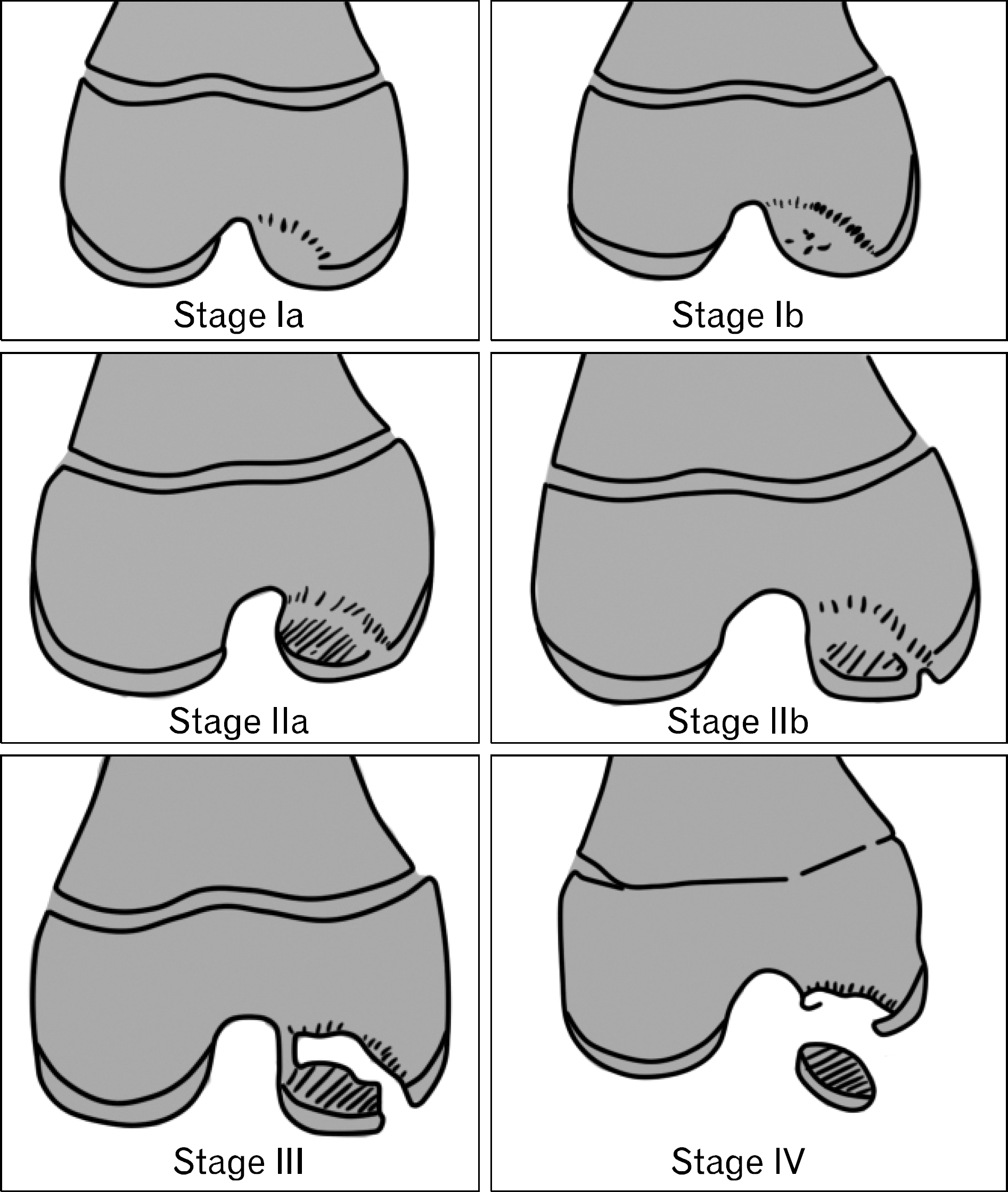 | Fig. 1.This illustration shows Bedouelle's radiologic classification (Data from Hayan et al. Child Orthop 2010;4: 39–448)). |
 | Fig. 2.A 16-year-old male patient with osteochondritis dissecans in the left trochlear lesion. (A) Arthroscopic multiple drilling with K-wire was done. (B) Preoperative coronal and sagittal T1 weighted magnetic resonance (MR) images are shown that the presence of a low signal line beneath the lesion and a breach of the articular cartilage with 10 mm×20 mm size. (C) Postoperative 3 months follow-up MR images are shown the trace of multiple drilling. (D) Postoperative 12 months follow-up MR images are shown that there are decrease of low signal intensity rim and the size of lesion. |
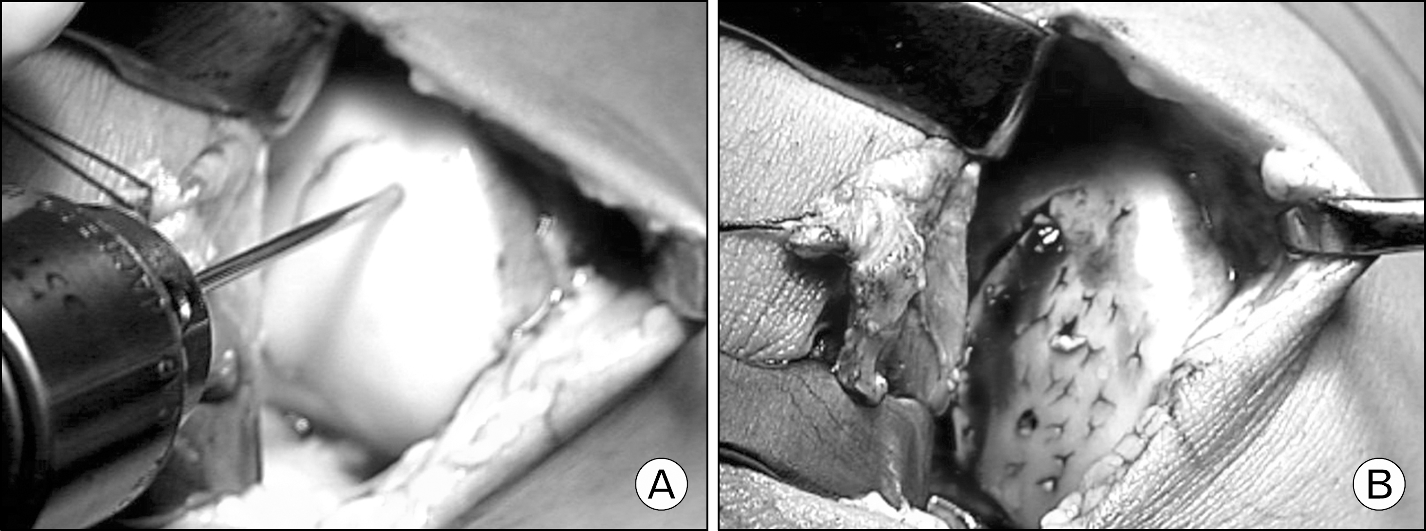 | Fig. 3.A 15-year-old male patient with unstable osteochodral lesion. (A) Vertical incision was made along the patella border and open multiple drilling was done. (B) Unstable osteochondral lesion was fixed with bioabsorbable pins after multiple drilling. |
Table 1.
Hughston clinical scale
 XML Download