Abstract
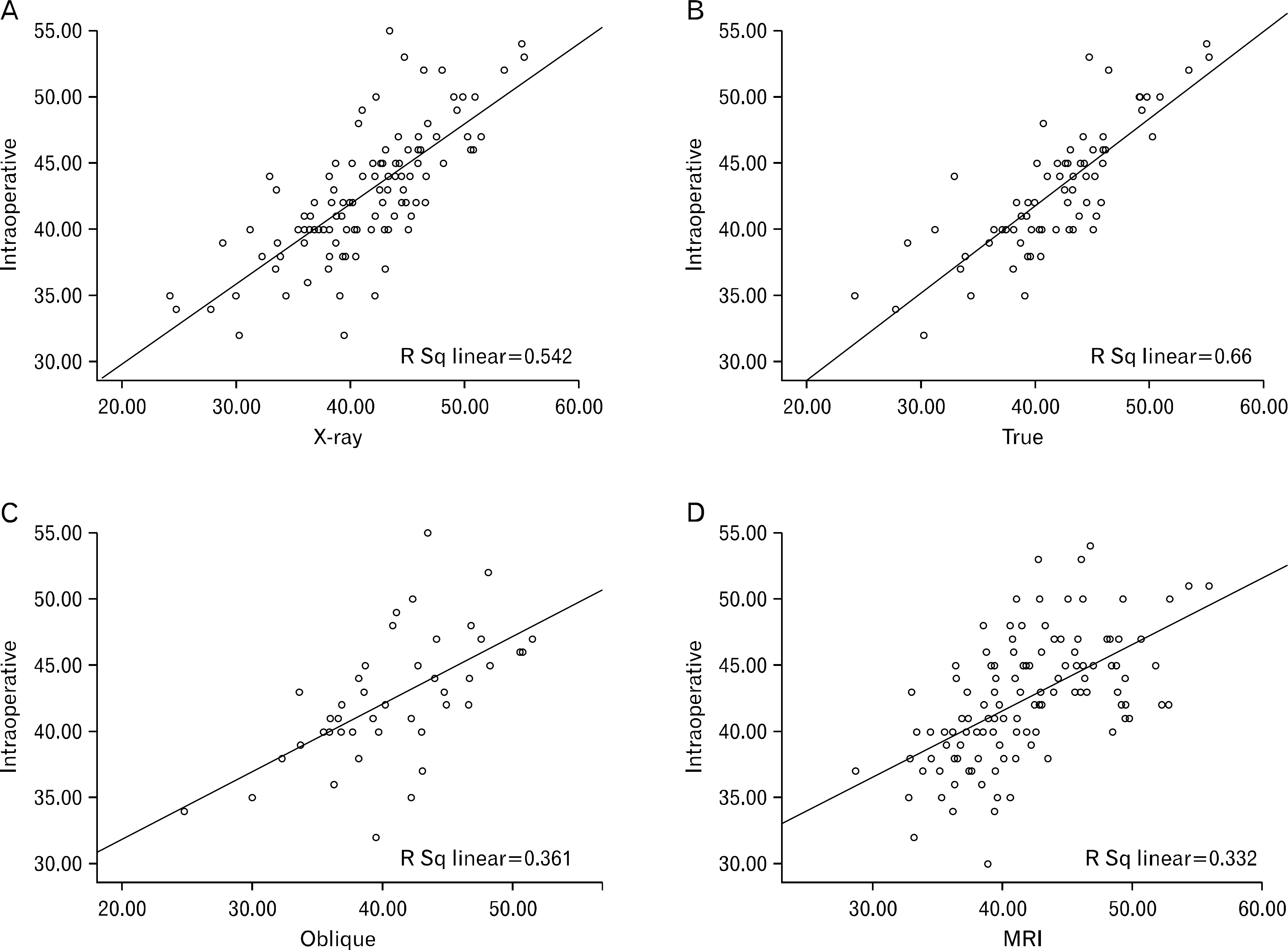
Preoperative prediction of patellar tendon length is important during anterior cruciate ligament (ACL) reconstruction using bone-patellar tendon-bone (BPTB) autograft. Three methods of imaging analysis to predict patellar tendon length were compared in this study. One hundred and twenty-three patients who underwent ACL reconstruction using BPTB autograft by single surgeon during October 2002 through April 2011 were included. We measured the patellar tendon length from true and oblique lateral simple radiographs (classified according to degree of rotation) and magnetic resonance image (MRI). These values were compared with actual length measured during operation and assessed accuracy by calculating the coefficient of determination. The mean length of patellar tendon measured during operation and by true lateral and oblique lateral radiographs and MRI were 42.4±0.45 mm (range, 32.0–54.0 mm), 41.7±0.61 mm (range, 24.2–55.3 mm), 40.7±0.57 mm (range, 24.8–51.5 mm), and 41.7±0.52 mm (range, 28.7–56.0 mm), respectively. The correlation of patellar tendon length was the most strong between actual length and value from true lateral radiograph (coefficient of determination, r2=0.660) according to simple linear regression analysis. R2 values were 0.361 and 0.332 for oblique lateral radiograph and MRI compared to actual value, respectively. In conclusion, Patellar tendon length measured on true lateral radiograph was the best method to coincide with actual patellar tendon length among various preoperative prediction methods.
Go to : 
References
1. Lee JH, Bae DK, Song SJ, Cho SM, Yoon KH. Comparison of clinical results and second-look arthroscopy findings after arthroscopic anterior cruciate ligament reconstruction using 3 different types of grafts. Arthroscopy. 2010; 26:41–9.

2. Prodromos CC, Joyce BT, Shi K, Keller BL. A metaanalysis of stability after anterior cruciate ligament reconstruction as a function of hamstring versus patellar tendon graft and fixation type. Arthroscopy. 2005; 21:1202.

3. Delay BS, Smolinski RJ, Wind WM, Bowman DS. Current practices and opinions in ACL reconstruction and rehabilitation: results of a survey of the American Orthopaedic Society for Sports Medicine. Am J Knee Surg. 2001; 14:85–91.
4. Verma NN, Dennis MG, Carreira DS, Bojchuk J, Hayden JK, Bach BR Jr. Preliminary clinical results of two techniques for addressing graft tunnel mismatch in endoscopic anterior cruciate ligament reconstruction. J Knee Surg. 2005; 18:183–91.

5. McAllister DR, Bergfeld JA, Parker RD, Grooff PN, Valdevit AD. A comparison of preoperative imaging techniques for predicting patellar tendon graft length before cruciate ligament reconstruction. Am J Sports Med. 2001; 29:461–5.

6. Choi YJ, Lee KW, Ahn HS, et al. The length of the patellar tendon in normal adults. J Korean Knee Soc. 2010; 22:39–45.
7. Odensten M, Gillquist J. Functional anatomy of the anterior cruciate ligament and a rationale for reconstruction. J Bone Joint Surg Am. 1985; 67:257–62.

8. Gil YC, Park JA, Yang HJ, Lee HY. Anatomy of the femoral attachment site of the anterior cruciate ligament and the posterolateral structures related to the stability of the knee joint. Korean J Anat. 2008; 41:57–65.
9. Bowers AL, Bedi A, Lipman JD, et al. Comparison of anterior cruciate ligament tunnel position and graft obliquity with transtibial and anteromedial portal femoral tunnel reaming techniques using high-resolution magnetic resonance imaging. Arthroscopy. 2011; 27:1511–22.

10. Yoon KH, Bae DK, Ha JH, Park HK, Kwon BK. Revision anterior cruciate ligament reconstruction: cause of graft failure and results of revision surgery. J Korean Sports Med. 2005; 23:246–50.
11. Bin SI, Kim JC, Kim CM, Youn DJ. Clinical application of N+7 method for the solution of graft: tibial tunnel discrepancy in anterior cruciate ligament reconstruction. J Korean Knee Soc. 2001; 13:62–6.
12. Chang CB, Seong SC, Kim TK. Preoperative magnetic resonance assessment of patellar tendon dimensions for graft selection in anterior cruciate ligament reconstruction. Am J Sports Med. 2009; 37:376–82.

13. Shabshin N, Schweitzer ME, Morrison WB, Parker L. MRI criteria for patella alta and baja. Skeletal Radiol. 2004; 33:445–50.

14. Yoo JH, Yi SR, Kim JH. The geometry of patella and patellar tendon measured on knee MRI. Surg Radiol Anat. 2007; 29:623–8.

15. Wang H, Hua C, Cui H, et al. Measurement of normal patellar ligament and anterior cruciate ligament by MRI and data analysis. Exp Ther Med. 2013; 5:917–21.

16. Li X, Xu CP, Song JQ, Jiang N, Yu B. Single-bundle versus double-bundle anterior cruciate ligament reconstruction: an up-to-date metaanalysis. Int Orthop. 2013; 37:213–26.

17. Alentorn-Geli E, Samitier G, Alvarez P, Steinbacher G, Cugat R. Anteromedial portal versus transtibial drilling techniques in ACL reconstruction: a blinded cross-sectional study at two- to five-year follow-up. Int Orthop. 2010; 34:747–54.

18. Wang H, Fleischli JE, Zheng NN. Transtibial versus anteromedial portal technique in single-bundle anterior cruciate ligament reconstruction: outcomes of knee joint kinematics during walking. Am J Sports Med. 2013; 41:1847–56.
19. Chalmers PN, Mall NA, Cole BJ, Verma NN, Bush-Joseph CA, Bach BR Jr. Anteromedial versus transtibial tunnel drilling in anterior cruciate ligament reconstructions: a systematic review. Arthroscopy. 2013; 29:1235–42.

20. Lim HC, Yoon YC, Wang JH, Bae JH. Anatomical versus non-anatomical single bundle anterior cruciate ligament reconstruction: a cadaveric study of comparison of knee stability. Clin Orthop Surg. 2012; 4:249–55.

21. Mandal A, Shaw R, Biswas D, Basu A. Transportal versus transtibial drilling technique of creating femoral tunnel in arthroscopic anterior cruciate ligament reconstruction using hamstring tendon autograft. J Indian Med Assoc. 2012; 110:773–5.
22. Alentorn-Geli E, Lajara F, Samitier G, Cugat R. The transtibial versus the anteromedial portal technique in the arthroscopic bone-patellar tendon-bone anterior cruciate ligament reconstruction. Knee Surg Sports Traumatol Arthrosc. 2010; 18:1013–37.

23. Hantes ME, Zachos VC, Liantsis A, Venouziou A, Karantanas AH, Malizos KN. Differences in graft orientation using the transtibial and anteromedial portal technique in anterior cruciate ligament reconstruction: a magnetic resonance imaging study. Knee Surg Sports Traumatol Arthrosc. 2009; 17:880–6.

24. Bedi A, Raphael B, Maderazo A, Pavlov H, Williams RJ 3rd. Transtibial versus anteromedial portal drilling for anterior cruciate ligament reconstruction: a cadaveric study of femoral tunnel length and obliquity. Arthroscopy. 2010; 26:342–50.

25. Reeboonlap N, Pongpatarat W, Charakom K. A comparison of lateral radiograph of the knee in extended weight bearing and 30 degrees flexion to predict a patellar tendon length. J Med Assoc Thai. 2012; 95(Suppl 10):S158–62.
Go to : 
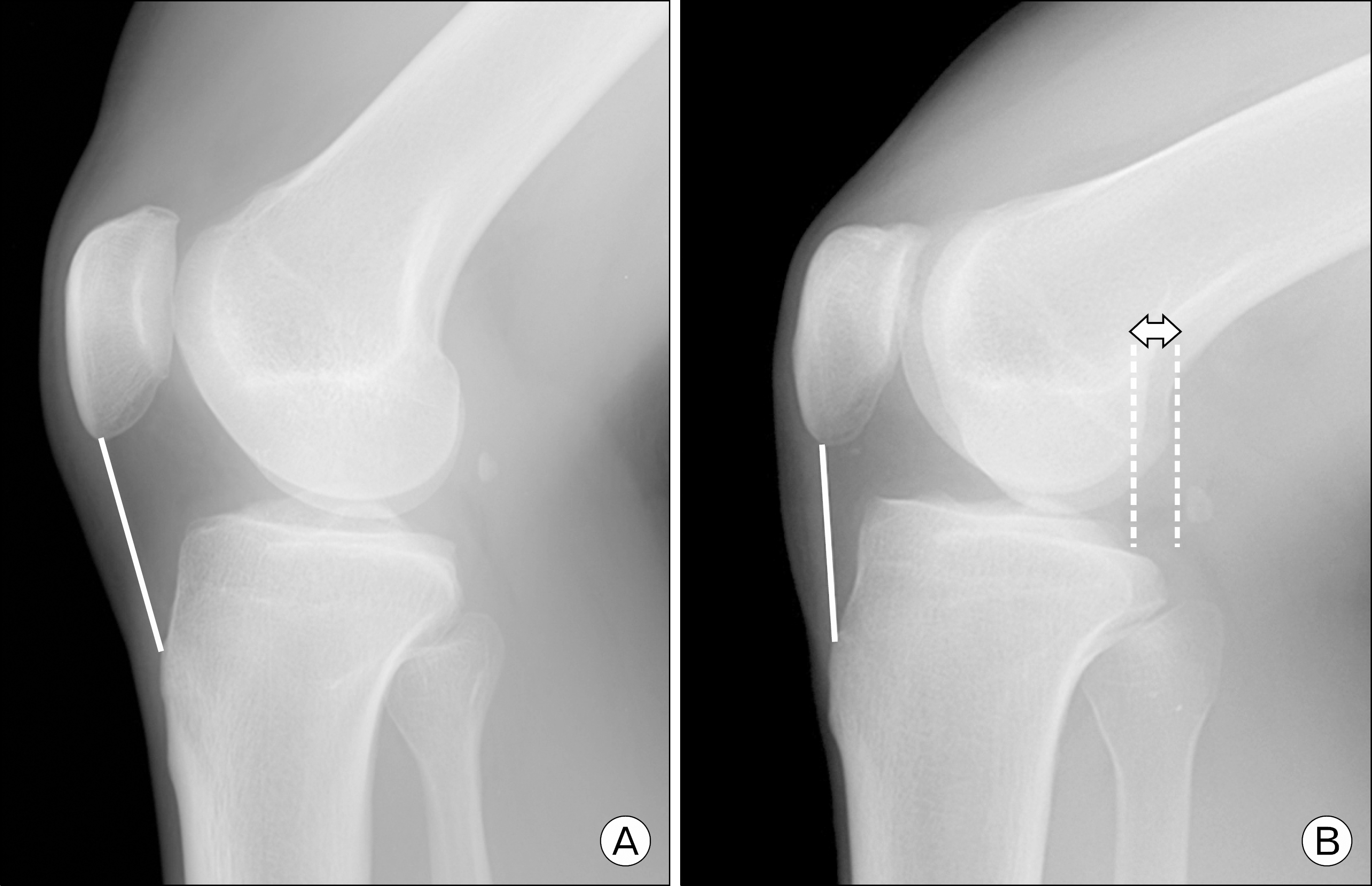
 | Fig. 2.The patellar tendon length was measured on both true lateral (A) and oblique lateral (B) radiograph. Oblique lateral radiograph was defined when there was distance over 5 mm (left right arrow) between medial and lateral posterior condyles. |
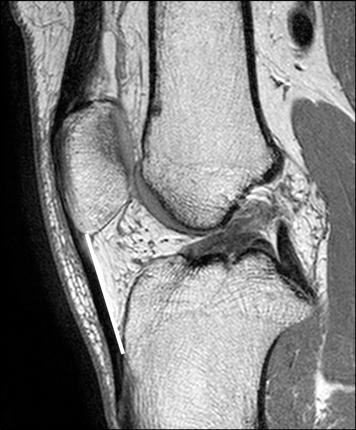
 | Fig. 3.Proton density weighted sagittal magnetic resonance image demonstrate the measurement of patellar tendon length which is measured the inner aspect of patellar tendon from inferior pole to tibial tuberosity. |
 | Fig. 4.The relationship between intraoperative and true lateral radiograph was most proportional, but between intraoperative and oblique lateral, magnetic resonance image (MRI) was less. The correlations between intraoperative data and the length from (A) X-ray including true lateral and oblique lateral view. (B) True lateral X-ray view. (C) Oblique lateral X-ray view, and (D) MRI view were drawn. |
Table 1.
The patellar tendon length measured with various methods
Table 2.
The simple linear regression analysis between patellar tendon lengths measured during operation and preoperative prediction methods
Table 3.
Inter-observer and intra-observer reliability (intraclass correlation coefficients value)


 PDF
PDF ePub
ePub Citation
 Print
Print


 XML Download