Abstract
Calcific tendinitis is most common seen within the rotator cuff of the shoulder, although it may develop around the hip, wrist, elbow, knee, forefoot, and neck. However, there has been no report in the medical literature regarding calcific tendinitis of the common extensor tendon. We present a case of a 26-year-old woman who had calcific tendinitis of the common extensor tendon. Intraoperatively, partial rupture and calcific deposit at the insertion of the common extensor tendon were seen. We were removed calcific deposit and ruptured tissue of common extensor tendon, and then ruptured common extensor tendon was sutured. The patient showed excellent result two years postoperatively with return to range in a degree of activity levels.
References
1. Cox D, Paterson FW. Acute calcific tendinitis of peroneus longus. J Bone Joint Surg Br. 1991; 73:342.

2. Murase T, Tsuyuguchi Y, Hidaka N, Doi T. Calcific tendinitis at the biceps insertion causing rotatory limitation of the forearm: a case report. J Hand Surg Am. 1994; 19:266–8.

3. Kim JS, Yoo JH, Yoo SO. Arthroscopic treatment of chronic calcific tendinitis of the shoulder. J Korean Shoulder Elbow Soc. 1998; 1:6–11.
4. Brinsden MD, Wilson JH. Acute calcific tendinitis of the peroneus longus tendon. Injury Extra. 2005; 36:426–7.

5. Uhthoff HK, Sarkar K, Maynard JA. Calcifying tendinitis: a new concept of its pathogenesis. Clin Orthop Relat Res. 1976; 118:164–8.
6. Hamada J, Ono W, Tamai K, Saotome K, Hoshino T. Analy-sis of calcium deposits in calcific periarthritis. J Rheumatol. 2001; 28:809–13.
7. Raghavendran RR, Peart F, Grindulis KA. Subcutaneous calcification following injection of triamcinolone hexacetonide for plantar fasciitis. Rheumatology (Oxford). 2008; 47:1838.

8. Uhthoff HK, Loehr JW. Calcific tendinopathy of the rotator cuff: pathogenesis, diagnosis, and management. J Am Acad Orthop Surg. 1997; 5:183–91.

9. Ark JW, Flock TJ, Flatow EL, Bigliani LU. Arthroscopic treatment of calcific tendinitis of the shoulder. Arthroscopy. 1992; 8:183–8.

10. Kim MK, Bae JH, Jeon YS. Conservative and early arthroscopic treatment of calcific tendinitis. J Korean Arthrosc Soc. 2009; 13:149–54.
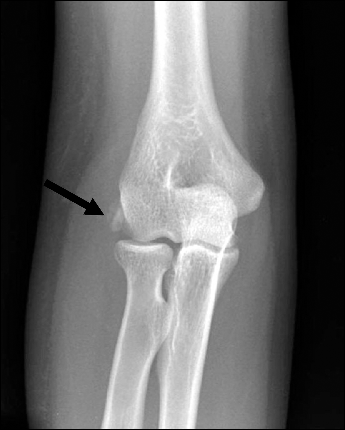
Fig. 1.
Anteroposterior radiograph of the right elbow shows rounded calcific deposit at the insertion of the common extensor tendon (black arrow).

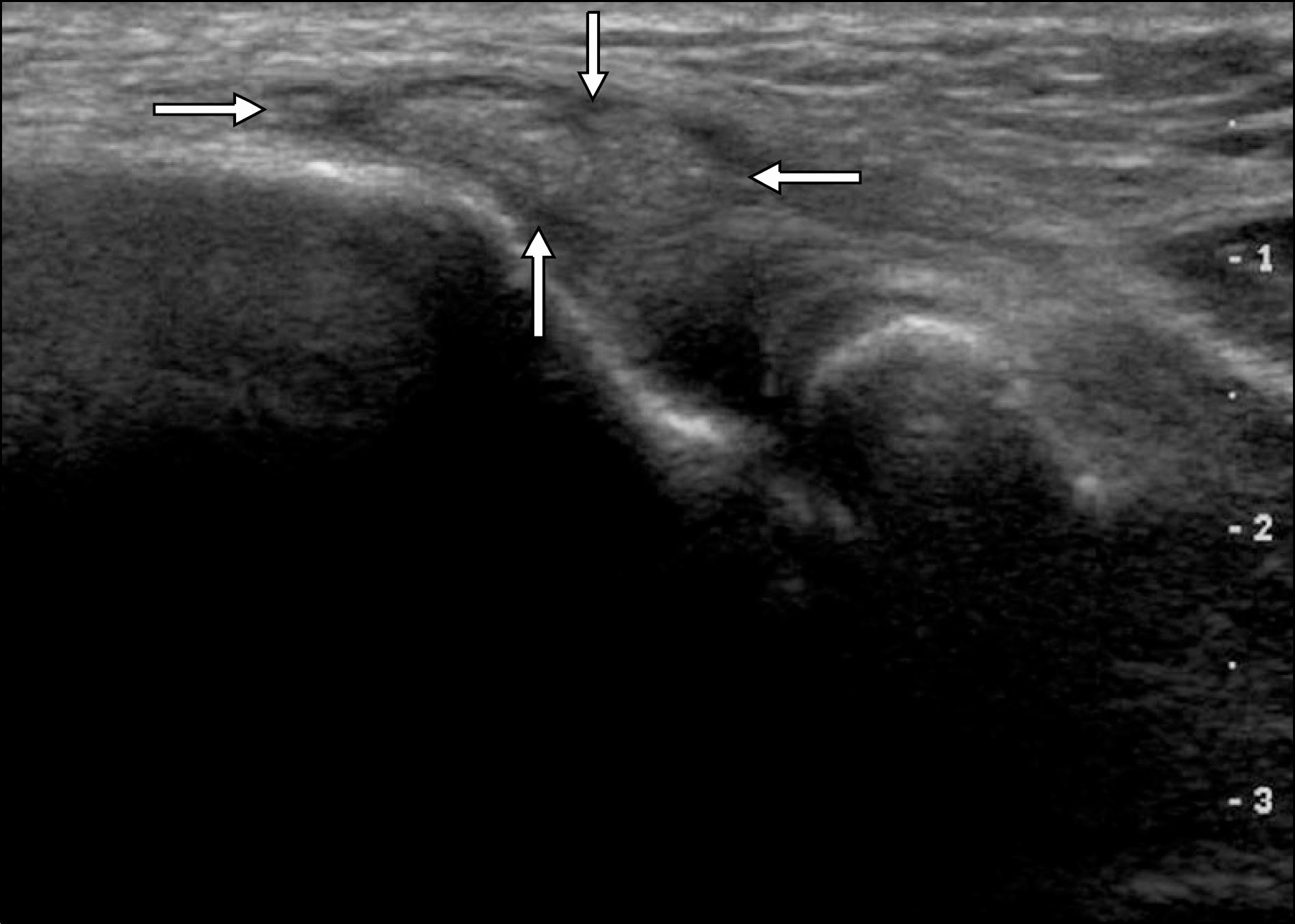
Fig. 2.
Sonographic image of the right elbow shows swel-ling and calcification at the insertion of the common ex-tensor tendon (white arrow).


 PDF
PDF ePub
ePub Citation
 Print
Print


 XML Download