

 PDF
PDF ePub
ePub Citation
 Print
Print


Abstract
The purpose of this study was to evaluate the change in three-dimensional (3D) ankle joint kinematics on the arch pad. Ten male subjects performed running trials on treadmill, and ten motion capture system were used to record a position data of the reflected markers on the lower limb. Two conditions (no support and support: arch pad) were compared with the joint angles which were analyzed by the Cardan method. As a result, ankle joint eversion angles with support shoe (−6.5o±2.5o) were significantly lower than that with no support shoe (−10.2o±3.2o) (p<0.01). Nevertheless, differences in ankle joint dorsiflexion and abduction angles were not significantly different between support (20.1o±3.1o, −2.5o±1.9o) and no support shoe (20.4o±3.4o, −2.4o±2.1o) (p>0.25, p>0.11). Shoes with medial arch support or using special shoe inserts may help correct one's running form by reducing pronation and may reduce risk of running injury.
Go to : 
References
1. Paffenbarger RS Jr, Hyde RT, Jung DL, Wing AL. Epidemiology of exercise and coronary heart disease. Clin Sports Med. 1984; 3:297–318.

2. Kohrt WM, Snead DB, Slatopolsky E, Birge SJ Jr. Additive effects of weightbearing exercise and estrogen on bone mineral density in older women. J Bone Miner Res. 1995; 10:1303–11.
3. Kim HY, Doyo W, Kozakai R, Aizawa H, Ando F, Shimokata H. The relations between bone mineral density (BMD) and mechanical loads acting on the lower limbs: using the gait analysis in middle-aged and elderly Japanese. J Biomech. 2007; 40:S485.
4. Neumann DA. Ankle and foot. Neumann DA, editor. Kinesiology of the musculoskeletal system: foundations for physical rehabilitation. 2nd ed.St. Louis (MO): Mosby;2011. p. 573–626.
5. Nigg BM. Biomechanical aspects of running. Nigg BM, editor. Biomechanics of running shoes. Champaign (IL): Human Kinetics Publishers;1986. p. 1–61.
6. Kim HY, Sakurai S, Ahn JH. Modifications of the shoe produce excessive pronation of the subtalar joint during stance phace of the running. Res J Health Sport Sci Chukyo Univ. 2007; 48:25–36.
7. James SL, Bates BT, Osternig LR. Injuries to runners. Am J Sports Med. 1978; 6:40–50.
8. Bates BT, Osternig LR, Mason B, James LS. Foot orthotic devices to modify selected aspects of lower extremity mechanics. Am J Sports Med. 1979; 7:338–42.
9. Nigg BM BA, Denoth J. Factors influencing kinetic and kinematic variables in running. Nigg BM, editor. Biomechanics of running shoes. Champaign: Human Kinetics Publishers;1986. p. 139–59.
10. Mueller MJ. The ankle and foot complex. Levangie PK, Norkin CC, editors. editors.Joint structure and function: a comprehensive analysis. Philadelphia (PA): F.A. Davis Company;2005. p. 437–75.
11. De Wit B, De Clercq D, Aerts P. Biomechanical analysis of the stance phase during barefoot and shod running. J Biomech. 2000; 33:269–78.
12. Hallemans A, D'Aout K, De Clercq D, Aerts P. Pressure distribution patterns under the feet of new walkers: the first two months of independent walking. Foot Ankle Int. 2003; 24:444–53.
13. Winter DA. Biomechanics and motor control of human movement. Hoboken (NJ): Wiley and Sons;1990.
14. Areblad M, Nigg BM, Ekstrand J, Olsson KO, Ekstrom H. Three-dimensional measurement of rearfoot motion during running. J Biomech. 1990; 23:933–40.
15. Kim HY, Sakurai S, Ahan J. Errors in measurement of center of pressure (CoP) computed with force plate affect on 3D lower limb joint moment during gait. Intl J Sport Health Sci. 2007; 17:71–82.
16. Nigg BM. The role of impact forces and foot pronation: a new paradigm. Clin J Sport Med. 2001; 11:2–9.
17. Nigg BM, Stergiou P, Cole G, Stefanyshyn D, Mundermann A, Humble N. Effect of shoe inserts on kinematics, center of pressure, and leg joint moments during running. Med Sci Sports Exerc. 2003; 35:314–9.
18. Park JY, Lee HJ, Moon GS. The analysis of electromyography pattern according to foot orthotic type. Korean J Sports Med. 2010; 28:50–6.
19. Ahn JH, Sakurai S, Kim HY. Comparison of muscular activity of the lower limb while running on inclined surfaces. Jpn J Phys Fit Sports Med. 2007; 56:167–8.
20. Perry J. Ankle foot complex. Perry J, editor. Gait analysis: normal and pathological function. Thorofare (NJ): Slack;1992. p. 52–87.
21. Hamill J, Knutzen KM. Angular kinematics. Hamill J, Knutzen KM, editors. editors.Biomechanical basis of human movement. 2nd ed.Philadelphia (PA): Lippincott Williams & Wilkins;2003. p. 309–36.
22. Otsuki S, Ookubo M, Tanaka K, Tsuji S, Kato T, Taguchi Y. Effects of sole support-plate (Am-fit) in the leg sports failure. Clin Sports Med. 1997; 14:469–75.
23. Konda S, Jinji T, Yanai T, Sakurai S, Shimizu T. A method for estimating three-dimensional scapular rotation with the use of optical motion capture system during humeral elevation. Jpn J Biomech Sports Exerc. 2007; 11:276–88.
24. Doyo W, Kozakai R, Kim HY, Ando F, Shimokata H. Spatiotemporal components of the 3-D gait analysis of communitydwelling middle-aged and elderly Japanese: age- and sexrelated differences. Geriatr Gerontol Int. 2011; 11:39–49.
25. Elftman H. Dynamic structure of the human foot. Artif Limbs. 1969; 13:49–58.
26. Stacoff A, Luethi SM. Special aspects of shoe construction and foot anatomy. Nigg BM, editor. Biomechanics of running shoes. Champaign (IL): Human Kinetics Publishers;1986. p. 117–37.
Go to :
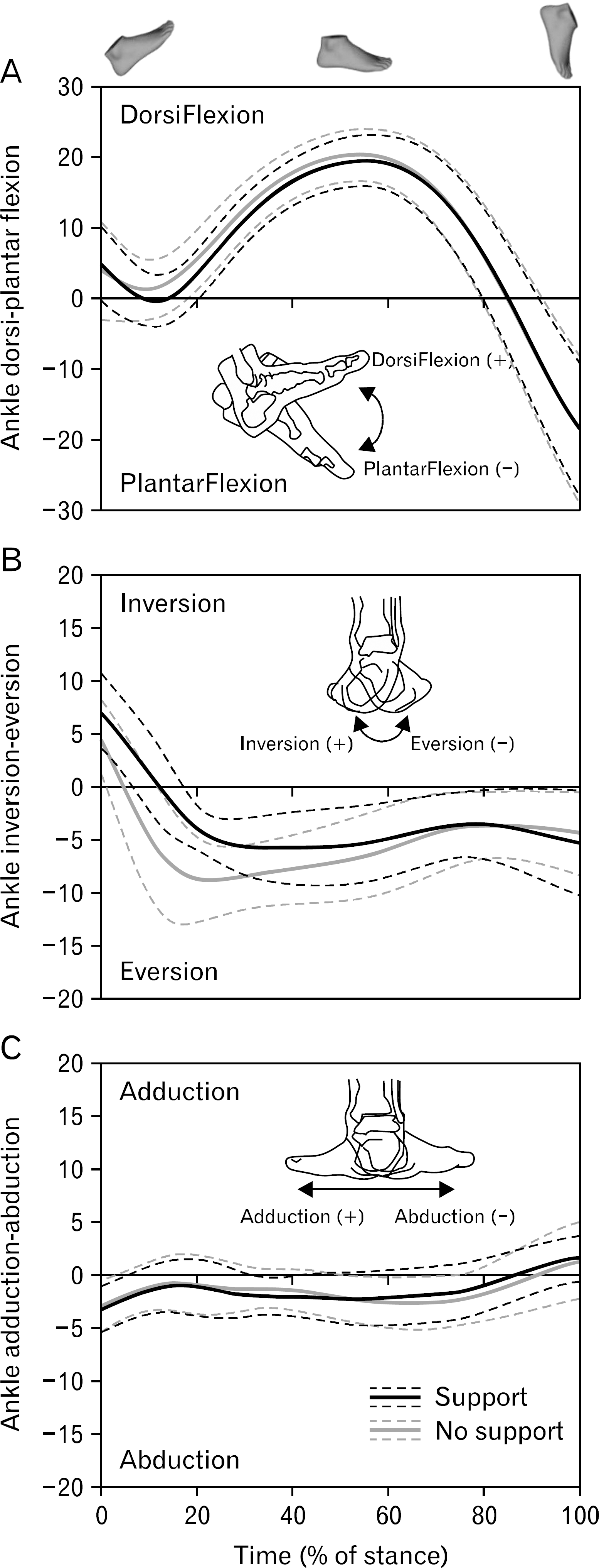 | Fig. 3.Changes of 3D ankle joint angles (n=10) during stance phase of treadmill running. Running time in each subject is normalized as 100%. Lines of each graph are mean and standard deviation. |
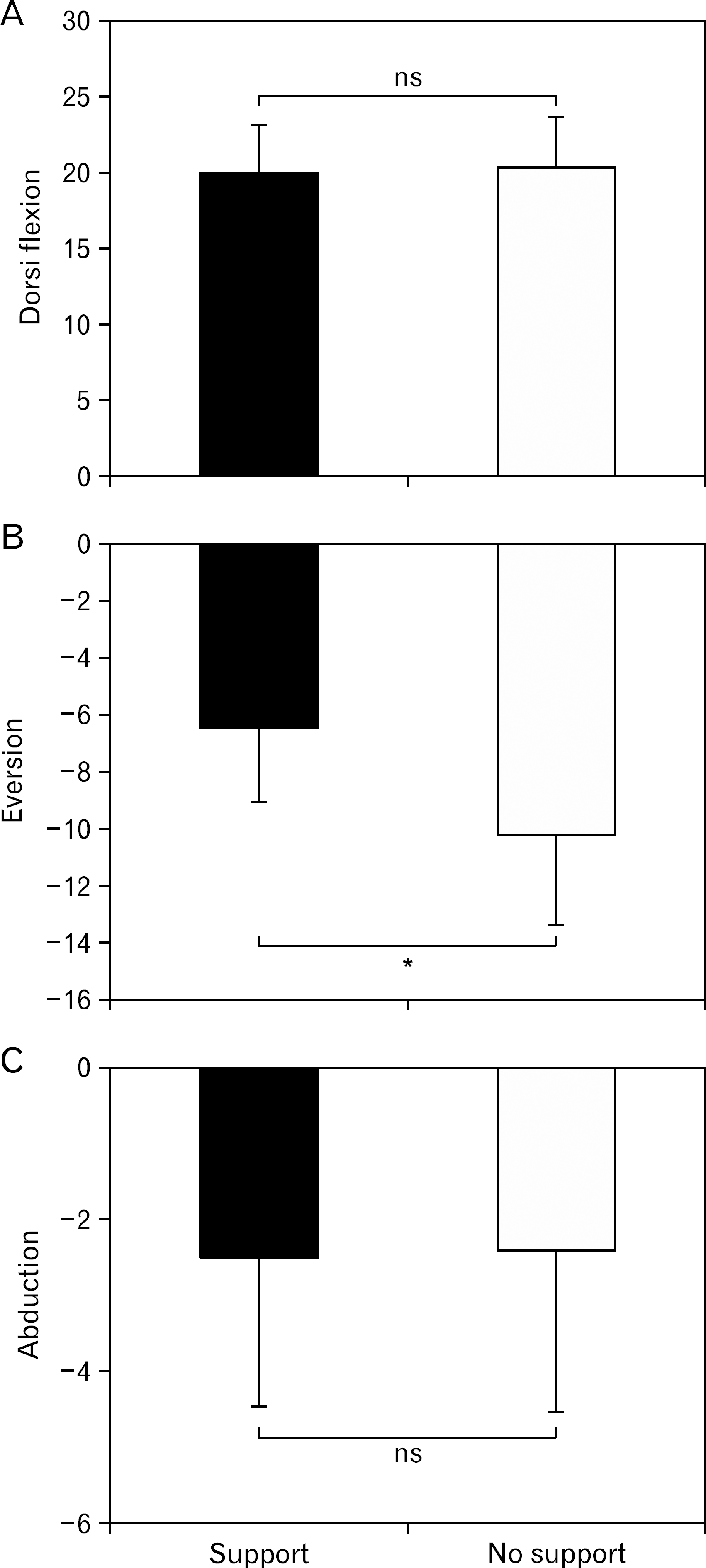 | Fig. 4.Comparisons of 3D ankle joint angles (dorsiflexion (A), eversion (B), and abduction (C)) in the shoes with a medial arch support and no support. ns: not significant.∗p<0.01. |
Table 1.
Physical characteristics and three-dimensional ankle joint angles of subjects (n=10)
| Subject (n) | Hight (cm) | Weight (kg) | DorsiFlexion (°) | Eversion (°) | Abduction(°) | |||
|---|---|---|---|---|---|---|---|---|
| No-support | Support | No-support | Support | No-support | Support | |||
| 1 | 178.5 | 66.8 | 24.8 | 22.6 | –11.1 | –8.8 | –3.5 | –2.7 |
| 2 | 169.3 | 57.1 | 21.0 | 20.8 | –7.4 | –5.9 | –4.4 | –4.6 |
| 3 | 167.4 | 60.6 | 22.0 | 23.6 | –8.5 | –4.1 | –1.6 | –1.8 |
| 4 | 169.3 | 58.3 | 16.7 | 16.3 | –4.3 | –3.0 | 2.9 | 2.5 |
| 5 | 166.6 | 68.1 | 16.5 | 16.9 | –13.7 | -11.7 | –4.8 | –4.2 |
| 6 | 172.1 | 64.4 | 14.0 | 14.2 | –9.0 | –3.8 | –4.2 | –3.9 |
| 7 | 175.5 | 69.5 | 20.7 | 18.9 | –11.1 | –5.8 | –2.6 | –3.5 |
| 8 | 165.1 | 53.8 | 23.0 | 23.9 | –8.3 | –5.9 | –2.2 | –2.2 |
| 9 | 170.2 | 58.2 | 22.1 | 21.6 | –15.0 | –7.5 | –2.4 | –3.0 |
| 10 | 167.1 | 57.7 | 23.6 | 21.9 | –13.8 | –8.7 | –1.2 | –1.7 |
| Mean | 170.1 | 61.5 | 20.4 | 20.1 | –10.2 | −6.5∗ | −2.4 | –2.5 |
| Standard deviat | tion 4.0 | 5.1 | 3.4 | 3.1 | 3.2 | 2.5 | 2.1 | 1.9 |
 XML Download