

 PDF
PDF ePub
ePub Citation
 Print
Print


INTRODUCTION
Many dentists are interested in using aesthetic treatments to meet patient demands.1 For this reason, anterior teeth receive more focus than posterior teeth do, due to the degree of visual exposure. Various analyses of the shape of anterior teeth have been conducted, which compare the shape of the anterior teeth with the golden ratio.2345 The golden ratio was devised by Euclid of Alexandria. This proportion was found to be visually harmonious, with a mathematical ratio of approximately 1.618.2 In 1973, Lombardi3 used the golden ratio to explain visual perception of aesthetics and Levin4 also applied this concept to the maxillary anterior teeth. Richetts5 reported that the golden ratio was useful for aesthetic restoration of the maxillary anterior teeth. However, previous reports have stated that the golden ratio was not readily observed in maxillary anterior teeth.67891011121314 In 1993, Preston6 reported that only 17% of maxillary anterior teeth met the golden ratio. Gillen et al.7 concluded that the golden ratio did not exist at all in the maxillary anterior teeth. In a study by Jordan, the maxillary anterior teeth of 375 dental students were analyzed according to the golden ratio, but this proportion was found to be present in only 31.3% of males and 27.1% of females.8 Thus, it is necessary to construct an additional technique, apart from the golden ratio, to analyze the aesthetics of anterior teeth.15
Since there is no absolute aesthetic standard for anterior teeth, an analysis of the relative size of the anterior teeth can serve as a starting point for the aesthetic analysis of anterior teeth. This type of study would be both objective and easily accessible, which is quite advantageous.16 Thus, studies on the aesthetic analysis of anterior teeth between different races have been reported.1217181920 These studies were mostly limited to research on young patients or dental college graduates.21521
Also, when analyzing anterior teeth, the apparent size of the teeth in the frontal view is more significant than the actual size of the teeth.15 Thus, most studies on aesthetic analysis have been done by analyzing 2D or 3D images observed from the frontal view, rather than analyzing the actual size of the teeth.1722 The lengths of the anterior teeth length were studied in regards to aesthetic treatment, such as anterior width ratio analysis.232425 This factor was associated with wear on the teeth, which differed according to age and gender, but these data were not sufficient to draw concrete conclusions, especially in the Korean population.
The purpose of this study was to analyze the differences in the width ratio of the six maxillary anterior teeth in the Korean population of various age groups. Another purpose of this study is to discover the differences in attrition on the basis of age and gender through the evaluation of the width to length (W/L) ratio of the maxillary central incisors. The first null hypothesis is that there is no difference in the width of the maxillary anterior teeth by age. The second null hypothesis is that there is no difference in the W/L ratio of the maxillary central incisor by age and gender.
Go to : 
MATERIALS AND METHODS
This study included maxillary dental casts of patients admitted to the Department of Prosthodontics, Chonnam National University Dental Hospital, from May, 2014 to April, 2015. Ninety-three volunteers were selected based on the following criteria10: I) all six maxillary anterior teeth present, II) no periodontal disease, III) no crowding or spacing of the anterior teeth, IV) no intrusion, extrusion, or rotation of the anterior teeth, V) no anterior restoration, and VI) no history of orthodontic treatment. Ethical approval was obtained from the Institutional Review Board of Chonnam National University Hospital (CNUDH-2015-017). All volunteers who provided a dental cast and participated in the research signed an informed consent form prior to participation in the study. The sample dental casts were divided into subgroups according to age: Group I (20 to 39 years), Group II (40 to 59 years), and Group III (over 60 years). Table 1 shows the age and gender distribution of the study subjects.
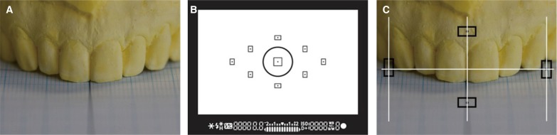
Irreversible hydrocolloid (Cavex impressional, Cavex Holland BV, Haarlem, The Netherlands) impressions of the maxillary arches were made using metal stock impression trays (Osung Industrial, Kimpo, Korea) and poured with Type III dental stone (Neoplumstone, Mutsumi Chemical Industry, Yokkaichi, Japan). Standardized photographs in the anterior view were taken using a digital single lens reflex (DSLR) camera (EOS 100D, Canon Inc., Tokyo, Japan) equipped with a 90 mm f/2.8 Di 1 : 1 Macro lens (Tamron Corp., Saitama, Japan) and a macro ring flash (Sigma EM-140DG, Sigma Corp., Kanagawa, Japan). Casts were positioned perpendicular to the floor with reference to the central incisor on a graph paper, and the DSLR camera was used with a photographic stand to maintain the same distance to each cast. Dental casts were taken in a central line on the auto-focusing mode screen. As a result, the baseline and midline of the graph paper was constantly in the center of the focus of the camera (Fig. 1). This method can be used to control the locations of the central incisor's mesial contact surface and occlusal plane.
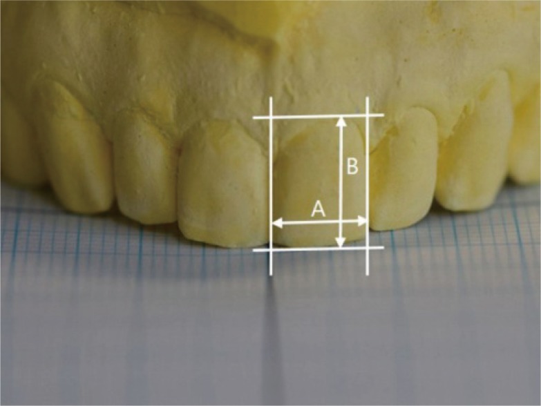
The digital images were saved in a Joint Photographic Experts Group (JPEG) format with a resolution of 300 dpi and evaluated with a ruler tool in photo editing software (Photoshop CS3, Adobe system Inc., San Jose, CA, USA). Tooth mesio-distal width was determined by measuring the maximum distance between the mesial and distal contact points of the tooth on a line perpendicular to the longitudinal axis (Fig. 2).10 After the mesio-distal width of the anterior teeth were computed in pixels, the records were collected and the analyses of LI/CI ratio and CA/LI ratio were carried out using calculating program (Excel, Microsoft Crop., Redmond, WA, USA). All values were compared to the golden ratio and compared among all groups. Right and left ratio values were measured independently.

In addition to this analysis, maxillary central incisor length was evaluated by the same program. Central incisor width was calculated using the aforementioned method, and the length of the central incisors was defined as the length from the incisal edge to the highest point of the gingiva line (Fig. 3). W/L ratios were evaluated based on age and gender.
Statistical analysis was performed using SPSS Version 21.0 (SPSS Inc., Chicago, IL, USA). One-sample t-tests were used to compare the tooth ratios to the golden ratio. One-way ANOVA (Analysis of Variance) was used to analyze the comparisons among the 3 groups by age. The difference in central incisor W/L ratio according to age was analyzed by one-way ANOVA and the post-hoc Tukey test. To investigate the effect of gender in each group, an independent t-test was used. P < .05 was taken to indicate statistical significance.
Go to :
RESULTS
The LI/CI ratio and the CA/LI ratio were statistically significant in comparison to the golden ratio in all groups (P < .001). The left CA/LI average ratio values were the greatest, 0.85 in all ages, and the smallest values were the LI/CI ratios, 0.69 in Group III. Table 2 displays the tooth ratio values, the golden ratio, and a comparison of these ratios.
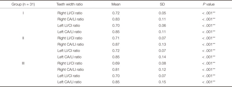
Table 2
One-sample t-test for comparison with the golden ratio
![]()
When we performed one-way ANOVA to compare the ratios among the groups, no statistically significant difference was observed (P > .05) (Table 3).
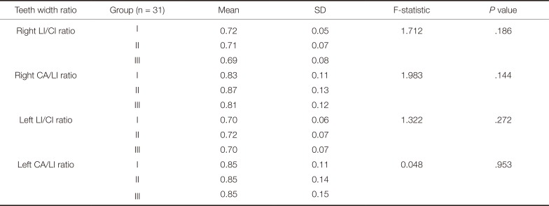
Table 3
Statistical analysis of LI/CI, CA/LI ratios by age using one-way ANOVA
![]()
The Tukey test confirmed a significant difference between the W/L ratio of Group III versus those of Groups I and II (Table 4). An independent t-test done to compare the W/L ratio values with respect to gender in each group confirmed a significant difference in the average values for Group II (men: left 0.86, right 0.85/women: left 0.78, right 0.78) (P < .05) (Table 5).
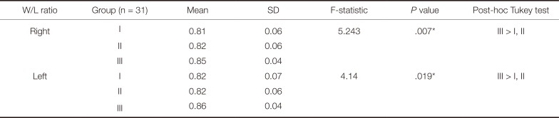
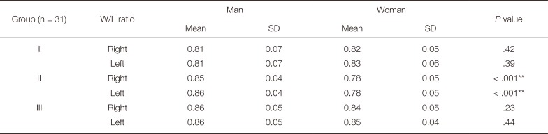
Table 4
One-way ANOVA for W/L ratios of maxillary central incisor
![]()
Table 5
Independent t-test for gender differences in W/L ratio
![]()
Go to :
DISCUSSION
The aim of this study was to compare maxillary anterior teeth width ratio according to age and gender in the Korean population. Another aim of this study was to evaluate the central incisor W/L ratios by age and gender. The first null hypothesis was supported because there was no significant difference among the 3 groups. A higher central incisor W/L ratio was observed in Group III, and a significant difference in values was observed according to gender in Group II, so the second null hypothesis was rejected.
This study utilized patients' maxillary dental casts in standard digital image analysis. In previous studies,1722 the values were often compared by 2D digital photography, and this method has been considered reliable for a long time. We chose to photograph stone casts to allow for a more constant distance between the cast and the digital camera.
For a long time, the golden ratio has been used to analyze the aesthetics of human maxillary anterior teeth. However, this concept is inconsistent with the actual ratios of the anterior teeth. The golden ratio was not observed in 100 Turkish students' maxillary anterior teeth,10 and a study of the anterior teeth of forty-nine Malaysians by Al-Marzok et al.17 also did not follow the golden ratio. Ward13 asserted a seventy percent LI/CI ratio as an aesthetic anterior tooth ratio, which is in line with the results of this study. Our study's findings of LI/CI ratios and CA/LI ratios were seventy or eighty percent; especially, CA/LI ratios were larger than LI/CI ratios. The reason for this result may be a difference of arch form based on race. Numerous Asians have a square arch, unlike Western population, so the canine ratio in the front view appears to be greater in Asians.1819
In a previous study,9 aesthetic harmony with the golden ratio was recognized only in long teeth and was not accepted in very short, short, normal height, or long teeth. Therefore, when the ratio of anterior teeth is researched, it is necessary to first analyze the anterior teeth width ratio according to age, gender, and race rather than to compare it with a uniform criterion. But in this study, maxillary anterior teeth width ratios were not significantly different according to age in a Korean population. Cooper et al.23 observed that patients' judgments of aesthetic treatment were less sensitive than those of dentists or dental technicians, the reason being a lack of significant difference in anterior teeth among all age groups in the study. Therefore, it would be advisable to carry out research on anterior teeth treatment by patient satisfaction, regardless of patient age.
The central incisor W/L ratios are another consideration for aesthetic treatment. In an evaluation of maxillary central incisor tooth W/L ratios in Caucasians, Sterrett et al., in 1999,11 identified a male ratio of 0.85 and a female ratio of 0.86. Also, W/L ratio ranged from 65% to 85% in a study by Peixoto,22 which is similar to that found in our study. In this study, the oldest age group was observed to have a higher W/L ratio than the other groups did. This may be attributed to attrition of teeth associated with prolonged use.24 Two potential reasons for the differences in W/L ratios according to gender in Group II were considered. The first reason was associated with a greater tendency for tooth wear in males. However, previous studies found that gender did not affect tooth wear.2025 The second reason involved hormonal changes in elderly women, which could potentially destroy periodontal tissues.26 Further study will be necessary for the analysis of this phenomenon.
The results of this study demonstrated that anterior tooth appearance was not consistent with the golden ratio and was similar in all age group in the Korean population. Finally, this study found that various morphologies of the maxillary central incisor existed. The limitations of this study include a lack of a sufficient number of patients and age groups, and an absence of classification according to arch form. If such issues are resolved in the future, stronger comparisons can be made and better guidelines can be developed for aesthetic treatment.
Go to :
CONCLUSION
Given the limitations of this study, the following conclusions can be drawn:
Maxillary anterior teeth width ratios do not follow the golden ratio in the Korean population. Maxillary anterior teeth width ratios are similar regardless of age. The average W/L ratio of maxillary central incisors is the greatest in patients over sixty years of age. In patients aged forty to fifty years old, the maxillary central incisor W/L ratio of males is higher than that of females.
Go to :
 XML Download