

 PDF
PDF ePub
ePub Citation
 Print
Print


INTRODUCTION
Since July 1, 2014, treatment with two dental implants as well as complete removable denture has been covered by Korea national health insurance in old people over 75 years old. As a result, the number of patients treated with such prostheses increases gradually, and therefore, detailed knowledge on anatomical structure is required. In particular, the mandible has a much faster absorption rate than the maxilla, and the residual bone height in posterior mandible is insufficient for implant placement compared with that of anterior mandible.1 Therefore, in a severely atrophied mandible, it is recommended to be treated with complete removable prostheses with a two-implant-supported overdenture which is placed between the mental foramen.2
Around the mental foramen, the mandibular canal divides into the mental and incisive canals and continues on to the region of the anterior teeth.3 The mental canal curves upward, backward, and lateral to reach the mental foramen, which is located below the second premolar,45 and the incisive canal continues to run toward the anterior teeth in a slightly downward direction, eventually reaching the chin.6 This means that the mandibular canal runs along the lingual cortical plate at the mandibular ramus and body, and then continues toward the mental canal with three dimensional (3D) complex course to exit the buccal side toward the mental foramen.78
At this ramification point, the canal forms an anterior loop and transitions back posteriorly to the mental canal; the mandibular and mental canals exist as two canals simultaneously.9 Therefore, even though the mental foramen is well known to clinician and the bone thickness on anterior mandible is thickest in canine distal region,10 at the interforaminal region between the mental foramen and the point at which the anterior loop develops, special care should be required during surgical procedures such as dental implant placement and genioplasty to avoid damaging the neurovascular bundle.579
In addition, the mandibular canal still includes the mental and dental nerves simultaneously at the interforaminal region across the mental foramen.11 The incisive nerve is totally separated from the surrounding epineurium of the mental nerve in the premolar region, being located lingually and inferiorly thereto, and continued into the incisive canal, which is incompletely formed and has a smaller diameter than the mandibular canal.3121314 Thus, on panoramic imaging, which is used widely for preoperative evaluation of the jaw, the mandibular canal and mental foramen are reportedly readily visible in 49% of images, while the anterior loop is readily visible in only 3%.12 And, although cone beam computed tomography imaging was recently found to enhance the visualization of the mandibular anatomical structures,515 detailed evaluation remains difficult due to low cortical bone density and an incomplete bony canal at the interforaminal region.
Micro-computed tomography (microCT) can produce high-resolution images and is an effective method for detailed evaluation of the internal structure of bones.1617 Furthermore, since 3D reconstructions are possible with these acquired high-resolution images, it is a very effective tool for examining the small facial canals.18 Therefore, the purposes of this study were to identify using 3D reconstruction of microCT images and to provide the diagram for clinicians to help them understand the complex course of the mandibular canal at the interforaminal region.
MATERIALS AND METHODS
Twenty-six hemimandibles from 19 cadavers that had been donated to the Department of Anatomy, School of Medicine, Chosun University for educational purposes were examined in this study. They were comprised of 16 males and 3 females, with a mean age at death of 54.4 years (range, 29 – 75 years), harvested all dentulous specimens which were from the first molar to the lateral incisor, and were subjected to microCT scanning. This study followed the Declaration of Helsinki with respect to the medical protocol and ethics.
The specimens were placed onto the holder so that the inferior border of the mandible was touching and perpendicular to the floor, and were scanned using microCT (TVXIMT225CT Dual type Micro CT, Techvalley, Seongnam, Korea) with a focus size of 1 µm. The obtained serial images were three-dimensionally reconstructed using 3D Doctor Software (3D-Doctor V 3.5 demo version, Able Software Corporation, MA, USA). Every fifth image was used for reconstruction because the three-dimensionally reconstructed results did not affect the original morphology of the mandible.
On the three-dimensionally reconstructed images, the mandibular and mental canals, mental foramen, and tip of the anterior loop, which coincides with the starting point of the mental canal, were identified. According to the result of Yu et al.19 that the mean diameters of the mandibular and mental canals are 2.80 and 2.63 mm, respectively, the tip of anterior loop was set a cutoff point of 2 mm for its minimum diameter. After that, at both the midpoint of mental foramen and the tip of anterior loop, the bucco-lingual distances from the mandibular canal to external cortex and the height from the inferior border of the mandibular canal to the inferior border of the mandible were measured. At the midpoint of mental foramen, the vertical distance from the inferior border of the mandibular canal to the superior border of the mental foramen was also measured. And, the horizontal distance between the midpoint of mental foramen and the tip of anterior loop, like as the anterior loop length of previous researches, was investigated. The positions of the midpoint of mental foramen and the tip of anterior loop were also examined relative to tooth site.
After the mandibular canal was set as the horizontal line, the specimens were classified into three types according to the divergent shape of the mental canal from the mandibular canal in the posterior-superior direction. And the angle that the mental canal diverges from the mandibular canal was also measured in lateral-superior direction.
After microCT scanning, the specimens were decalcified for 3 days in 10% nitric acid and then neutralized in distilled water for 12 hours. While conserving the outer shape of the mental foramen, the buccal cortical and cancellous bone were carefully removed with the aid of a surgical microscope (OPMI-FC, Carl Zeiss, Oberkochen, Germany) to prevent damage to the inferior alveolar neurovascular bundle. The shape of the divergence of the mental canal from the mandibular canal and the positions of the mental foramen and the anterior loop of the mental canal relative to tooth site were re-examined on the dissected specimens.
One-way ANOVA was used to analyze differences between observers, bucco-lingual distances at each measured locations, and buccal distance and height between two locations using IBM SPSS Statistics (version 23.0, IBM Corporation, Somers, NY, USA). The significance level was set at P < .05.
RESULTS
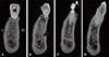
The mandibular and mental canals were observed running anteriorly past the mental foramen on the serial, coronalplane microCT images (Fig. 1). While the cortical wall that surrounds the mandibular canal was well formed, the cortical wall of the mental and incisive canals was partially broken down, rendering it difficult to identify the canal borders.
The buccal distance from the mandibular canal was significantly shorter than lingual distance at both the mental foramen and the tip of anterior loop (P < .05). The mandibular canal at the tip of anterior loop was significantly located closer to buccal side (P < .05) and higher (P = .101) than at the mental foramen (Table 1). And the mean vertical distance from the mandibular canal to the mental foramen was 8.38 ± 2.16 mm.
The mean horizontal distance between the mental foramen and the tip of anterior loop was 5.19 ± 1.48 mm, ranging from 2.98 to 9.45 mm. The mental foramen was observed most commonly below the second premolar (54.6%, n = 12), and the tip of the anterior loop was observed most commonly below the first premolar (45.5%, n = 10; Table 2).
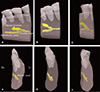
As a result of 3D reconstruction, the divergent shape of the mental canal, which runs a curved course posteriosuperiorly from the mandibular canal, was classified into three types according to the angle of that curvature. Type 1 (0 – 30°) was observed in 18% (n = 4) of the specimens, with the mental canal lying almost parallel to the mandibular canal. Type 2 (30 – 60°) was the most common type, occurring in 59% (n = 13) of the specimens; the mode was 50°. Type 3 (60 – 90°) appeared in 23% (n = 5) of the specimens, and since the mental canal in these cases was almost perpendicular to the mandibular canal, the anterior loop of the mental canal was very short or absent (Fig. 2). And the mean angle that the mental canal diverges from the mandibular canal was 41.6 ± 9.22° in lateral-superior direction.
DISCUSSION
The mandibular canal crosses the mental foramen, then forms the anterior loop at the interforaminal region, and separates into the mental canal and the incisive canal.20 When the clinicians give a surgical treatment in the interforaminal region including the parasymphyseal area, the mental canal with its complex course, the anterior overextension of the anterior loop beyond the mental foramen, and the large size of the incisive canal should be carefully taken into account.6721 Repetitive and empirical surgeries performed without an accurate preoperative understanding of these anatomical structures may result in discomfort and postoperative pain for patients.
As the residual bone height is not sufficient for implant placement, the bucco-lingual distance from the mandibular canal is more important.8 In previous research, the mandibular canal courses close to the lingual side in molar region leans to the buccal side toward the premolar region,781122 and then runs close to the middle portion in incisor region.11 In the present study, at both the mental foramen and the tip of anterior loop, it was also located close to the buccal side. Moreover, at the tip of anterior loop more anteriorly, it was significantly closer to buccal side than the mental foramen. In particular, at the mental foramen, the height from the inferior border of the mandibular canal to the inferior border of the mandible was 13.11 mm and the vertical distance to mental foramen was 8.38 mm. The combined value was approximately 21 mm similar to the value of Liang et al.'s study.1 The distance from the superior border of the mandibular canal to the cementoenamel junction was approximately 19 mm.22 Therefore, it was thought that the height of alveolar bone above the mental foramen was only about 10 mm in normal patient without periodontal diseases.
As the anterior loop length in previous researches, the horizontal distance from the mental foramen to the tip of the anterior loop varies in the range 0.2 – 7.6 mm,19 and its prevalence has been reported an wide range of 0 – 88%.5 In the present study, the midpoint of the mental foramen was most commonly found below the second premolar and the tip of the anterior loop was most commonly found below the first premolar, with a mean length of 5.19 mm ranged from 2.98 to 9.45 mm. Therefore, careful attention needs to be paid to the width of a premolar tooth, which is approximately 7 mm,23 anteriorly from the mental foramen in older edentulous patients.
The mandibular canal has direction with 67.2° superior, 39.4° lateral, and 80.2° posterior based on the mental foramen, forms the anterior loop in 61.5%, and transitions into the mental canal.713 In the present study, the mental canal from the mandibular canal diverged into an angle of approximately 50° posterior-superior and 41° lateral-superior similar to previous research. Therefore, de Freitas et al.24 recommended that the needle be inclined to around 55° from back to front and around 40° from outward to inward when blocking the mental nerve. During implant placement, Krekmanov et al.25 described that if the fixture is tilted 25 – 35° from the anterior loop, an average distance of 6.5 mm can be earned for prosthetic support.
The mental canal was almost parallel (0 – 30°) with the mandibular canal in type 1 cases, meaning that there may be sufficient alveolar bone height superior to the alveolar crest to enable a relatively stable implant placement. However, in type 3 the mental canal was almost perpendicular to the mandibular canal (60 – 90°), and so no anterior loop was formed; this could have a negative effect on implant placement around the mental foramen, but since the anterior mandible at the interforaminal region is relatively extended horizontally, it could have a positive effect on genioplasty. Hu et al.26 noted a vertically formed anterior loop of the mental nerve in 15.4% of cases, and a straight form toward the anterior teeth in 23.1% of the cases. Hence, additional research is needed to evaluate the correlation between the divergent angle of the mental canal relative to the mandibular canal and the length from the mental foramen to the anterior loop, and to identify cases in which the mental canal continues toward the anterior teeth with an angle of more than 90°.
CONCLUSION
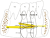
Since the mandibular canal divides into two terminal canals, the mental canal including the anterior loop presents a Y-shaped or delta-shaped divergence.2027 In the present study, the mental canal diverged from the mandibular canal below the first premolar by approximately 50° posteriorsuperior and 41° lateral-superior direction, in which was located about 5 mm in front of the mental foramen, and exited to the mental foramen below the second premolar. Therefore, it could form a hazardous tetrahedron space at the interforaminal region as diagrammed in Figure 3 in which there is the potential for simultaneous damage to the mental and dental branches. The clinician need to pay attention to the width of a premolar tooth from the mental foramen anteriorly during dental implant placement and genioplasty at the interforaminal region.


 XML Download