

 PDF
PDF ePub
ePub Citation
 Print
Print


INTRODUCTION
Natural dentition represents a balanced position between the surrounding muscles and the tongue, with the dental arch balanced with craniofacial structures.1 The position of the anterior tooth and rearmost molar, ridge width, length, and height can be important standard criteria in prosthodontic treatment for the production of a natural profile and functional restoration for edentulous and dentate patients. Understanding the natural dental arch form is important, as the average values of the anatomical shape and the size of the arch are useful for prosthetic restoration.2 For this reason, many studies have addressed the shape and size of the dental arch.
Race and ethnicity influence the form of the human craniofacial complex,345 and the dental arch form is correlated with the craniofacial skeletal pattern.678 In addition, the morphological characteristics of the size and shape of the dental arch, and the arrangement of dentition differ between race, culture, and region.910111213
Several studies have addressed the dental arch in Koreans. The dental arch in Koreans is different in form compared with that in Caucasians; Koreans tend to have larger inter-canine and inter-molar widths.141516171819 Several reports identified the type of dental arch using the sum of the width of six anterior teeth20 or presented the arch form as a parabola using a second-degree polynomial,2122 with U, V, and O arch shapes.23 However, these studies focused only on the arch shape and were concerned with dentition for teeth arrangement without considering the alveolar bone, or lacked sufficient measurement sites that did not include the arch height. There is a lack of information about standardized figures and simplified division in the dental arch of Koreans that can be easily applied in clinical practice.
Dental arch shape and size are related to eating habit and culture.111213 This emphasizes the need for information about the dental arch in Koreans, given the changed diet and culture of Koreans in the recent past,242526 with existing data being collected more than 10 years ago.14151617181920212223
This study investigated relatively young Korean adults and measured a variety of reference points for both dentition and alveolar bone. Based on the results, a simplified classification of dental arch shape is proposed to facilitate better understanding of the dental arch shape in Koreans and clinical application in prosthodontics through accurate data analysis and a statistical approach.
MATERIALS AND METHODS
Fifty individuals who visited Kyung Hee University Dental Hospital at Gangdong in Seoul, Korea, were selected. Inclusion criteria were complete permanent dentition without 3rd molars in both arches, normal occlusion (Class I molar), clinically normal arch shapes with no more than 4 mm of dental crowding, no anterior or lateral crossbite, no extensive restorations, and no previous orthodontic treatment. This study protocol was approved by IRB 2016-08-025 and informed consent was obtained from all participants. The participants ranged in age from 24 to 32 years. The study casts were made using irreversible hydrocollidal impression material (alginate; Aroma Fine DFII, G-C. Co., Tokyo, Japan) and dental stone (New Plastone, G-C. Co., Japan). Measurements were made by the same investigator.
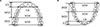
A special measuring device was designed and manufactured (Fig. 1A) for use with digital vernier calipers (Mitutoyo Co., Kawasaki, Japan) and a digital depth gauge (Mitutoyo Co., Kawasaki, Japan) to measure a three-dimensional value up to 0.01 mm. For the reference plane, a T-shape template was fixed in the measuring device and casts were fixed to an adjustable surveyor table (Ney parallerometer) to be on the reference plane. The base of the three-dimensional measuring device and the T-shape standard plane template were maintained parallel to each other within an error range of 0.01 mm. The contact point between incisor edges of the central incisors was positioned in the narrow, small T-shaped window with a standardized plane template. Both 2nd molars were located in the same position anteroposteriorly using a ruler for the perpendicular plate of the standardized plane template (Fig. 1B).
Then, the plane, on which the adjustable table was attached to enable easy reach of the standardized plane template from the incisors and both 2nd molars, was set up as the reference occlusal plane.23
For the incisor-canine distance (ICD), the anteroposterior distance between two mediolateral lines was determined. One line was a tangent through the most forward points on the incisal edge of central incisors. The other line passed passing through the tips of the canine cusps. For the incisor-1st molar distance (IM1D), the anteroposterior distance between two mediolateral lines was determined. One was a tangent through the most forward points on the incisal edge of central incisors. The other passed through the tips of the mesio-buccal cusps of the 1st molars. For the incisor-2nd molar distance (IM2D), the anteroposterior distance between two mediolateral lines was determined. One line was a tangent through the most forward points on the incisal edge of central incisors. The other passed through the tips of the disto-buccal cusps of the 2nd molars. For the intercanine distance (CW), the mediolateral distance between the tips of the canine cusps was determined. For the inter 1st molar distance (M1W), the mediolateral distance between the tips of the mesio-buccal cusps of the right and left first molars was determined. Finally, for the inter 2nd molar distance (M2W), the mediolateral distance between the tips of the disto-buccal cusps of the right and left first molars was determined (Fig. 2).
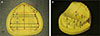
Using the measurements, the following ratios were calculated: ICD/CW, IM1D/M1W, IM2D/M2W, and IM1D/IM2D. Also, the maxillary cast was measured in 12 sites (four arch widths, six arch heights and two arch lengths). The mandible was measured in 13 sites (four arch widths, seven arch heights, and two arch lengths) (Fig. 3).
For the upper dental arch, the following measurements were made (Fig. 3). The mediolateral distance was measured between the basement of buccal part of the alveolar ridge below interproximal contact between the 1st and 2nd molars (BM1,2). The mediolateral distance was measured between the disto-buccal cusp tips of 2nd molars (M2D). The mediolateral distance was measured between the buccal cusp tips of 2nd premolars (P2). The mediolateral distance was measured between the base lines of buccal part of alveolar ridge below point angle of mesio-bucco-occlusal 2nd premolars (BP2). The vertical distance was determined between the midpoint of a line passing through the buccal cusp tips of 1st premolars and palate directly below that of midpoint (PVP1). The vertical distance was measured between the midpoint of a line passing through the distobuccal cusp tips of 2nd molars and palate directly below that of midpoint (PVM2). The vertical distance was determined between the plane passing through the incisal edge of central incisors and the buccal origin of the labial frenum (I-La.F). The vertical distance was measured between the canine cusp tips and the deepest part of the buccal vestibule directly below the canine cusp tip (C-V). The vertical distance was determined between a virtual plane through buccal cusp tips of 1st and 2nd premolars and the deepest part of buccal vestibule below that interproximal contact between 1st and 2nd premolars (P-BV). The vertical distance was measured between a virtual plane through buccal cusps of 1st and 2nd molars and the deepest part of buccal vestibule below that interproximal contact between 1st and 2nd molars (M-BV). The anteroposterior distance was determined between two mediolateral lines; one was a tangent through the most forward points on the incisal edge of central incisors, and the second was a line passing through the most forward point of the buccal frenum origin (I-BF). Finally, the anteroposterior distance between two mediolateral lines was determined; one was a tangent through the most forward points on the incisal edge of central incisors, and the other was a line passing through the most posterior point of 2nd molars (I-M2).
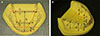
For the lower dental arch, the following measurements were made (Fig. 4): the distance between the base lines on the lingual side of the alveolar ridge below the distal surface of the 2nd molars (LM2), the distance between the disto-lingual cusp tips of the 2nd molars (M2D), the distance between the lingual cusp tips of the 2nd premolars (P2), the distance between the base on the lingual side of the alveolar ridge below the interproximal contacts between the 1st and 2nd premolars (LP2), the vertical distance between the deepest part of lingual frenum origin and the virtual plane passing through the incisor edges of the central incisors (I-Li.F), the vertical distance between the line passing through buccal cusp tips of 2nd premolars and the base on the lingual side of alveolar ridge below that of line (P-V), the vertical ditance between the line passing through the mesiobuccal cusp tips of the 2nd molars and the base on the lingual side of alveolar ridge below that of line (M2-V), the vertical distance between the plane passing through the incisal edge of central incisors and the deepest part of the labial frenum (I-La.F), the vertical distance between the canine cusp tip and the deepest point of the buccal alveolar ridge below the canine cusp tip (C-V), the vertical distance between the virtual plane passing through buccal cusp tips of the 1st and 2nd premolars and the deepest part of buccal vestibule below interproximal contact between the 1st and 2nd premolars (P-BV), the vertical distance between the virtual plane passing through buccal cusp tips of the 1st and 2nd molars and the deepest part of buccal vestibule below interproximal contact between the 1st and 2nd molars (M1,2-V), the anteroposterior distance between two mediolateral lines with one line tangent through the most forward points on the incisal edge of central incisors and the other line passing through the most forward point of the buccal frenum origin (I-BF), and the anteroposterior distance between two mediolateral lines with one tangent through the most forward points on the incisal edge of central incisors and the other line passing through the most posterior point of 2nd molars (I-M2).
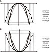
Each of the five determined ratios was classified into three groups through the K-means clustering method in SPSS 12.0 (SPSS Inc., Chicago, IL, USA). Then, the significance among the three groups in each ratio was evaluated by one-way analysis of variance in SAS 9.1 software (SAS Institute Inc., Cary, NC, USA). Relative deviations in each ratio for the three groups from mean values in the overall data are presented in graphically in Fig. 5. The dental arch shape was classified as a V-shape, U-shape, or O-shape based on these data using the least squares method, with R-square values calculated based on the ratio of each measurement.
RESULTS
Measurement of arch dimensions for the classification of dental arch forms are presented in Table 1 and the mean and standard deviations values for arch width, length, and height are presented in Table 2. The relative proportions of each ratio of the three classified dental arch form to the overall mean values are presented in Fig. 5. In the V-shaped dental arch, the ICD/CW value in the anterior part was significantly different from those in other groups in both the maxilla and mandible. The U-shape dental arch exhibited higher differences in IM1D/M1W and IM2D/M2W for the posterior part compared with the other groups. Each ratio for the O-shape dental arch showed relatively similar results compared with the overall average values. The O-shape was the most common in both the maxilla (52%) and mandible (56%). The V-shape appeared 28% of the time in both the maxilla and the mandible, while the U-shape appeared 20% of the time in the maxilla and 16% of the time in the mandible (Table 3). The curves for the three dental arch types obtained from five ratios in each group are presented in Fig. 6.
DISCUSSION
Many statistical researches about the dimensions and morphology of the oral anatomy have been done in dentistry. This fact reflects the importance and value of the information for clinical treatment procedures and outcomes. The means of examining the dental arch form include direct measurement or cross section copying using models, and measurements obtained from pictures and radiographs.272829 In this study, we directly measured a model using a precise measuring device and obtained most of the required data for the intraoral structure.
Dental arch dimensions in Koreans differ from those in other ethnic groups despite individual variations. The dental arch width in Koreans is wider and the arch length is shorter than in the French,15 the inter-canine and inter-molar widths in the arch of Koreans are larger than in Caucasians,1819 and the intermolar width in the maxilla of Koreans is wider than those of Japanese and Taiwanese subjects.19 Ready-made trays made using statistical data obtained from non-Koreans have proven to be unsuitable for the Korean dental arch shape.141617
The present results are not consistent with previous studies; the overall measured values for the arch dimensions exceed those in previous studies involving Koreans.15181922 As well, the arch width and the overall measured values are similar or larger than those of Caucasians described in previous studies.15181922 Koreans are characterized by a relatively large arch width and length when compared with other populations. The present data could indicate changes in the arch dimensions of Koreans in recent years resulting from changing diet habits and culture,242526 which have been reportedly related with dental arch shape and size.111213 The subjects in the present study were < 32 years of age and so had lived through the period of changing diet and culture. In previous studies, direct comparison with other races was hampered by the different ages of the subjects.
A recent comparative study reported that Vietnamese people tend to have deeper and wider arches than Korean people.30 The depth of the mandibular arch in the buccal and lingual aspect was significantly smaller than the value reported in a previous study17 using the same measurement points. These differences may have reflected different impression techniques. This study used pre-existing trays with one-step border molding using irreversible hydrocholloid. The previous study used customized individual trays with a sequential border molding procedure and a modeling compound. The different border molding techniques and tongue movements could have yielded different depth values of the lingual and buccal vestibules.
The proportions of the three arch type distribution ratios differed compared with a previous study about Koreans.23 However, in both studies, O-shape dental arches comprised the highest proportion for both arches. The R-square value in the regression line for the three dental arch groups was greater than 0.90, indicating high compatibility between the three groups and the entire sample.
This study obtained statistically valid results by measuring the dentition and basal bone site of the dental arch in the maxilla and mandible of Koreans. It bolsters the findings of a recent investigation that reported the changing diet and culture in Korea in young adults as the subjects.
Although the subjects in this study are considered a special group as they visited a hospital, the participants can be regarded as a randomized sample.5 Therefore, these results can help understand the current general characteristics of dental arch dimensions and shapes in Koreans. This can help with clinical dental treatment and contribute to satisfying the gradually increasing need for quality treatment procedures and the development of general clinical criteria.
This study included only normal occlusions in young adults, thus further investigations involving different types of malocclusions and classification by ages are required for more interesting insights. A comparative study among different races would also be illuminating.


 XML Download