

 PDF
PDF ePub
ePub Citation
 Print
Print


INTRODUCTION
Different attachment systems can be used in the treatment of partial edentulism.123 These attachment systems reversibly connect teeth or implants to dentures.4 Double-crown systems are alternative attachment systems for the treatment of partial edentulism, and these systems provide great patient satisfaction and a high survival rate.56
Double-crown systems consist of a primary crown (patrix, male), which is cemented to the teeth or the implant abutment, and a secondary crown, which is attached to the prosthesis.467 In cylindrical double crowns, all surfaces are prepared in parallel so that a piston-cylinder effect occurs. In conus crowns, parallelism is constructed only between the contact surfaces of the primary and secondary crowns, and a pressing effect is achieved via the geometry of these surfaces. One of the most important factors for the success of double-crown systems is setting the optimum retentive force, which requires technical skill, ability, and experience.8910 Körber has recommended that the retentive force per abutment in double crowns be 5 - 10 N.11 And added that when a precious alloy is used, these values can be obtained with 6-degree cone angles, whereas when a non-precious alloy is used, the angle of the cone must be lowered.
Conventionally, double-crown systems are produced with precious or non-precious alloys via the lost wax technique.8 Various materials, such as zirconia, can be used in double-crown systems due to the advancement of CAM/CAM technology.8111213 In the last few years, rapid prototyping has emerged as an alternative to CAD/CAM methods. Direct metal laser sintering (DML), a fast method of prototype production, can be defined as an additive fabrication technique that involves binding a powdered metal together to form a solid structure using a laser as a power source.14151617 To our knowledge, there are no available studies that investigated the use of laser sintering technology with the production of both primary and secondary crowns in double-crown systems. The purpose of this study was to evaluate retention force changes on double crowns fabricated with various materials and techniques over long-term use. The null hypotheses of this study are as follows: 1. Cone angle affects retention forces. 2. DML can be used for fabricating double crowns. 3. At the end of the 5,000 insertion and separation cycles, retention forces changes and wear will be evident.
MATERIALS AND METHODS
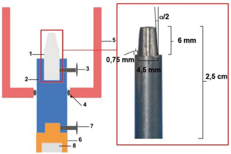
The design and production of the dies that were intended to imitate tooth or implant abutments were made in a Computer Numerical Control (CNC) machine (Hi-Eco 10, Hwacheon, Kwangju, Korea) from stainless steel. The dies had the following dimensions: 6 mm in height and 4.5 mm in base diameter (Fig. 1). The mechanism of the experiment is shown in a technical drawing (Fig. 1).
A total of ten groups were evaluated; each group contained six samples with either 4° or 6° cone angles.
Groups: 1. Casting gold alloy primary crown - casting gold alloy secondary crown (AA) 2. Laser sintering primary crown -laser sintering secondary crown (LL) 3. Casting Cr alloy primary crown - casting Cr alloy secondary crown (CC) 4. Zirconia primary crown - electroformed gold secondary crown (ZA) 5. CAD/CAM titanium alloy primary crown - CAD/CAM titanium alloy secondary crown (TT)
The primary crowns of all groups were designed with the same CAD unit (DV3, Dental Wings, Montreal, Canada).
AA groups: The primary crowns were produced by casting plastic blocks (CopraPlex, Whitepeaks, Essen, Germany). The models were invested (Bellavest SH, Bego, Bremen, Germany) and cast in gold alloy (Ceramic 74, Novametal Europa, Torino, Italy) according to the directions of the manufacturer. After the casting traces were removed from inside the primary crowns, they were placed on the dies without force to check for irregularities that might prevent the seating of the gold alloy primary crowns on the dies, and a complete fit was achieved. The primary crowns were checked and cone angles of 4 or 6 degrees were set for each group. Secondary crown production was also accomplished via the traditional casting technique.
LL groups: The primary crowns were fabricated with a DML machine (EOSINT M 270, EOS, Münih, Germany). They were milled with a milling device (Exacto 1, Heimerle-Meule), and 4° and 6° conus angles were set. Rubber polishers were applied, and the procedure was completed. The primary crowns were placed on the dies, and a complete fit was achieved. The primary crowns were scanned, and the secondary crowns were designed. Secondary crowns were fabricated using the same direct metal laser sintering machine and milling and polishing procedures.
CC groups: The primary crown samples were produced by casting plastic blocks (CopraPlex, Whitepeaks, Essen, Germany). The models were invested (Bellavest SH, Bego, Bremen, Almanya) and cast in alloy (Wirobond Easy, Bego, Bremen, Germany) according to the directions of the manufacturer. After casting, the traces inside the primary crowns were removed. They were milled with a milling cutter, and 4° or 6° conus angles were set. The secondary crowns were prepared on the primary crowns and produced using the same procedure.
ZA groups: The primary crowns were produced from zirconia blocks (DD BİO ZW ISO, Dental Direkt, Spenge, Germany) and sintered in a special furnace (ThermoStar M2 Plus Sintering Furnace, Thermo Star GmbH, Aachen, Germany) at 1,500℃ for 12 hours. The primary crowns were milled, set at 4° or 6° cone angles, polished with a special bur kit for zirconia (sets 4430 and 4431, Komet), and mounted with water cooling. Secondary crowns were produced in the electroforming machine (Trend galvano genius perfect, Binder Dental, Georgsmarienhütte, Germany). A thin layer of silver lacquer was applied with a special brush. The amounts of liquid, activator, and time required were automatically calculated by the system, and the secondary crowns were produced to receive 250 µ of width.
TT groups: The primary crowns were produced from titanium blocks (Coprati - 5, White Peaks Dental Solutions, Wesel, Germany), and samples were milled with a milling device (Exacto 1, Heimerle-Meule), and 4° and 6° conus angles were set. Rubber polishers were applied, and the procedure was completed. The primary crowns scanned, and the secondary crowns were designed. The secondary crowns were produced by following the same steps as with the primary crowns.
All primary crowns were cemented to dies with resin cement (BisCem, Bisco Inc., Schaumburg, IL, USA) according to the manufacturer's instructions.
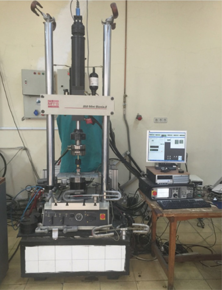
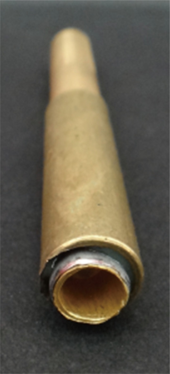
5,000 insertion - separation cycles were performed, and the retention force were measured after every 500 cycles with a Mini Bionix II machine (Mini Bionix II, MTS Systems Corporation, Eden Praire, MN, USA). (Fig. 2) The secondary crowns were connected to the machine with a cylindrical brass connector. (Fig. 3) After the primary and secondary crowns were placed in the test device, ten insertion - separation cycles were performed to provide retention vertically but freedom laterally, with a magnet being located at the bottom of the experimental mechanism. Retentive force measurements were repeated three times.
A special program was used to conduct the 5,000 insertion-separation cycles for the retention measurements. The insertion and the separation of the secondary crown on top of the primary crown were set to occur at 300 mm/min and a frequency of 2.2 Hz. When the retention forces were measured, the descending speed was set to 10 mm/min, the ascending speed to 20 mm/min, and vertical displacement of the secondary crown was set to 2 mm. The secondary crowns were seated on the primary crowns with a load of 50 N. All insertion - separation cycles and retention force measurements were performed in artificial saliva, which was produced according to Shannon's procedure.18
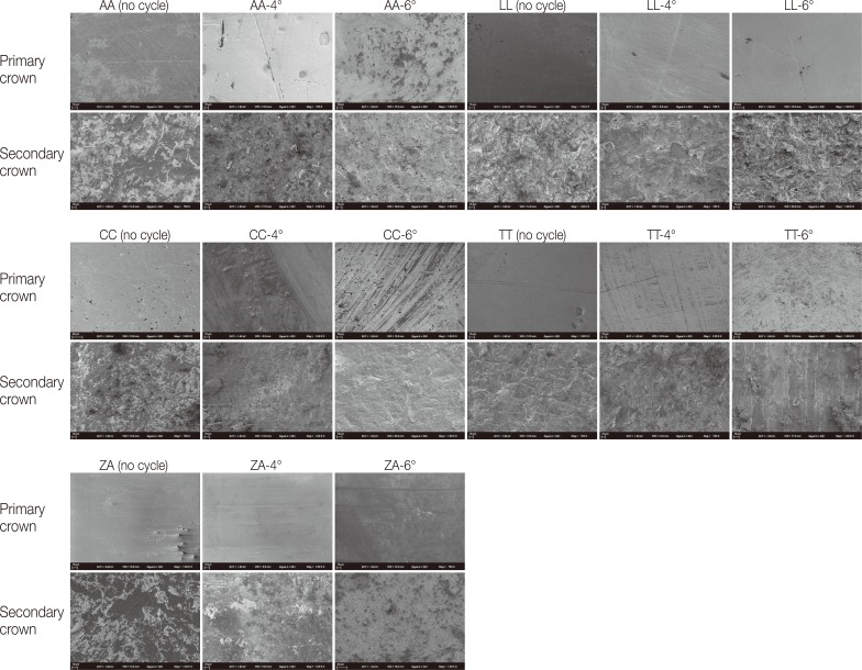
At the end of 5,000 cycles, for each group, one sample that was not subjected to the test procedure and one sample that had the retention force value nearest to the group mean were chosen for scanning electron microscopy (SEM) (EVO LS 10, Zeiss, Göttingen, Germany). The secondary crowns of all samples were cut into two pieces vertically, with applying low pressure. Then, the surfaces of both the primary and secondary crowns could be examined.
For the statistical analyses, the IBM SPSS Statistics 22 (IBM SPSS, Istanbul, Turkey) program was used. A Shapiro-Wilks test, two-way ANOVA, Student's t-test, and one-way ANOVA test were used for the statistical analyses, and a Tukey HSD test and/or Tamhane's T2 test were used for post-hoc analyses (P < .05).
RESULTS
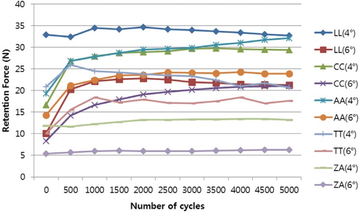
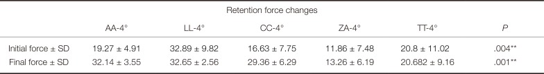
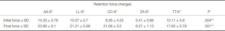
Table 1 and Table 2 show the initial and final retention force values for all groups. The LL-4° group had the highest initial and final retention forces (32.89 N - 32.65 N). The ZA-6° group had the lowest initial and final retention forces (5.41 N - 6.27 N). The retention force values were statistically significantly increased in the AA-4°, AA-6°, LL-6°, CC-4°, CC-6°, ZA-6°, TT-6°, and LL-4° groups. Decreases in retention force values were observed in the ZA-4° and TT-4° groups, but they were not statistically significant. Retention forces increased when the conus angle decreased in all groups (Fig. 4).
The SEM images of one sample from the AA, LL, CC, ZA, and TT groups and one control sample can be seen in Fig. 5.
Wear traces are more detectible in the primary crowns in groups AA, LL, CC, and TT than those in ZA group. The traces seen on the ZA primary crowns were because of the grinding and polishing process.
DISCUSSION
There is no standard protocol for in vitro studies in which double-crown systems are evaluated. For this reason, the methods used in this study were chosen with great care. Our study tooth and implant abutments were imitations of stainless steel dies that were 6 mm in height. The dimensions were determined based on measurements in the literature 4121319 as well as most implant company standards.
Engels et al.20 state that patients remove their removable dentures two times per day. Bayer et al.21 state that patients remove their removable dentures 2.74 times in a day. Thus, 5,000 insertion - separation cycles correspond to a clinical service time of 6.5 to 7 years.
In a number of studies222324 evaluating the clinical complications involving double-crowns, decementation is the most commented. For this reason, the primary crowns were cemented to the dies with a resin cement before the experimental procedures were applied.
Artificial saliva can affect the tribological properties of the production material and remove debris that develops due to wearing.4 Bayer et al.25 state that the absence of saliva changes the frictional wear due to retention forces. We used artificial saliva in the experimental procedure to simulate clinical conditions.
Clinically, the secondary crown seats the primary crown with a chewing force. This chewing force value can vary based on the opposite arc situation, the location of the tooth in relation to the holder, the occlusion relationship, and personal factors. Ohkawa et al.19 evaluated the effect of retention forces with various preloads in double crowns. They state that increasing preloads up to 50 N increases retention but that increasing preloads to above 50 N will not change retention forces. Turp et al.13, Güngör et al.26, Beuer et al.7 used 50 N in their study. For these reasons, we have decided to use this most common preload value of 50 N in our study.
Several separation speeds were applied to double-crown systems in the literature as follows: Weigl4 20 mm/min, Turp13 20 mm/min, Shimakura12 5 mm/min, Bayer21 120 mm/min, and Güngör26 0.5 mm/min. In our study, the separation speed was set to 20 mm/min.
In our study, retention force increased when the conus angle decreased in all samples. This finding is similar to other findings in the literature.111321 The reason for this is that in double-crown systems with a cone angle, compressive stress is generated because the primary crown, with an occlusal force, acts like a wedge.89 The retention mechanism of conus crowns is based on this wedge effect. As the cone angle between the primary and secondary crown surfaces increases, compressive forces decrease.
Retention forces were more stable in the ZA groups than in the other groups. Weigl et al.4 state that double-crown assemblies with electro-formed secondary crowns have more stable retention forces than double-crown assemblies with cast secondary crowns. Researchers explain this via the fact that the retention mechanism of electroformed secondary crowns is based on adhesion, not the wedge effect. Our SEM findings support this notion. The use of a hard and wear-resistant primary crown material against a less hard secondary crown material, as in the ZA material coupling, may be advantageous because no wear traces are seen on the surfaces.
We reviewed in vitro studies in which the long-term retention values of double-crown systems were examined. There are studies2127 that show an increase in retention values after insertion-separation cycles, as well as studies1926 showing a decrease in retention force values. Such increases are explained by mechanical adaptation28 at the occlusal gap level between the primary and secondary crowns, and such decreases are explained by metal abrasion that occurs on the surfaces of the primary and secondary crown surfaces after long-term use.19 Weigl et al. report that clamping points are most commonly observed between the primary and secondary crowns when double crowns are produced via the casting method. Chewing forces may cause excessive pressures at the clamping points, and cold welding with the opposite metal or plastic deformation at the surface structure may occur. When this does occur, adhesive strength and adhesion force are increased. In our study, we found increases between the initial and final retention values. We believe these to be due to mechanical adaptation and cold welding.
Wagner et al.29 compared the initial retention values of the double-crown systems produced by the Co-Cr milling and CO-Cr casting methods and found the retention force values to be similar. In terms of our study results, we believe that the reason for the different retention values for LL and CC groups is differences in the dimensions of the dies, cone angles, and experimental procedures.
Besimo et al.27 evaluated double-crown systems produced by casting chromium alloys, gold alloys, and titanium with two cone angles, 5.5° and 6.5°. Similar to our study results, the investigators observed increasing retention force values in all sample groups.
To our knowledge, no current studies investigate the use of laser sintering technology in the production of both primary and secondary crowns. In our study, we find that the LL groups' retentive forces were higher than the clinically suggested values. However, the retentive force values are similar among all groups except for the ZA groups. It will be useful to perform new studies on the use of the laser sintering method with double-crown systems.
The amount of retention values should be at a level that will not damage the abutment teeth. Retention force is important, but the retention values should be at a level that will not damage abutment teeth. Korber has recommended that the retentive force per abutment in the double crowns should be kept within 5 - 10 N.11 A zirconia primary crown and an electroformed secondary crown with a cone angle of 6 degrees reach this goal. A zirconia primary crown and an electroformed secondary crown with a cone angle of 4 degrees were close to reaching these values.
Wear was seen in all primary and secondary crowns, but mostly in the primary crowns in the AA, LL, CC, and TT groups. There were no wear traces seen in the ZA groups. The traces seen on the ZA primary crowns were because of the grinding and polishing process. Our findings are similar to those of Weigl et al..4 Signs of wear did not appear at joined ceramic and electroformed surfaces, and after 10,000 insertion - separation cycles, wear was observed on the precious alloy primary and conventionally cast secondary crowns. Weigl et al. explain wear differences between the groups due to the fact that the retention mechanism of electroformed secondary crowns is based on adhesion, not the wedge effect, and that cold welding does not occur between different alloys.
The following limitations apply to this study: 1. Just one type of artificial saliva was tested. 2. Horizontal forces were not simulated; only vertical forces were simulated. 3. Implant abutments and teeth of only one height were imitated.
CONCLUSION
According to the results and considering the limitations of the study, the following conclusions can be drawn.
The retentive force increases when the cone angle decreases for all samples. Retention forces were more homogenous and no wear traces were observed in the zirconia primary crown - electroformed secondary crown groups than in the other groups. The zirconia primary crown - electroformed secondary crown group with a cone angle of 6 degrees is the most suitable double-crown system in terms of achieving optimum retentive force. Double crowns produced with laser sintering technology have high retention values, and these values are similar to those of gold double crowns.
 XML Download