

 PDF
PDF ePub
ePub Citation
 Print
Print


INTRODUCTION
Current digital workflow technologies for manufacturing dental restorations include both the digital impressions formed by intraoral scanning and the synthesis of 3D objects by additive or subtractive manufacturing. In particular, the process of designing restorations by intraoral scanning (CAD) and subsequent manufacturing with subtractive technologies (CAM)1 is much more comfortable for patients than conventional impressions, not only allowing easy preparation modification in real time2 but also enabling the use of advanced ceramic materials with refined composition and microstructure.3 Furthermore, when using in-house milling units, defective teeth can be restored in a single appointment.4
However, despite such improvements of additive manufacturing, subtractive technologies remain the most advanced and common manufacturing process of dental ceramics.5 Notably, lithium disilicate and zirconia blanks are common materials for restoration manufacturing processes using subtractive technologies and are widely used by dentists and dental technicians. Lithium disilicate ceramics were problematic because they initially displayed irreparable microcracks, low chemical resistance, and undesirable transparencies,6789 but current restorations made with lithium disilicate ceramics have many advantages such as high flexural strength, exceptional aesthetic design, and tolerable wear resistance.101112 Furthermore, the status of pre-crystallized lithium disilicate ceramics is receptive to subtractive technologies and is thus very useful.13 Zirconia ceramic is mechanically the most desirable ceramic in all dental fields.13 Restorations manufactured from zirconia ceramics are made from partially sintered zirconia blanks, soft milled by CAD/CAM technology.14 The use of partially sintered zirconia blanks ensures quicker and easier milling and also lowers the wear and tear of milling tools.13
Restoration manufacturing using CAD/CAM technology partially replaces the currently popular lost-wax technique. In particular, this technology, by subtractive manufacturing, removes the contraction of wax materials, burn outs, deflection during casting, and other factors affecting the proficiency of manufacturing and ultimately restoration reproducibility.5 However, restoration manufacturing using CAD/CAM systems still has errors such as inaccuracy, tool wear, and tool transformation.15 In addition, if digital impressions by intraoral scanning are applied, errors will arise from the variables used to obtain data.16 Therefore, although digital workflow using CAD/CAM technology has clear advantages over current methods, its restoration reproducibility, which is a measure of fabrication reliability, is still unclear. Moreover, there have been no qualitative or quantitative analyses of the reproducibility of restorations made by digital workflow.
The purpose of this in vitro research was to estimate the reproducibility of digitally manufactured ceramic single crowns. This was achieved by analyzing the differences between 3D representations of each crown. The null hypothesis was that there is no significant difference between the reproducibility of single crowns made of lithium disilicate and zirconia blank.
MATERIALS AND METHODS
A maxillary right first molar typodont tooth prepped with a 1.5 mm occlusal height reduction, 1.5 mm axial reduction, and at least 1.0 mm wide deep chamfer was prepared (ANKA-4 V CER; Frasaco GmbH, Tettnang, Germany). The prepared tooth was mounted on a typodont model, and then the typodont was fixed in a simulated patient position on a phantom head. Digital impressions were obtained using a noncoating video intraoral scanner (CEREC Omnicam; Sirona Dental Systems GmbH, Bensheim, Germany), carried out by a single dentist proficient with the systems; the dentist performed scans following the scanning protocols of the manufacturer at a temperature of 23 ± 2℃. Digital impressions obtained were sent by a data upload program (CEREC Connect Software 4.3; Sirona Dental Systems GmbH, Bensheim, Germany) to a dental design software (CEREC inLab software; Sirona Dental Systems GmbH, Bensheim, Germany) used to manufacture restorations.
The ideal direction for inserting a restoration and the preparation margin were established for the abutment tooth of the digital impression by using the dental design software. Subsequently, the parameters provided by the manufacturer for the smallest thickness needed for the anatomical crown shape in the program database were applied, and the CAD design for the restoration was completed. The finished CAD dataset was used for fabricating both lithium disilicate (IPS e.max CAD; Ivoclar Vivadent AG, Schaan, Liechtenstein) and zirconia blank (Sirona inCoris TZI; Sirona Dental Systems GmbH, Bensheim, Germany) single crowns. Fouraxis milling machines (inLab MC XL; Sirona Dental Systems GmbH, Bensheim, Germany) were used to fabricate presintered zirconia and pre-crystallized lithium disilicate crowns. After the milling, pre-sintered zirconia crowns were carefully removed from their holders by diamond disks, and the attached areas were ground to a smooth finish with tungsten carbide burs. Pre-crystallized lithium disilicate crowns were also removed from their holders by diamond disks, and the attached areas were ground with fine-grained diamond (< 60 µm). Firing schedules for the sintering of pre-sintered zirconia crowns and crystallization of pre-crystallized lithium disilicate crown are listed in Table 1.
All manufactured ceramic crowns were digitized using a reference scanner (Smartscan R5; Breuckmann GmbH, Meersburg, Germany). This apparatus obtained data from two cameras set at nonsymmetric angles such as 10°, 20°, and 30°; thus the apparatus could measure previously difficult areas and has an accuracy of 7 µm. Both outer and inner surfaces of all ceramic crowns were computed by the STL dataset format by the reference scanner.
For each digitalized zirconia and lithium disilicate crown dataset, all possible combinations of the two scanned files were chosen, and the files were superimposed (i.e., 12C2, n = 66) on each other (Control; Geomagic GmbH, Rock Hill, SC, USA). For each combination, the root mean square deviation and color-coded difference images were used as quantitative and qualitative dimensional differences between ceramic datasets, respectively. Additionally, areas showing the highest and lowest dimensional difference in 3D analysis were observed using a digital microscope (KH-7700; Hirox, Hackensack, NJ, USA).
The IBM SPSS 21 software package (IBM SPSS Inc., Chicago, IL, USA) was used in calculating the mean (RMS), standard deviations, and 95% confidence intervals of the reproducibility of zirconia and lithium disilicate crowns. Saphiro-Wilk test and Levene's test were used to confirm the normality and homoscedasticity, and independent samples t-test was used to analyze significant differences in the RMS values of zirconia and lithium disilicate crowns. Statistical significance was set at P < .05. Power analysis was performed to calculate the sample size needed for the experiment. Among two experimental groups with an effect of 1.25, an alpha of 0.05, and a power of 0.80, 24 samples (12 per group) were needed altogether.
RESULTS
The ceramic crown materials showed no statistically significant difference in the reproducibility on the inner surface, but the difference was statistically significant on the outer surface (Table 2).
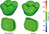
The prominent color-coded difference images obtained from the qualitative reproducibility analysis of each crown group (Fig. 1) showed that, on the outer surface of lithium disilicate crown, an over-contouring of +50 to +150 µm occurred primarily on the buccal surface and an under-contouring of -50 to -100 µm on the inner occlusal surface. The outer surface of zirconia crown had both an over-contouring of +50 to +150 µm and an under-contouring of -50 to -150 µm on the buccal surface, whereas the inner surface had an under-contouring of -50 to -100 µm on the marginal area.
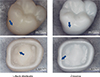
In addition to the qualitative reproducibility analyses of the 3D data of each crown group, the crowns were directly observed using a digital microscope (Fig. 2). Over-contoured and under-contoured areas were observed to be identical to the areas seen in the color-coded difference images.
DISCUSSION
This in vitro study provides information about the reproducibility of manufacturing lithium disilicate and zirconia single crowns by digital workflow. Data of the reproducibility of restoration manufacturing were gathered by applying a method for evaluating the accuracy of digitizing devices.1718 The reproducibility of lithium disilicate single crowns and that of zirconia blank single crowns differed in a statistically significant way, thereby disproving the null hypothesis.
In a previous study on the accuracy of restoration manufacturing, the trueness of partial crowns manufactured by chairside CAD/CAM milling processes were also analyzed.19 The study, similarly to this one, also used digital workflow and intraoral scanning to manufacture restorations using CAD/CAM milling. However, the past study did not analyze the accuracy of the whole digital workflow but only the process of CAD/CAM milling by 3D superposition of the data from digitizing restorations. In addition, the intraoral scanner used to digitize the restorations was imprecise, and errors that occurred in trueness values and precision (reproducibility) due to digitization were not measured. Therefore, this study used a scanner with high precision to reduce errors in digitization, and reproducibility could be inspected over the whole digital workflow manufacturing process by studying intraoral scanning. Thus, the accuracy of the whole workflow was analyzed.
The intraoral scanner used in this study not only takes continuous images and immediately renders them in three dimensions, but also shows the images using real color because no powders are needed.20 The CAD/CAM milling unit used for milling was used to minimize damage to the ceramic block by an intermittent-touch process using a water-soluble lubricant in an environment of constant water-cooled sprays.21 The previously mentioned two procedures are commonly used for chairside CAD/CAM milling processes.
A recent study analyzed the reproducibility of heat-pressed ceramics.22 The study used the lost-wax technique and heat-pressing technique, both of which are commonly used, to manufacture a lithium disilicate partial crown. The crown's marginal reproducibility and internal reproducibility were reported to be 23 µm and 14 µm, respectively, and the results were similar to those of the reproducibility values of the inner surface of lithium disilicate crown in this study.
The over-contouring on the outer buccal surface of the lithium disilicate crown where it was attached to the holder (Fig. 1) was presumably caused by errors occurred when the holder was detached. This was confirmed by a microscopic analysis of the real model (Fig. 2). The outer surface of zirconia crown similarly had over- and under-contouring where the holder was affixed (Fig. 1); the cause was again the detachment of the holder, as confirmed by the microscopic analysis (Fig. 2). The detachment of the holder was done by the technician, and therefore these errors produced in the crowns are not attributable to digital workflow. The under-contouring on the inner occlusal surface of the lithium disilicate crown (Fig. 1) seemed to be a milling error; this was confirmed by the microscopic analysis, which showed clear uneven milling in the real model (Fig. 2). The small errors in the inner marginal area of the zirconia crown (Fig. 1) were attributed by the microscopic analysis to chipping near the acute edges (Fig. 2). This chipping phenomenon of zirconia crown marginal area is thought to originate from the initiation and propagation of cracks and fissures during milling.23 On the other hand, the marginal area of the lithium disilicate crown is less affected by the ceramic's chipping. Thus, the marginal area of a crown can be recreated more accurately using lithium disilicate.
This study had several limitations. Although the acrylic resin tooth of the typodont model had similar light reflection property as natural dentition, the chemical composition, surface structure, and physical properties differed. Furthermore, the in vitro environment in which the impression was obtained was not similar to the actual mouth. Ultimately, only reproducibility, which is a measure of precision of digital workflow, was analyzed, whereas trueness was not.


 XML Download