

 PDF
PDF ePub
ePub Citation
 Print
Print


INTRODUCTION
The reconstruction of multiple missing teeth with dental implants is a predictable and proven treatment technique for edentulous patients in both anterior and posterior regions.12 However, the molar teeth are often shown to be a troublesome area for implant restorations from mechanical and biological aspects,3 which is due to complicated and complex factors of implant prosthetic components and the load-related bone contact area.4 The strong occlusal forces exert harmful effects on an implant prosthesis and alveolar bone in the posterior area,56 which results in marginal bone loss and decreased implant stability, and can lead to complications in an implant fixture and its suprastructure.78
Natural teeth are traditionally splinted in order to decrease the stress and increase the stability of prosthesis. This can also result in a smaller horizontal load being transferred to the supporting teeth, and can compensate for the crown-root ratio increasing in various alveolar bone-loss regions and periodontally compromised patients.39 Several studies have recommended that adjacent implants should be splinted with the fixed retained prosthesis.3 When off-axis forces are applied to an implant, they induce an adverse loading that can cause mechanical failure of a restored implant and biological failure of the surrounding bone that could lead to implant failure.101112 The aim of a splinted implant restoration is to favorably distribute the stress between the implants in order to minimize the transmission of horizontal forces to the bone-implant contact area.31314 In particular, two-implant splinting (2-IS) in the posterior region can promote the stability in the mesiodistal direction and relieve the stress in the buccolingual direction.9 2-IS can also be considered as an important treatment option in patients without anterior guidance or with parafunctional oral habits.15 Nevertheless, several procedures of multiple-implant restoration splinting are highly technique-sensitive, and the accuracy of the final prosthesis is mainly limited.15
Alveolar bone loss is common in patients with periodontitis, which will lead to an unfavorable crown-implant ratio (C/I ratio). An off-axis force acting on an implant restoration with an increasing C/I ratio and crown height space (CHS) of the implant-defined as the distance from the alveolar bone crest to the occlusion plane-can induce a detrimental load at the implant restoration neck area, and result in surrounding bone loss and eventual prosthetic failure.1617
According to Grossmann et al.,3 the splinting technique can be an appropriate treatment option for periodontitis patients who have an impaired occlusal relationship due to the loss of alveolar bone and multiple teeth.
In spite of splinted implant restoration being beneficial for periodontitis patients with severe alveolar bone loss and excessive occlusal forces in the posterior region, most of the studies have been theoretical, with insufficient clinical analyses and negative long-term results.3 Several studies of splinted prostheses have involved short-term investigations and shown limited efficacies and controversial results, and so further investigations and long-term studies are required.151819
The aim of this retrospective study was to determine the efficacies of 2-IS, and compare them with those of single implant restoration (1-IR) in the first and second molar regions, which has been demonstrated to produce good results in previous studies. This study has also identified the appropriate clinical considerations for splinted implant restoration of the molar region.
MATERIALS AND METHODS
This study was approved by the Institutional Review Board of the Ilsan Hospital, National Health Insurance Service (NHIS) (Approval no. 2015-02-006). All surgical treatment procedures were performed by periodontists at the Department of Periodontology, Ilsan Hospital, NHIS. The study was limited to the posterior region, including patients with missing first and second molars only, in order to minimize the effects of position and occlusal force. The internal connection implant fixtures comprising a sand-blasted, large-grit, acid-etched surface (Implantium, Dentium, Seoul, Korea; Straumann, Institut Straumann, Basel, Switzerland) were placed using a one- or two-stage surgical procedure as the manufacturer's protocol. The prosthodontic procedures were started after a healing period of more than 3 months. The prosthesis type [i.e., occlusal screw (OS), lateral screw (LS), cementation (CM), and screw-cement-retained prosthesis (SCRP)] was selected depending on the condition of the patient and the preferences of the prosthodontist, and occlusal adjustment was carried out to obtain the optimal centric and eccentric contact forces. The final prostheses were installed more than 4 months after the implant surgery. Maintenance care that emphasized scaling and oral hygiene instruction was provided every 3 - 6 months, and intraoral periapical or panorama radiographs were obtained every 12 months.
Patients who had undergone implant surgery at the Department of Periodontology, Ilsan Hospital, NHIS during 2005 - 2014 were reviewed over a mean functional loading period (FLP) of 40 months. The following inclusion criteria were applied: sex ratio of 1 : 1, aged 20 - 80 years (mean age 58.5 years), and good systemic health condition (including well-controlled systemic diseases). The implant prosthesis had been functioning for 1.1 - 102.8 months, with a mean loading period of 41.4 months. One or two implant placements in the first and second molar regions were included. Single-implant restorations in the first or second molar region were classified into the 1-IR group, while restorations involving two splinted implants in the first and second molar regions were classified into the 2-IS group. Patients with severe systemic disease, advanced or aggressive periodontitis, or parafunctional oral habits (e.g., excessive occlusal force, heavy clenching, or bruxism) were excluded from this research.
In total, 408 implants in 234 patients who conformed to the inclusion and exclusion criteria were investigated. All data related to these patients with implant treatments were based on the clinical treatment records, clinical photographs, and radiographs of the patients. The following clinical factors of the patients were considered: sex, mean age, implant location, FLP, bone grafting, clinical C/I ratio, CHS, horizontal distance (HD) between the two implants (HDI, the first and second molar positions), and HD between the natural tooth in the mesial position and the implant in the distal position (HDNI, the first or second molar position).2021 Based on previous studies, the clinical C/I ratio was defined as the distance ratio measured from the clinical crown to the implant fixture (standard fulcrum located at the marginal bone), and CHS was measured as the distance from the alveolar bone crest to the occlusion plane.22 HDI and HDNI were measured as the distance at the marginal bone level on the day of implant placement. A PACS workstation (Centricity GE Healthcare, Waukesha, WI, USA) was used to calculate the clinical C/I ratio, CHS, HDI, and HDNI on the radiographs, and distortion caused by magnification was corrected using a calibration based on the known interthread pitch of the implant (Implantium, Dentium: 0.6 mm; Straumann, Institut Straumann: 1.25 mm) as a reference. Mechanical complications [i.e., SL, screw fracture (SF), CF, and repeated SL] and biological complications [i.e., peri-implant mucositis (PM) and periimplantitis (PI)] were evaluated for each patient. The biological complications were examined based on mobility, suppuration, probing depth, bleeding on probing, and alveolar bone loss. A reversible inflammation of peri-implant mucosa was diagnosed as PM, and loss of alveolar bone was diagnosed as PI.23 All measurements were performed using a UNC periodontal probe (Hu-friedy, Chicago, IL, USA).
In comparisons and analyses of two groups, the chi-square test and Student's t-test (two-tailed with independent samples) were used to identify the relationships between the clinical factors (i.e., sex, mean age, implant location, FLP, clinical C/I ratio, CHS, HDI, and HDNI) and the complication rates (i.e., mechanical and biological complications). The chi-square test was applied to noncontinuous variables and Student's t-test (two-tailed with independent samples) was applied to continuous variables. The optimal cutoff value for FLP related to the complications was evaluated using receiver operating characteristics (ROC) analysis. The results obtained in all of the investigations were analyzed using SPSS software (version 19.0, SPSS, Chicago, IL, USA). The cutoff for statistical significance was set at P < .05. All measured data are presented as mean ± SD values.
RESULTS
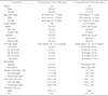
The subjects of this study comprised the 1-IR group, which contained 124 patients (69 males, 55 females) with a mean age of 56.26 years (range, 23 - 77 years), and the 2-IS group, which contained 110 patients (53 males, 57 females) with a mean age of 59.01 years (range, 34 - 91 years).
The implants in the 1-IR group were distributed in the posterior region as follows: maxillary first molar, n = 22 (15.9%); maxillary second molar, n = 10 (7.3%); mandibular first molar, n = 43 (31.2%); and mandibular second molar, n = 63 (45.6%). Totals of 32 (23.2%) and 106 (76.8%) implants were positioned in the maxilla and mandible, respectively. In the 2-IS group, 134 (67 pairs, 49.6%) and 136 (68 pairs, 50.4%) implants were positioned in the maxilla and mandible, respectively.
The mean FLP was 42.87 months (range, 1.84 - 101.25 months) for the 1-IR group and 39.86 months (range, 1.08 - 102.75 months) for the 2-IS group. In addition, bone grafting was performed for 32 (23.3%) implants in the 1-IR group and for 108 (54 pairs, 40%) implants in the 2-IS group, showing an intergroup difference of about threefold. For the 1-IR group, the clinical C/I ratio was 1.11 ± 0.47, CHS was 9.65 ± 1.98 mm, and HDNI was 2.80 ± 1.17 mm; the corresponding values for the 2-IS group were 1.07 ± 0.21, 9.62 ± 1.75 mm, and 3.26 ± 1.30 mm, respectively. These results are consistent with a previous study finding that in order to minimize bone loss, HDI (i.e., interimplant distance) should be longer than HDNI (i.e., distance from the adjacent natural tooth to the implant).24 Most of the data represents a mean value of normal distribution curve; the deviation is observed in individual clinical situation of a patient. Since periodontist performed the treatment in the controlled clinical setting, it can be said that the position of implant is appropriate in this study (Table 1).
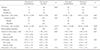
Mechanical complications were found in 31 (22.6%) of the 138 implants in the 1-IR group, with SL (n = 23, 16.7%) being the most common complication. This was followed by SF and CF (n = 3, 2.2%), and then repeated SL (n = 2, 1.5%). Mechanical complications were found in 30 (11.1%) of the 270 implants in the 2-IS group, corresponding to approximately half the rate in the 1-IR group. CF (n = 14, 5.2%) was the most common complication, followed by SL (n = 10, 3.7%) and then SF (n = 6, 2.2%), while repeated SL did not occur in any of the implants. The rate of mechanical complications differed significantly between the two groups (P = .020), with only SL showing a clearly significant increase in the 1-IR group (P < .001).
The rate of biological complications was markedly higher in the 2-IS group than in the 1-IR group. Only PI (n = 5, 3.6%) was found in the 1-IR group. In contrast, out of the 44 (16.3%) implants with biological complications in the 2-IS group, PI was found in 26 (9.6%) implants, followed by PM, which was found in 18 (6.7%) implants. The rate of biological complication differed significantly between the two groups (P < .001), with significantly elevated incidence rates of PM (P = .002) and PI (P = .046) in the 2-IS group.
In 1-IR group, SL, SF, and CF occurred simultaneously on one implant. Also, SL and CF occurred simultaneously on another implant, and SL and PI occurred simultaneously on the other implant.
In 2-IS group, SL and SF occurred simultaneously on one implant, and SL, CF, and PI occurred simultaneously on another implant (Table 2).
Patients in the 2-IS group who did or did not experience complications at least once were classified into the complication and success groups, respectively.
Mechanical and biological complications showed no statistically significant associations with sex, age, implant location in the jaw, and bone grafting.
The FLP was the only clinical factor to show a statistically significant difference with complications and success in the 2-IS group (P = .049). 2-IS remained relatively successful up to a mean of 38 months, while biological and mechanical complications arose after mean time periods of approximately 49 and 56 months, respectively.
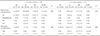
Implant-supported fixed dental prostheses (ISFDPs) located in the posterior region can induce stress in the implant and marginal bone when there is an unfavorable C/I ratio (anatomical and/or clinical C/I ratio of ≥ 2).25 In the present study, the overall clinical C/I ratio in the 2-IS group was 1.07 ± 0.21 (first molar region = 1.06 ± 0.18, second molar region = 1.09 ± 0.23), and the maximum value was 1.84 (Table 1), confirming that a favorable C/I ratio had been achieved. Moreover, the clinical C/I ratio was close to 1 : 1 in both the mechanical complication and success groups (1.05 ± 0.14 and 1.08 ± 0.21) and the biological complication and success groups (1.05 ± 0.23 and 1.08 ± 0.20), demonstrating favorable clinical C/I ratios.25 Correspondingly, there were no statistically significant differences in the clinical C/I ratios.
Recent studies262728 have found that CHS values exceeding 15 mm indicate an increased risk of implant prosthesis failure due to a vertical cantilever effect. In this study, the overall CHS in the 2-IS group was 9.62 ± 1.75 mm (first molar region = 9.82 ± 1.65 mm, second molar region = 9.43 ± 1.84 mm), with a maximum value of 14.28 mm (Table 1). Given that this is within the acceptable CHS range (8 - 12 mm) and below 15 mm, CHS is not expected to have a negative effect on the prognosis of the implant prosthesis. CHS did not differ significantly among the mechanical complication and success groups (9.67 ± 1.31 and 9.62 ± 1.80 mm, respectively) and the biological complication and success groups (10.04 ± 1.94 and 9.53 ± 1.70 mm, respectively).
HDI also did not differ significantly among the mechanical and biological complication groups (3.36 ± 1.27 and 3.51 ± 1.51 mm, respectively) and the mechanical and biological success groups (3.25 ± 1.30 and 3.21 ± 1.25 mm, respectively) (Table 3).
LS was the most commonly used type of prosthesis in the 1-IR group (n = 84, 60.9%), and was associated with the following rates of mechanical complications: SL, n = 14 (16.7%); repeated SL, n = 1 (1.2%); SF, n = 2 (2.4%); and CF, n = 2 (2.4%). The next most common type of prosthesis was CM (n = 23, 16.7%; SL, n = 5, 21.7%; repeated SL, n = 0, 0%; SF, n = 1, 4.4%; CF, n = 1, 4.4%), followed by SCRP (n = 19, 13.8%; SL, n = 2, 10.5%; repeated SL, n = 0, 0%; SF, n = 0, 0%; CF, n = 0, 0%) and OS (n = 12, 8.7%; SL, n = 2, 16.7%; repeated SL, n = 1, 8.3%; SF, n = 0, 0%; CF, n = 0, 0%). SL was the most frequent mechanical complication, and was associated with the prosthesis types as follows: OS, n = 2 (16.7%); LS, n = 14 (16.7%); CM, n = 5 (21.7%); and SCRP, n = 2 (10.5%). Although SL occurred proportionally the most often in CM, in terms of absolute numbers it occurred the most often in LS. However, there were no statistically significant associations between prosthesis types and mechanical complication rates (P = .304).
LS was the most commonly used type of prosthesis in the 2-IS group (n = 150, 55.6%), and was associated with the following rates of mechanical complications: SL, n = 8 (5.3%); repeated SL, n = 0 (0%); SF, n = 4 (2.7%); and CF, n = 6 (4.0%). The next most common type of prosthesis was CM (n = 35, 25.9%; SL, n = 2, 2.9%; repeated SL, n = 0, 0%; SF, n = 2, 2.9%; CF, n = 4, 11.4%), followed by SCRP (n = 17, 12.6%; no complications) and OS (n = 8, 5.9%; no complications). The overall mechanical complication rates were lower for all prosthesis types in the 2-IS group than in the 1-IR group, with SL being particularly rare. OS and SCRP, which were used proportionally less, showed no complications at all. As with the 1-IR group, there were no statistically significant differences (P = .425) (Table 4).
There was no case of PM for any of the prosthesis types in the 1-IR group. PI occurred in OS (n = 3, 25.0%), LS (n = 1, 1.2%), and CM (n = 1, 4.4%), but was not observed in SCRP.
Conversely, biological complications occurred for all prosthesis types in the 2-IS group, with the following rates: OS (PM, n = 0, 0%; PI, n = 4, 25.0%), LS (PM, n = 4, 2.7%; PI, n = 18, 12.0%), CM (PM, n = 12, 17.1%; PI, n = 4, 5.7%), and SCRP (PM, n = 2, 5.9%; PI, n = 0, 0%). PM was most common in CM, while PI showed the highest rate in OS but the highest absolute frequency in LS.
In both groups, there were no statistically significant associations between prosthesis types and biological complications (P = .385 and 0.385, respectively) (Table 4).
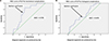
The results listed in Table 3 indicate that FLP was the only variable that had an important impact on mechanical and biological success. The ROC curve for FLP of mechanical and biological complications is shown in Fig. 1. The area under the ROC curve (AUC) for FLP of mechanical complications is 0.725, which indicates a reliable result since the value exceeds 0.5. The optimal cutoff value was 46.57 months (95% confidence interval, 0.61 - 0.84), which gave a sensitivity of 69.2% and a specificity of 69.7%. In addition, the AUC for FLP of biological complications was 0.615, which is also a reliable result (i.e., > 0.5). The optimal cutoff value was 39.80 months (95% confidence interval, 0.49 - 0.74), which gave a sensitivity of 54.5% and a specificity of 54.9%.
DISCUSSION
While 1-IR has been predictable treatment modality in the posterior edentulous region, mechanical and biological complications occur frequently.2930 These complications include SL, CF, implant fixture fracture, de-cementation, PM, and PI, and also, they often occur in multiple-implant restoration splinting.31 In addition, the implant success rate of 1-IR (94.3%) was not significantly lower than that of multiple-implant restoration splinting (97.1%).32 Therefore, these two types of the implant restoration have been reported to have similar success rates.
However, there were some differences between the 1-IR and 2-IS groups in the present study in the characteristics of mechanical and biological complication rates related to clinical factors.
There was no significant association between the implant position (first or second molar region) in the 1-IR group and the occurrence of mechanical and biological complications (P = .243 and P = .746, respectively). These results were identical to those of a previous study.33 Also, prosthesis types were not associated with complication rates in the 1-IR and 2-IS groups, and did not affect the comparison of the two groups (P = .276).
Compared to 2-IS, 1-IR showed a higher rate of mechanical complications (P = .020). SL was reported as the most common mechanical complication of implant-supported single crowns (ISSCs),2934 and also in the present study this was the only major complication associated with a statistically significant increase in occurrence (P < .001). Sex was the only clinical factor exerting an important influence on the occurrence of mechanical complications in 1-IR (P = .040). Finally, 91.3% (21 implants) of all SL cases occurred in male patients, which is possibly due to the biting force and occlusal contact area both being greater than in female patients.3536
Conversely, in 2-IS, there was a significant increase in the rates of biological complications (P < .001). FLP (P = .049) played an important role in this pattern. One possible explanation is that although regular dental hospital visits following the completion of the final implant prosthesis enabled examinations during the early stages, the intervals between the dental hospital visits became longer over time, resulting in a reduced awareness about oral hygiene management. Similarly, the significant increases in PM (P = .002) and PI (P = .046) are due to the difficulty of performing adequate oral hygiene management in splinted implant restorations. Several studies have found that nonsplinted implant restoration was advantageous for oral hygiene management.3 These results emphasize the need to provide patients with specific instructions about the use of dental floss and interdental brushes, especially for 2-IS.
Previous studies2737 found that SL induced changes in centric and eccentric contact forces and nonideal occlusion, and that this was a major cause of SF and CF. Moreover, food impaction on the inferior aspect of the implant prosthesis was caused by SL, and the resulting gingival redness and swelling not only increased the occurrence rates of PM and PI but also decreased the survival and success rates of the implants.38 In the present study we also observed the simultaneous occurrence of SF, CF, and PI with SL in three implants from the 1-IR group and in two implants from the 2-IS group.
The superiority of 1-IR in the first and second molar regions has been reported previously.3940 2-IS was advantageous over 1-IR in terms of mechanical complications but disadvantageous in terms of biological complications. The decreased incidence of mechanical complications is probably attributable to the improved stress distribution resulting from the splinting technique reducing the transfer of excessive forces to the implant fixture and surrounding bone31314. In addition, the splinting technique is able to promote the retention and resistance of the prosthesis, and successful outcomes can also be expected under certain limiting conditions such as insufficient abutment length, abnormal loading, or long treatment period.3 The increased incidence of biological complications is probably due to structural aspects of the splinting technique. It is often difficult to ensure the formation of appropriate paths and distances when placing fixtures in patients with periodontal disease. This makes it difficult to form an appropriate contour and embrasure between the inferior aspect of the splinted implant restoration and the interproximal region, which is a limitation in oral hygiene management. Given that biological complications cause inflammation and the loss of tissue and bone in the surrounding implant, and increase the risk of implant failure, these complications need to be managed carefully. Conversely, the disconnection and reconnection are more convenient for 1-IR, and examining and ensuring the hygiene of the prosthesis are easier than for the splinted implant restoration.4142
The only clinical factor that strongly affected the mechanical and biological complications associated with 2-IS was the FLP. In 2-IS, the biological complications occurred earlier in the investigations of FLP values. Relatively successful outcomes were maintained for up to 38 months on mean after the placement of the implant prosthesis. However, biological and mechanical complications occurred after mean FLP of approximately 49 and 56 months, respectively. These findings contrast with a previous study finding that mechanical complications usually occurred sooner than biological complications during follow-up periods of 1 - 2 years.43 We attribute this difference to our clinical protocol of implant treatment for preventing mechanical complications, since patients were encouraged to visit the dental hospital continuously at short intervals after completing the treatment so that their implant prostheses could be examined regularly. PM and PI were previously reported to occur in 50% and 10 - 43% of all implants, respectively.4445 However, the incidence of biological complications was considerably lower in the present study, with PM and PI occurring in 0% and 3.6% of 1-IR, respectively, and in 6.7% and 9.6% of 2-IS. These results also demonstrate that the periodontal checkups were thorough. The dental hospital visits occurred regularly at intervals of 3 - 6 months during the 3 years after implant placement in the present study. However, after 3 years the interval between the dental hospital visits increased to 1 year, and some cases were lost to follow-up due to cancelled appointments. This led to neglect of oral hygiene management, which resulted in increased rates of biological complications due to the deposition of plaque and calculus and of mechanical complications due to the lack of regular examinations approximately 1 - 2 years later.
Therefore, within the limitations of this study, the optimal cutoff values for FLP with significant effects on the mechanical and biological success of 2-IS were calculated using ROC analysis. Those for the biological and mechanical complications were 39.80 and 46.57 months, respectively. This information can be used to determine the optimal period for follow-up by predicting the time at which complications might occur. Such a strategy will help prevent complications, while making it possible to explain the importance of regular dental hospital visits to patients and also providing them with motivation.
In the previous study, an excessively long HDNI (> 3.7 mm) was a significant factor affecting the prognosis of ISSCs located in the first and second molar regions.33 However, in the present study, HDI was not an important cause of the complications in 2-IS. This is probably due to HDI has to be longer than HDNI in order to reduce the alveolar bone loss,24 and the reduction in the cantilever effect due to dispersal of the occlusal forces when the implant prosthesis is splinted. Moreover, careful consideration should be given to a certain degree of HDI needing to be established for an interproximal design in order to simplify the use of oral hygiene products such as dental floss and interdental brushes.
The results of this study need to be analyzed carefully because they were limited by the lack of investigations of various clinical factors that could affect implant restoration, such as the individual physiological characteristics, occlusal relationships, and parafunctional oral habits of each patient.464748 Therefore, more reliable results can be expected from future investigations of restorations involving two or more splinted implants to analyze the effect of the above factors on mechanical and biological complications and from long-term evaluations. In addition, a careful prospective study of FLP should be performed, since the present study found FLP to be an important factor affecting mechanical and biological complications in 2-IS.
CONCLUSION
This study found 2-IS to be more advantageous than 1-IR from mechanical aspects. However, the biological aspects of 2-IS need careful investigation. FLP are the most significant clinical factor for the mechanical and biological complication rates of 2-IS. Biological complications come about in FLP of 39.80 months and mechanical complications in FLP of 46.57 months. Therefore, clinical consideration of FLP will help to prevent the mechanical and biological complications of 2-IS.

 XML Download