

 PDF
PDF ePub
ePub Citation
 Print
Print


Introduction
Difficulties to achieve a natural appearance with metal ceramic restorations have led to the use of all ceramic restorations.1 Among various all ceramic restorations, the use of zirconia restorations has been increased in restorative dentistry due to biocompatibility and excellent mechanical properties of zirconia cermics.2345678 Besides, advanced computer-aided design and computer-aided manufacturing (CAD/CAM) systems have been well established in prosthodontics to fabricate zirconia restorations. From the esthetic point of view, metal ceramic crowns have a disadvantage compared to zirconia crowns, caused by the metal margin and its show above the gingiva.19
Generally two types of zirconia crowns can be applied: zirconia-based crowns and full zirconia crowns. In zirconiabased crowns, CAD/CAM-fabricated zirconia cores are layered by porcelain veneers, while CAD/CAM-fabricated full zirconia crowns are monolithically prepared from zirconia ceramics without layering. Natural zirconia is white in color and optically a semi-translucent material.1 Therefore, partial light transmission through zirconia ceramic structure may be expectable. Some factors may affect the color of zirconia-based restorations including: dental substrate,10 cement,11 zirconia core,12 porcelain veneer,1314 and glaze.15 Despite the fact that cements play a significant role in the color of all ceramic restorations,161718 the effect of cement on the color of zirconia-based restorations has not been clearly understood. Fazi et al.11 reported that in zirconia-based restorations, a 0.5 mm thickness of porcelain veneer was not sufficient to prevent the color shift from cements.11 Chang et al.19 investigated the optical effect of composite luting cements on all ceramic crowns and revealed that cemented Katana zirconia crowns created perceptible color changes in the cervical thirds of the crowns. Choi and Razzoog20 evaluated the masking ability of zirconia ceramic with and without porcelain veneer and concluded that the zirconia ceramic without veneer was rather capable to mask the different tested substrates. As zirconia ceramic is a semi-translucent material,1 it may not completely mask its background.
Investigators have employed spectrophotometry to determine color attributes and color changes in CIELab color system.2122 In this system L*, a*, and b* are defined as lightness, red-green value, and yellow-blue value, respectively. ΔE color difference is usually measured by this formula: ΔE*ab = [(L*2 − L*1)2 + (a*2 − a*1)2 + (b*2 − b*1)2]1/2, which is the most commonly used and feasible formula for ΔE.1 Spectrophotometers can detect even small amounts of the color differences that human eyes cannot percept.23 A perceptional threshold for ΔE has been determined by researchers to analyze the data of the spectrophotometric measurements.24 If the ΔE color difference is more than the perceptional threshold, a color mismatch is accepted. Some in vivo studies determined the perceptional threshold of ΔE = 2.6 - 3.7 as a guide.2526 The perceptional threshold for in vitro studies has been considered to be less than in vivo studies (ΔE = 0.4 - 1) due to better optical conditions.27
Conventional luting agents can be used to cement zirconia-based restorations.11 Two types of luting agent can be applied, including permanent cements or temporary cements. Cementing a restoration on a temporary basis may be occasionally suggested so that the clinician can evaluate its appearance and function over more time.28 Furtheremore, implant-supported zirconia-based restorations may be cemented by temporary cements for a long-term use.29 Permanent cements are mostly used on a definitive basis in the long term. However, from an esthetic point of view, no current consensus has been published to the more proper cementing option.
Considering the zirconia core with a normal thickness of 0.5 mm for zirconia-based crowns,30 optical characteristics of zirconia ceramic,1 and limited masking ability of porcelain veneers especially in cases with a limited restorative space or an improper veneering procedure,11192031 the impact of cement type on the color of zirconia ceramic has to be clearly determined to achieve better clinical results. Therefore, this study aimed to evaluate the effects of four different cements on the color attributes of a zirconia ceramic. The null hypothesis was that the cement type would not affect the color attributes of zirconia ceramic.
MATERIALS AND METHODS
Considering results of a previous study, an 80% power and a 0.05 level of significance, this study assigned ten specimens in each group. Therefore, a total of 40 zirconia disk specimens (N = 40) were prepared and divided into 4 groups of 10. The disk specimens were cemented to composite substrates using four different types of cement. Spectrophotometric measurements were performed on the specimens before and after cementation. The procedure was conducted precisely as follows:
A CAD/CAM system (CORITEC 250i, imes-icore GmbH, Eiterfeld, Germany) was used for milling zirconia blanks (Luminesse High Strength 98 mm Discs #5113, Talladium, Valencia, CA, USA) to prepare zirconia disks with a specific design. The disks were 0.5 mm in thickness and 10 mm in diameter. The disks had two sides: an outer side with a flat surface and an inner side with a hollow space. The hollow space was 0.1 mm in depth and 8 mm in diameter, designed for a cement space. The inner side had an external edge width of 1 mm around the hollow space. Three grooves were prepared on the external edge of the inner side as vents for excess cements. The zirconia disks were dipped in a coloring liquid shade A2 (DD Bio ZX2 monolithic zero LZDD, Dental Direkt GmbH, Spenge, Germany) for 10 seconds. The colored disks were dried by a lamp for 45 minutes. All the zirconia disks were sintered at 1500℃ for a 12-hour process in a sintering furnace (iSINT HT, imes-icore GmbH, Eiterfeld, Germany). A digital micrometer (293 MDC-MX Lite, Mitutoyo Corporation, Tokyo, Japan) with the accuracy of 0.002 mm was employed to measure the thicknesses of the disks. The disks were adjusted to have the thickness of 0.5 ± 0.01 mm. An adjustment and polishing kit (BruxZir, Glidewell Direct, Irvine, CA, USA) was used to reduce the thicknesses to the mentioned range. In case of lack of the acceptable thickness, the disk was excluded from the study. The zirconia disks were polished and were then cleaned in an ultrasonic bath (Elmasonic S-30, Dentec, North Shore, Australia) containing 98% ethanol for 15 minutes and dried.
In order to fabricate composite substrates, a cylindrical wax pattern was initially formed with 10 mm diameter and 5 mm height. A putty silicone impression (Speedex, Coltene, Altstatten, Switzerland) was taken from the wax pattern to prepare a mold. A light-polymerized composite resin of shade A3.5 (Z100 Restorative, 3M ESPE, St. Paul, MN, USA) was applied in layers to the mold. A light-polymerizing unit (Elipar FreeLight 2, 3M ESPE, St. Paul, MN, USA) was used to polymerize the composite resin incrementally for 40 seconds with an intensity of 800 mW/cm2. The composite substrates were polished with 800 grit silicon carbide abrasive papers for 10 minutes. Then, they were cleaned in the same ultrasonic bath containing 98% ethanol for 15 minutes. A total of forty composite substrates were prepared according to the number of zirconia disks.
In order to cement the zirconia disks to the composite substrates, four different cements were used including: Glass Ionomer (GC Gold Label, GC Corporation, Tokyo, Japan), Panavia F2.0 resin cement (Panavia F 2.0, Kuraray, Tokyo, Japan), Zinc Phosphate (Phosphate cement, Hoffmann Dental Manufaktur GmbH, Berlin, Germany), and TempBond (TempBond, Kerr, Salerno, Italy). Each zirconia disk specimen was cemented to a composite substrate. A clean glass slide was placed onto the zirconia disk and a 9.8 N compressive force16 was applied for 5 minutes. The cementation process was performed according to the manufacturer's instruction for each cement.
A spectrophotometer (SpectroShade Micro, MHT, Verona, Italy) was employed for spectrometric measurements.32 A putty silicone material (Speedex, Coltene, Altstatten, Switzerland) was adapted to the mouth piece of the spectrophotometer to match the conditions of spectrophotometry for all specimens and to prevent external lights. The specimens were located at the center of this putty mold. Before each measurement, the spectrophotometer was calibrated with the white and green calibration plates, respectively. The color attributes of L*, a*, and b* in the CIELab color system were measured in three situations:
1. Substrate alone (S)
2. Disk on substrate before cementation (SD)
3. Disk cemented to substrate after cementation (SDC)
All color measurements were conducted at the center of the specimens marked by a pen device on the monitor screen of the spectrophotometer, and the color attributes of L*, a*, and b* were recorded for each specimen in the three above mentioned situations. First, a substrate was located on the mold and the measurement was done (S). Second, a disk was placed on the substrate with a water drop in between to prevent the light refraction,19 and the measurement was performed (SD). Lastly, the substrate and the disk were dried for 15 seconds using air spray, and then the disk was cemented to the substrate, after which the unit was located on the mold for the measurement (SDC). Additionally, ΔE was measured by the spectrophotometer to determine the color differences between the situations, including: S-SD, SD-SDC, S-SDC. This formula was employed by the device to measure ΔES-SD, ΔESD-SCD, and ΔES-SCD: ΔE*ab = [(L*2 − L*2)2 + (a*2 − a*2)2 + (b*2 − b*2)2]1/2. The perceptional threshold of ΔE = 3.3 was hypothesized in this study.112527
A normal distribution of the data was accepted in all groups by the Kolmogorov-Smirnov test (P > .05). A software (SPSS 21, SPSS Inc., Chicago, IL, USA) was used to analyze the data. Effects of the type of cement, type of situation, and their interaction on the color attributes of L*, a*, and b* were evaluated using Repeated Measures ANOVA. One-way ANOVA assessed the ΔE values of the cement groups in situations of S, SD, and SDC. Pairwise comparisons of the groups were accomplished by Tukey Post Hoc and Bonferroni tests. A software (STATA, StataCorp LP, Lakeway, TX, USA) was used to compare the ΔE values of the groups with the predetermined perceptional threshold of 3.3 using One-sample t-test. All tests were carried out at the 0.05 level of significance.
RESULTS
The results were explained according to the measured parameters of L*, a*, b*, and ΔE in four sections.
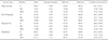
The means and standard deviations of the L* values for the four groups in three situations of S, SD, and SDC were demonstrated (Table 1). Repeated Measures ANOVA was used to evaluate the effects of cement type, situation type, and their interaction on the L* values. The results of this test showed that the cement type (P < .0001), situation type (P < .0001), and their interaction (P < .0001) affected the L* values. Pairwise comparisons of the four groups in the SDC, using Tukey Post Hoc and Bonferroni tests, showed significant differences among all the cement groups (P < .0001), except between Zinc Phosphate and Tempbond (P = .145).
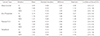
The means and standard deviations of the a* values for the four groups in three situations of S, SD, and SDC were demonstrated (Table 2). Repeated Measures ANOVA was used to evaluate the effects of cement type, situation type, and their interaction on the a* values. The results of this test showed that the cement type (P < .0001), situation type (P < .0001), and their interaction (P < .0001) affected the a* values. Pairwise comparisons of the four groups in the SDC, using Tukey Post Hoc and Bonferroni tests, showed no significant differences between Zinc Phosphate and Glass Ionomer (P = 1), and between Panavia F2.0 and TempBond (P = .88). The differences between Glass Ionomer and Panavia F2.0 (P < .0001), between Glass Ionomer and Tempbond (P < .0001), between Zinc Phosphate and Panavia F2.0 (P < .0001), and between Zinc Phosphate and Tempbond (P = .043) were statistically significant.
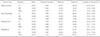
The means and standard deviations of the b* values for the four groups in three situations of S, SD, and SDC were demonstrated (Table 3). Repeated Measures ANOVA was used to evaluate the effects of cement type, situation type, and their interaction on the b* values. The results of this test showed that the cement type (P < .0001), situation type (P < .0001), and their interaction (P < .0001) affected the b* values. Pairwise comparisons of the four groups in the SDC, using Tukey Post Hoc and Bonferroni tests, showed no significant difference between Zinc Phosphate and Panavia F2.0 (P = 1). The differences among the other cement groups were statistically significant (P < .0001).
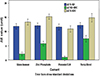
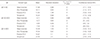
The means and standard deviations of the ΔES-SD, ΔESD-SDC, and ΔES-SDC values for the four groups were demonstrated (Table 4) (Fig. 1). One-way ANOVA test showed no significant difference among the four groups in ΔES-SD values (P = .55). Also, this test showed a significant difference among the four groups in ΔESD-SDC values (P < .0001). Pairwise comparisons of the four groups, using Tukey Post Hoc test, represented significant differences among all the cement groups in ΔESD-SDC values (P < .0001). One-way ANOVA showed a significant difference among the four groups in ΔES-SDC values (P < .0001). Pairwise comparisons of the four groups, using Tukey Post Hoc test, represented significant differences among all the cement groups in ΔES-SDC values (P < .0001), except between Zinc Phosphate and TempBond (P = .88).
In order to compare the means of the ΔES-SD, ΔESD-SDC, and ΔES-SDC values for the four groups with the predetermined perceptional threshold of ΔE = 3.3, One-sample t-test (one-sided) was employed. Considering the null hypothesis of µ ≤ 3.3, in ΔES-SD and ΔES-SDC, it was rejected for the four groups (P < .0001). In ΔESD-SCD the null hypothesis was accepted for Glass Ionomer (ΔE = 2.11) (P = .99) and Panavia F2.0 (ΔE = .94) (P = 1) and rejected for Zinc Phosphate (ΔE = 5.77) (P < .0001) and TempBond (ΔE = 7.50) (P < .0001).
DISCUSSION
The present study evaluated the L*, a*, b*, and ΔE values for zirconia disk specimens before and after cementation using four different cements of Glass Ionomer, Panavia F2.0, Zinc Phosphate, and TempBond. Statistical analysis indicated significant differences among the four cement groups in the L*, a*, b*, and ΔE values, comparing before (SD) and after cementation (SDC). The results showed that the cement type affected the color attributes of zirconia ceramic. Hence, the null hypothesis of the study was rejected. The results of this study can be interpreted based on the color attributes of L*, a*, b*, and ΔE as follows:
According to Table 1, comparison of the L* values for the cement groups before and after cementation (SD and SDC) showed that all the tested cements increased the L* values except Panavia F2.0. This may be due to the optical characteristics of Glass Ionomer, Zinc Phosphate, and TempBond. It seems that the higher L* values of these three cements compared to Panavia F2.0 are responsible for increasing the L* values in SDC. The three mentioned cements acted like bright backgrounds under zirconia ceramic. However, this effect of Glass Ionomer was less than those of Zinc Phosphate and TempBond.
As indicated in Table 2, comparison of the a* values for the cement groups before and after cementation (SD and SDC) showed that Panavia F2.0 and TempBond increased the a* values, while Glass Ionomer and Zinc Phosphate decreased the a* values. This result may be related to the natural a* values of Panavia F2.0 and TempBond. The a* value for TempBond at situation SDC showed a higher standard deviation compared to the other color attributes of the same group and also compared to the other cement groups. This may be due to a color difference between the accelerator and base pastes of the Tempbond cement, which affects the red color tendency of this cement. However, this had no significant influence on the standard deviation of the ΔE color change of this cement.
Based on the data in Table 3, comparison of the b* values for the cement groups before and after cementation (SD and SDC) demonstrated that all the tested cements except Glass Ionomer increased the b* value. This result may be caused by the yellow shade tendency of Panavia F2.0, Zinc Phosphate, and Tempbond, which does not exist in Glass Ionomer.
According to Table 4 and Fig. 1, comparison of the ΔESD-SDC values for the cement groups before and after cementation (SD and SDC) demonstrated that the color differences induced by Glass Ionomer and Panavia F2.0 cements were less than the predetermined perceptional threshold (ΔE < 3.3). Accordingly, Panavia F2.0 and Glass Ionomer induced acceptable color changes. The color differences induced by Zinc Phosphate and Tempbond were more than the predetermined perceptional threshold (ΔE > 3.3). Consequently, Zinc Phosphate and Tempbond cements caused perceptible color changes. In other words, overall changes of the L*, a*, and b* values created by Zinc Phosphate and TempBond led to the increase of ΔE values beyond the perceptible limit. This color shift may be due to the opaque optical properties of Zinc Phosphate and Tempbond. No significant differences were found among the tested groups in the ΔES-SD values (P = .55), which can be a reason for accurate and precise sample matching of the groups. The ΔES-SCD values represented significant differences among the groups (P < .0001), except between Zinc Phosphate and Tempbond (P = .88) due to their similar optical characteristics. The ΔES-SDC values displayed that the cement and zirconia created impressive color changes on the composite substrate (ΔE > 3.3).
Tracing the changes of L*, a*, and b* values (Table 1, Table 2, Table 3) indicated that the most amount of differences belonged to the L* attribute. Accordingly, the cement types had the highest impact on the L* attribute. Therefore, the ΔE changes have mainly been derived from this attribute (Table 4).
Fazi et al.11 reported that among the four tested cements of Fuji Plus resin modified glass-ionomer, Zinc-Phosphate, Rely X Unicem resin cement, and Maxcem resin cement, only Zinc-Phosphate caused perceptible color changes (ΔE > 3.3). This result on Zinc Phosphate cement was confirmed by the present study. Although the resin and glass ionomer cements used by Fazi et al.11 were different compared to the current study, both studies showed similar results on the cements, which created acceptable color changes (ΔE < 3.3). In the present study, the zirconia specimens were cemented to the substrates. However, in the study of Fazi et al.,11 the zirconia specimens were placed onto the cement disks without an interface. Obviously, the cementation procedure in the current research was more similar to a clinical setting. Additionally, the current study assessed TempBond cement, a temporary cement, which was not evaluated by Fazi et al.11
Chang et al.19 investigated the optical properties of resinbased composite cements and their effects on the color of Empress crowns and Katana zirconia crowns and concluded that the tested composite cements created perceptible color differences in the cervical and body regions with particular combinations of die material, cement, and ceramic crown. In their study, the color of luting cements could make color changes in the cervical thirds of Katana zirconia crowns. Although Chang et al.19 used pre-colored full zirconia crowns and luting cements different from the ones used in the current investigation, both studies revealed the possibility of cement-induced color changes for zirconia ceramics. A monolithic zirconia has a more translucency than a zirconia core in an equal thickness, however a zirconia core has a less thickness than a monolithic zirconia. This may be a reason for the similarity of the results.
Choi and Razzoog20 evaluated the masking ability of zirconia ceramic with and without porcelain veneer on four different substrates and reported that the zirconia without veneer had a degree of masking ability. On the other hand, the current study surveyed the effect of four cements on the color attributes of a zirconia ceramic, and according to its results, this degree of masking ability of the zirconia without veneer mentioned by Choi and Razzoog20 seems inadequate to prevent a cement-induced color mismatch.
Based on the results of this study, Zinc Phosphate (a permanent cement) and TempBond (a temporary cement) can change the color of zirconia core beyond the predetermined perceptional threshold. The color change due to TempBond cement in its short term use may lead clinicians to an incorrect clinical judgment like concerning the other affecting factors like zirconia core, veneer porcelain, and staining, while a proper choice of permanent cement can subsequently correct the color mismatch. Thus, in zirconiabased restorations, these cements should not be applied, or their negative effects should be reduced by sufficient porcelain veneers if possible. Moreover, Zinc Phosphate cement can be rationally substituted by the other cements like Panavia F2.0 and Glass Ionomer, especially in cases with limited restorative space in esthetic zones.
Considering the semi-translucent optical properties of zirconia ceramic and the thicknesses of zirconia cores (approximately 0.5 - 0.6 mm in a normal case), light transmission through the zirconia structure can be expected in zirconia-based restorations. Also, the underlying materials such as cements may affect the color. Thus, clinicians should realize color behaviors of the cements used for zirconia-based restorations to decrease the risk of a color mismatch. The color behaviors of four luting agents were evaluated in this study, and more investigations on various luting agents are recommended in zirconia-based restorations. On the other hand, the zirconia core overlaying materials such as porcelain veneers may influence the final color of the zirconia-based restorations, which were not evaluated in this study. As several effective factors play a role in the final color of zirconia-based restorations, the impact of each factor should be analyzed separately to determine its contribution to the final result and, ultimately, the cumulative effect of the factors should be determined. Therefore, the authors should aim to evaluate the other factors in future studies.
This study had some limitations such as using a specific brand of zirconia ceramic, a specific shade of composite substrate, one shade of coloring liquid, and four types of cement. More studies on the above mentioned subjects are suggested.
CONCLUSION
Within the limitations of this study, it was concluded that Zinc Phosphate and Tempbond cements affected the color attributes of the tested zirconia ceramic beyond the perceptional threshold. However, Glass Ionomer and Panavia F2.0 cements affected the color attributes of the tested zirconia ceramic within the perceptional threshold.
 XML Download