

 PDF
PDF ePub
ePub Citation
 Print
Print


INTRODUCTION
The stainless steel crowns (SSCs) are often the first choice for the repair of defects in primary dentition caused by caries, and have been one of the most effective and efficient methods of tooth restoration in pediatric dentistry since Humphrey first used them in pediatric patients in 1950.1 SSC placement is simple and economical with a high success rate for protecting remaining teeth weakened by excessive preparation.23 However, SSCs are disadvantaged by a non-aesthetic appearance,4 and this esthetic issue has become more and more important in patients with primary tooth problems. In a survey, esthetic concerns were confirmed to be the most critical issue for dental restoration in pediatric patients.5 Accordingly, new materials, such as, open-faced crowns, bonded strip crowns, and pre-veneered SSCs were developed to replace SSCs.67 These new materials improve esthetics, but have side effects that include poor gingival health, hemorrhage of gingival tissues, and exposure of dental margins, which are associated with the presence of metals.3 In particular, the veneer resin of preveneered SSCs frequently chips occurs and results in additional treatment and cost.78
The recently developed all-ceramic crown is now used as an esthetic restorative material for permanent teeth, and ready-made primary zirconia crowns are now being used for primary teeth. Zirconia, also known as "ceramic steel", has reasonable aesthetics and has excellent mechanical properties for the restoration of permanent teeth, and has been used for crowns, fixed partial dentures, implant abutments, fixtures, inlays, and onlays.9 However, zirconia crowns are thicker than SSCs and cannot be corrected, and thus, manufacturers recommend passive seating. Furthermore, due to their thicknesses, aggressive tooth preparation is required and pulp exposure is increased. Preveneered SSCs can still provide full coverage despite esthetic veneer chipping or fracture, but zirconia crowns must be replaced when fractured.10 This limitation can be overcome by lithium disilicate or leucite crown restoration for permanent teeth, but no study has been conducted as yet on the use of different ceramics for the repair primary teeth . Furthermore, no study has evaluated the wear resistance of primary teeth restored using different ceramic materials based on considerations of occlusal force in pediatric patients.
Primary tooth wear is common, and occurs due to the loss of enamel and dentin on the occlusal surface.1112 The abrasivities of primary and permanent teeth differ due to their different enamel strengths,13 morphological factors, such as, enamel and dentin thickness,14 and the different biting forces of adults and infants.15 In a comparative study conducted by Nose16 on molar enamels of primary and permanent teeth, the Vickers hardness number (HV) of primary teeth was 106 (HV) and that of permanent teeth was 126 (HV), which suggests poorer wear resistance of primary teeth. In a study by Mortimer,17 primary teeth were found to be less mineralized than permanent teeth, and Nelson et al.18 reported primary tooth enamel was thinner than permanent tooth enamel, and thus, more prone to wear. Proffit and Fields15 studied the bite forces of children and adults and found mean bite forces of 17.4 kg (mean age: 9.3) and 31.0 kg (mean age: 26.9) at a 2.5 mm opening; and 15.5 kg and 35.6 kg at a 6.0 mm opening, respectively, which confirmed that in children low bite forces reduced wear on the occlusal surfaces of posterior composite restorations.
Restoration materials affect enamel wear.19 Seghi et al.20 stated that the wear rates of dental restoration materials should be similar to that of the enamel. Previous studies on primary tooth abrasivity against restoration materials have focused on composite or glass ionomer restorative materials.212223 Due to growing interest in esthetics and the development of zirconia crowns, various studies have been conducted utilizing different restorative materials. In an evaluative study by Jung et al.24 on the abrasivity of natural teeth as compared with those of restoration materials, the natural tooth wear caused by zirconia was found to be much less than that caused by feldspathic dental porcelain. Sripetchdanond and Leevailoj25 confirmed that zirconia and composite resins wear enamel significantly less than glass ceramics and enamel. In pediatric dentistry, various cases of anterior tooth restoration using ready-made primary zirconia crowns have been reported.2627 However, few studies have conducted on primary tooth wear by ceramic restoration materials.212223 In this study, the associations between restoration materials, namely, zirconia crowns, lithium disilicate, and leucite and primary tooth wear were compared and evaluated to investigate the possibility of using ceramic restoration materials for primary tooth restoration.
MATERIALS AND METHODS
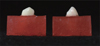
Primary canine teeth with no cusp wear, which were lost naturally during transition to permanent teeth, were used in this study. Teeth were washed in an ultrasonic cleaner and then placed in deionized water at 37℃ for 24 hours. The primary canine teeth were fixed in an acrylic resin (Orthodontic resin, DENTSPLY, Philadelphia, PA, USA) and approximately 5 mm of the cusps of the primary canine teeth was exposed (20 mm wide and deep and 10 mm high). Severely worn or fractured teeth and teeth with caries were excluded (Fig. 1).
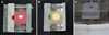
The 40 antagonistic specimens were assigned equally to a stainless steel (the Steel group; the control group), a leucite glass-ceramic (the Leucite group), a lithium disilicate glass-ceramic (the Lithium group), or a monolithic zirconia (the Zirconia group) group (Table 1). For the Steel control group, 11 mm-wide and -deep, and 13 mm-high, cubic specimens were prepared by crimping stainless steel (STS 304L) of SSCs (Kids crown®, Shinhung, Seoul, Korea). For the Leucite, Lithium, and Zirconia experimental groups, cylindrical metal specimens were prepared using a milling machine. Prepared specimens were scanned using a 3D scanner (Ceramill® Map400, Amanngirrbach Corp., Charlotte, NC, USA), and converted to STL files using design software (Ceramill® Mind, Amanngirrbach Corp., Charlotte, NC, USA). The materials used were; a monolithic zirconia block (Zirtooth Fulluster®, HASS, Kangneng, Korea), a lithium disilicate block (Rosetta SM®, HASS, Kangneng, Korea), and a leucite block (Rosetta BM®, HASS, Kangneng, Korea). Using a milling machine (Ceramill® Motion2, Amanngirrbach Corp., Charlotte, NC, USA), 11 mm-diameter and 13 mm-high specimens were prepared. Heat treatments were conducted in ceramics except Leucite group according to manufacturers' instructions, and specimens were subsequently washed using an ultrasonic cleaner (Table 2). Prepared specimens were fixed with an acrylic resin (Orthodontic resin, Dentsply, Philadelphia, PA, USA) using uniform molds by the same method used to prepare tooth specimens (Fig. 2). A single dental technician prepared all specimens.
Wear tests were conducted using a CS-4.8 masticator (SD Mechatronik, FeldKirchen-Westerham, Germany). In each chamber, restorative materials were placed on top and antagonistic teeth at the bottom using specimen holders (Fig. 3). A 3 mm vertical movement and a 2 mm horizontal movement were reproduced using two computer-controlled servomotors. Masticatory force was established using the results of previous studies that compared the abrasivities of dental materials and primary teeth.2223 In the present study, 50 N was used as the masticatory force, which was the middle value of the lowest and the greatest masticatory forces used in previous studies. To meet the 50 N condition, 5 kg was loaded on each chamber. In addition to the weight, a thermodynamic condition similar to the real oral environment was reproduced using a computer-controlled hot/cold water circulation system. Testing was conducted over 100,000 loading cycles (Table 3).23
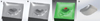
To measure the amount of the volume of tooth loss, teeth were scanned using a 3D scanner before and after testing.2829 The 3D data obtained before and after testing were overlapped using 3D software (Dentacian Software, EZplant, Seoul, Korea). Worn areas were then separated using Boolean operations and wear volumes were measured using 3D software (Dentacian Software, EZplant, Seoul, Korea) (Fig. 4). Wear losses of restoration materials after testing were determined using an electronic scale (PAG213, Ohaus, Seoul, Korea) accurate to ± 10-3 gram.
To qualitatively characterize wear patterns, antagonistic teeth and restorative materials were evaluated under a scanning electron microscope (S-3500, Hitachi Ltd., Tokyo, Japan) operated at 15 keV at magnifications of 40×, 100×, and 1000×.
The statistical significances of changes in tooth volumes and weights of restoration materials in each group were evaluated. Distribution normality and variance homogeneities were determined using the Shapiro-Wilk's test and Levene's test. The analysis was conducted by one-way ANOVA (α = .05) with Tukey's post-hoc test (α = .05). SPSS Ver. 21.0 (SPSS Inc., Chicago, IL, USA) was used throughout, and significance level was set at 5%.
RESULTS
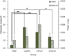
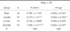
The means and standard deviations of the volume losses of antagonistic teeth (Vt) and the weight losses of restorative materials (Vm) in each group after wear testing are shown in Table 4 and Fig. 5. The Leucite group showed the greatest tooth volume loss at 2.670 ± 1.471 mm3, followed by the Lithium group with 2.042 ± 0.696 mm3, the Zirconia group with 1.426 ± 0.477 mm3, and the Steel group with 0.397 ± 0.192 mm3. Tooth volume losses in the Leucite and Lithium groups were significantly greater than that in the Steel group (P < .05), but no significant difference was observed between the Steel group and the Zirconia group (P > .05). In terms of weight losses of restorative materials after testing, the Lithium group showed most at 0.006 ± 0.002 g, followed by the Leucite group (0.003 ± 0.002 g) and the Steel and Zirconia groups (0.002 ± 0.001 g). Weight loss in the Lithium group was significantly greater than in the Steel and Zirconia groups (P < .05), but no significant difference was observed between the Steel, Leucite, and Zirconia groups (P > .05).
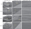
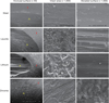
The SEM images of wear surface areas in enamel and tested materials are shown in Fig. 6 and Fig. 7. In enamel SEM images of the Steel group, smooth surfaces, fragmentation or particle chipping, and striated plow marks were observed. In tested material images, no notable surface changes or wear were observed other than slight stress marks and scratches in the sliding direction. Even after wear testing, dense and homogeneous surfaces were observed. In SEM images of the Leucite group, notable surface changes and wear were observed on the rough surfaces of tooth enamel with ploughing in the sliding direction. In particular, the presence of fragments chipped from enamel surfaces was confirmed by irregular concavities and gaps on worn surfaces. The Lithium group showed wear patterns similar to those of the Leucite group. Cracked and chipped fragments were observed on enamel wear surfaces. Striated wear grooves and rough surfaces, caused by lateral movements of the test instrument, were clearly observed on tested materials and on enamel surfaces. In enamel SEM images of the Zirconia group, surfaces were mostly smooth, and no chips or fractures were observed. No significant surface change or wear was observed for tested materials.
DISCUSSION
Comparative studies have been conducted on the abrasivities of different dental restorative materials and teeth. Due to growing interest in esthetics, the abrasivities of teeth and of ceramic materials, such as, leucite, lithium disilicate, and zirconia, which have excellent mechanical properties and esthetics, have been evaluated. However, these attempts have been limited to permanent teeth.3031 More recently, in pediatric dentistry, several manufacturers have started to produce ready-made primary zirconia crowns, and clinical applications of these products have already been reported. 2627 However, no comparative study has yet addressed the abrasivities of these ceramic materials when worn against primary teeth.
The clinical evaluation of abrasivity is costly and time-consuming. Moreover, accurate measurements are challenging because variables such as chewing forces and environmental factors cannot be completely controlled.32 Although in vitro studies are limited in terms of accurately reproducing mastication in the oral cavity, simple movements, such as, tooth grinding and clenching, can be reproduced, and the anti-wear mechanisms of various restoration materials can be evaluated at the pre-clinical stage.33 Because of the advantages of in vitro studies, various mastication simulators that reproduce wear process in the oral environment have been developed.3435 Various tests (e.g., pin-on-block, pin-on-disk, three-body wear, and toothbrush simulation) can be used to investigate the wear performance of dental materials, and antagonist wear has been confirmed to be closely associated with ceramic material types and testing conditions.36 Since the two-axe wear test device used in this study is practical, durable, and cost-effective, it has been widely used. The device has eight interior chambers and reproduces the mandibular closure of mastication through sliding movements after occlusal contact.30 In this study, a two-body wear test was conducted. This method is widely used to measure wear resistance, and reproduces the attrition caused by occlusal contact between restoration materials and teeth.34 This method can also reproduce the friction and fatigue wear caused by the direct contact between the maxillary and mandibular teeth during swallowing or non-functional dynamic occlusal movements.37 The 50 N force and 100,000 cycles used for the wear test in this study were determined based on the suggestions of previous studies that compared the abrasivities of various restoration materials and primary teeth.2223 Simultaneous thermal cycling with water to reproduce temperature changes in the oral cavity also removes wear particles generated during the wear test, and thus simulates aging.38 Suggestions made regarding the standardization of the enamel surface of natural teeth in the wear test are controversial.36 Krejci et al.39 suggested a non-standardized enamel cusp of natural teeth is the most appropriate antagonist. Kunzelmann et al.40 reported that unlike untreated enamel, polished enamel could show changes in the wear properties of enamel. Accordingly, in this study, the enamel surfaces of primary canine teeth that were naturally lost during the transition to permanent dentition were used as antagonist teeth. Magne et al.41 suggested height loss measurements provided a convenient method of measuring wear because they can be easily determined and are associated with clinical vertical measurements of occlusion. However, volumetric loss provides a more sensitive means than weight loss because measurements change linearly with time.42 In the present study, three-dimensional wear measurements with a laser scanner were used to measure the tooth wear. Using this method, teeth surfaces could be precisely and efficiently scanned three-dimensionally. 2829 On the other hand, in two-dimensional studies, it is difficult to overlap restoration material profiles before and after wear. Weight measurement has also been used to measure and quantify the amounts of wear in restoration material. 43 but it is difficult to remove moisture from the teeth after wear due to the presence of dentinal tubules. Despite drying, water can remain in teeth, and thus, moisture levels cannot be determined before testing. However, after testing restoration materials were completely dried, and thus, precise measurements of weight losses were obtained.
In this study, the abrasivity of primary teeth was compared with that of the four types of crown restoration materials: SSCs (Kids crown®), which has been widely used for the crown restoration of primary teeth; zirconia as represented by the recently developed ready-made primary zirconia crowns (Zirtooth Fulluster®); lithium-disilicate (Rosetta SM®), a particle-filled glass ceramic; and leucite (Rosetta BM®). The amount of wear of primary teeth was greatest in the Leucite group, followed in order by, the Lithium group, the Zirconia group, and the Steel group. In particular, amounts of wear in the Leucite and Lithium groups were significantly greater than that in the Steel group. However, no significance difference was observed between the Steel and Zirconia groups.
Surface hardness and friction factor are used to estimate the wear levels of dental restoration materials. Greater hardness is known to typically result in more enamel wear,24 and metal hardness has been shown to be associated with enamel wear.30 However, according to Oh et al.,44 enamel wear by ceramic is associated more with surface roughness, microstructure, fracture toughness, and environmental factors than with hardness per se. In the present study, enamel wear was greatest in the Leucite group because leucite has comparatively low flexural strength (120 MPa) and fracture toughness (1.42 MPa·m1/2). In contrast, zirconia has high flexural strength (1,200 MPa) and fracture toughness (5.5 MPa·m1/2), and showed less enamel wear than the Leucite group. This result agrees with that of a previous study that demonstrated an association between a low wear rate and high flexural strength and fracture toughness.29 If a restorative ceramic material does not have enough ductility fracture and/or chipping may occur. Furthermore, chips function as abrasives on ceramic surface and increase wear rates.45 The high enamel wears caused by glass particles in the Leucite and Lithium groups could be partly explained by the formation of wear debris. Glass particles freed by wear function as abrasives, a phenomenon referred to as the "three-body wear mechanism".46 The Steel group showed the lowest enamel wear rate because occlusal forces were absorbed by the ductility of the steel. In addition, as shown in the SEM images, smooth surfaces might reduce the wear rates of antagonist teeth. In the present study, enamel wear in the Zirconia group was comparatively small, as has been previously reported.24253031 This outcome is attributed to its excellent physical properties, such as, its hardness, flexural strength, density, and fracture toughness, which inhibit the formation of surface microfractures, and to the addition of yttria, which reinforces the crystal structure and prevents crack propagation.47 As a result, the smooth surface of zirconia was maintained during the wear test and only a small amount of enamel wear was observed after testing.37
The Lithium group showed the largest amount of restoration material wear, followed by the Leucite, Zirconia, and Steel groups. Wear in the Lithium group was significantly greater than that in the Steel and Zirconia groups, but no significantly difference was observed between the Leucite and Lithium groups. These outcomes concur with those of previous studies.34 The Leucite and Lithium group showed high amounts of wear because glass ceramics like leucite and lithium disilicate are sensitive to fatigue, which causes material defects.48 Actually, wear starts with crack formation on the ceramic surface, is propagated by repetitive loading, and eventually results in material loss.49
According to our findings, the amounts of wear of restoration materials and of antagonistic primary teeth tended to be positively related. Moreover, the flexural strength and fracture toughness of the ceramic materials (leucite, lithium disilicate, and zirconia) also seemed to be correlated with amounts of primary tooth wear.
The occlusal force, masticatory movement, and conditions used in this study differ from those in the oral environment. In addition, two-body wears were evaluated in this study, and three-body wear tests could result in different outcomes. Therefore, we suggest further long-term studies that simulate the oral environment be conducted to investigate the clinical implications of various ceramic material in primary teeth
CONCLUSION
In vitro measured volumetric losses in the Leucite and Lithium groups were significantly greater than in the Steel group, but no significant difference was found between the Steel and Zirconia groups. In this study, zirconia was the only material found to have an abrasivity that did not differ significantly from that of the steel used in conventional steel crowns. On the other hand, the Leucite and Lithium groups showed comparatively high primary-tooth abrasivity levels, which if confirmed in vivo may suggest that they should be used with caution in pediatric dentistry.


 XML Download