

 PDF
PDF ePub
ePub Citation
 Print
Print


INTRODUCTION
Synthetic elastomers, including polysulfide polymers, condensation and addition silicone, polyethers, and hydrocolloids, are used in dentistry to make impressions for reproducing oral conditions.1 The dimensional stability and accuracy of these impressions can vary under different conditions, but ensuring their consistency is crucial for the accuracy of the final prosthetic restoration. Nicholls2 noted that the dimensional stability of dental impression materials dictates the material's ability to maintain its accuracy over time; therefore, distortion results in loss of accuracy from the original dimension and can cause permanent deformation. The accuracy of dental impressions also depends on the correct choice of impression material.3
Various measuring techniques are used on dental casts and scanned models in clinical and academic dentistry . Manual measurements, performed with Vernier calipers or needle point dividers, are the standard method for evaluating the accuracy of the resulting dental casts.4 Manual measurement has many advantages, such as simple application, low cost, and immediate availability. However, the measurement accuracy can be adversely affected by operator fatigue and error.5 As an alternative, three-dimensional (3D) computer dental models, generated with optical or laser beam scanning, seem suitable for dental cast measurements in the clinical setting.6 For example, Tarawneh et al.7 reported that the accuracy for a single tooth is sufficient for practical use. However, inaccuracies of this method have been reported when calculating some orthodontic analyses and in academic works.68 Another 3D dental model technique is computed tomography (CT), where models are scanned using a 3D CT scanner. The use of CT measurement is advantageous because the whole object is scanned in a dimensionally stable manner and displayed without overlap.9
The storage time period is a crucial step before pouring of dental stone. Best results of accuracy were observed in immediate pouring after the removal of the impressions from patient's mouth. Due to time limitation in clinical condition, impressions are normally not poured immediately. Also, immediate pouring may not be possible if impressions are transferred to dental laboratory. Some manufacturers now claim that new impression materials have a longer storage time under suitable storage conditions. However, if this period is prolonged, the dimensional stability of the impression material will change and the impression will undergo distortion. An investigation is needed to support the manufacturers' claim. Thus, the aim of the present study was to use cone beam computed tomography (CBCT) scanning to evaluate the effect of storage time on the dimensional accuracy of impression materials. The null hypothesis was that storage time or type of impression material would not affect the dimensional accuracy of impression materials.
MATERIALS AND METHODS
This study used an ideal acrylic complete denture with no tooth malalignments as the master model. Two reference points were selected to standardize the measurements and were marked on the complete denture using a 0.1 mm stainless round bur. The reference points were made between the right and left first molar mesiobuccal cusp tips (Line 1) and between the right and left canine cusp tips (Line 2). The master model was tightened to the horizontal base of a dental surveyor.
Five impression materials were used to produce stone models from the master model: polyether impression material, IM (Impregum Penta Soft, 3M ESPE, St. Paul, MN, USA); two irreversible hydrocolloids, HY (Hydrogum 5, Zhermack Spa, Badıa Polesıne, RO, Italy) and AL (Alginoplast, Heraeus Kulzer GmbH, Hanau, Germany); a condensation silicone, ZF (Zetaflow, Zhermack Spa); and an addition silicone, HO (Honigum, DMG, Hamburg, Germany). In total, 200 impressions (n = 40) were taken from the master model. The impressions were divided into four equal groups and subjected to four different storage time intervals (n = 10). Following the manufacturers' instructions, all impression materials except for polyether were mixed manually and used in a traditional same-sized tray (Jescoform, Aesculap AG & Co. KG Am Tuttlingen, Germany). The polyether impression materials were mixed using an automatic mixing device (Pentamix, 3M ESPE, Platz Seefeld, Germany) and used with a special tray. Before making the impression, a thin layer of tray adhesive (Examix, GC America Inc., Alsip, IL, USA) was brushed onto the inside surfaces and edges of the special tray. The traditional and special trays were tightened to the vertical shaft of the dental surveyor. The use of the dental surveyor provided minimal tray movement for standardization. After mixing, the tray was first filled with the impression material, and then the impression was injected over the master model using an impression syringe. The Double Mixing technique was used for the condensation and addition silicone impressions.
The 200 impressions were divided into four groups according to the storage time interval as follows: immediate group: ten IM, ten HY, ten AL, ten ZF, and ten HO (50 impressions); one-day group: ten IM, ten HY, ten AL, ten ZF, and ten HO (50 impressions); three-day group: ten IM, ten HY, ten AL, ten ZF, and ten HO (50 impressions); five-day group: ten IM, ten HY, ten AL, ten ZF, and ten HO (50 impressions) (Table 1).
The 50 impressions in the immediate group were poured into stone within one hour using an improved Type IV dental stone (Moldano, Heraeus Kulzer), which was prepared on a vacuum mixer (Twister, Renfert GmbH, Hilzingen, Germany) to avoid the formation of air bubbles. If the gypsum could not be poured immediately, excess water was drained off the impression and the impression was stored in a hermetically sealed bag at room temperature (23℃/73°F) according to the manufacturer's instructions. A wet environment was maintained by wrapping the impression in a saturated moist paper towel. The same amount of moisture was ensured by wetting each paper towel with 25 mL distilled water and keeping the plastic bags sealed until the pouring time. By contrast, the polyether and two silicone impressions were stored under constant conditions (temperature 23℃/73°F, humidity 55 ± 5%). All impressions from the one-day group were poured after 24 hours; the impressions from the three-day group after 72 hours; and the impressions from the five-day group after 120 hours. All 200 dental cast stone models were scanned using CBCT.
In this study, the number of variables was reduced by using a master model, the same sized impression trays, and the same kind of dental stone. The measurements were also standardized using artificial reference points that were prepared on the complete denture using a 0.1 mm stainless round bur.
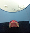
The dental stone casts were placed on a platform perpendicular to the horizontal plane, and the midsagittal plane was aligned with the midsagittal-positioning laser of the CBCT unit (Fig. 1). All dental stone cast models were scanned with the CBCT (NewTom 5G, QR Verona, Verona, Italy) system using the same parameters and standardized recording conditions. This system was operated in the high resolution denture scan mode: 110 kVp, 1 - 20 mA, scanning time of 36 seconds, exposure time of 7.3 seconds, axial thickness of 150 µm, and field of view (FOV) of 8 × 8 cm. The voxel size was 150 µm and isotropic (Fig. 2).
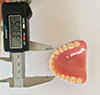
The master model was analyzed by three different researchers to achieve reliability. Each measurement was repeated five times. The master model was measured with an electronic caliper (Astor 150, Tok Ticaret Mak.San., İstanbul, Turkey) to an accuracy of 0.01 mm (Fig. 3). The mean of electronic caliper measurements was determined as follows: Line 1 (between right and left first molar mesiobuccal cusp tips): 49.72 mm; Line 2 (between right and left canine cusp tips measurement): 33.05 mm.
The normality of the data distribution was tested using the Kolmogorov-Smirnov test. The mean differences of the Line 1 and Line 2 between the CBCT scan differences and the master model were analyzed using two-way univariate analysis of variance (ANOVA) with impression materials and storage time as the main factors. A multiple comparison test was performed using the Duncan's complementary test with SPSS 16.0 for Windows statistical software (SPSS Inc., Chicago, IL, USA) at significance level of α=.05.
RESULTS
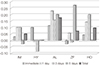
The mean differences in Line 1 and Line 2 were analyzed by two-way univariate analysis of variance (ANOVA) and the Duncan's complementary test (Table 2 and Table 3). For Line 1, the total mean differences of IM (0.05) and HY (0.00) were statistically different from that of AL (0.175) (P < .05). The impressions made with the condensation and addition silicones [ZF (0.071) and HO (0.096)] showed smaller discrepancies. The total difference of the AL (0.175) impression resulted in a greater difference than those of the other impressions materials (P < .05). For Line 2, IM (-0.061) showed a smaller total mean difference and was statistically different from the other impression materials (P < .05). The total mean difference of ZF (-0.182) showed the highest difference, followed by HO (-0.125), HY, and AL (-0.111) (P < .05).
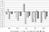
Significant differences were observed between Line 1 and Line 2 for the different storage periods (P < .05). The comparison to the master model value for Line 1 revealed that the IM impression specimens showed differences for the immediate (0.029), 1 day (0.100), 3 day (0.100), and 5 day (-0.029) groups, but the differences were not statistically significant. The HY impression specimens also showed no statistically significant differences for the immediate (0.100), 1 day (-0.014), 3 day (-0.086), and 5 day (0.000) groups. The AL impression specimens showed significant differences for the immediate (0.114), 1 day (0.229), 3 day (0.157), and 5 day (0.200) groups. The ZF impression specimens showed a significant difference for the 5 day group (0.272), but differences for the immediate (-0.029), 1 day (0.057), and 3 day (-0.014) groups were not statistically significant. The HO impression specimens showed significant differences for the 1day (0.143) and 3 day (0.200) groups, but not for the immediate (0.043) and 5 day (0.000) groups.
Comparison to the master model value for Line 2 revealed that the IM impression specimens showed a statistically significant mean difference for the 3 day group (-0.200), but not for the immediate (-0.071), 1 day (0.071), and 5 day (-0.043) groups. The HY impression specimens showed statistically significant mean differences for the 1 day (-0.171) and 3 day (-0.243) groups, but not for the immediate (-0.029) and 5 day (0.000) groups. The AL impression specimens showed statistically significant differences for the immediate (0.214), 1 day (-0.343), 3 day (-0.157), and 5 day (-0.157) groups. The ZF impression specimens showed statistically significant differences for the immediate (-0.286), 1 day (-0.186) and 3 day (-0.286) groups, but not for the 5 day group (0.029). The HO impression specimens showed a significant differences for the immediate (-0.300) and 5 day (-0.200) groups, but not for the 1 day (-0.029) and 3 day (0.029) groups.
DISCUSSION
The present investigation evaluated the mean difference of five dental impression materials based on different storage time intervals (Fig. 4 and Fig. 5). Significant differences were found among the total mean differences of impression materials for Line 1 (P < .05). The total mean differences of the HY and IM impression materials resulted in smaller differences, whereas the AL impression material showed the highest total mean difference. For Line 2, IM showed a smaller total mean difference and its mean difference was statistically different from those of the other impression materials (P < .05). The total mean difference was highest for ZF, followed by HO, HY, and AL (P < .05). Significant differences were seen for the storage time intervals between Line 1 and Line 2 (P < .05). Thus, the null hypothesis was rejected.
Dental irreversible hydrocolloids tend to undergo dimensional changes over time because they lose water, ultimately causing contraction of impressions. Conversely, irreversible hydrocolloid impressions expand when they absorb water.21011 Therefore, the best results are observed when irreversible hydrocolloid impressions are poured within 10 minutes of the removal from the patient's mouth and the impressions are poured within at least an hour, as this avoids distortion from irreversible hydrocolloid contraction or expansion.12 Dalstra and Melsen13 demonstrated the dimensional stability of irreversible hydrocolloid impressions after transportation for three to five days. Sending irreversible hydrocolloid impressions by mail did not affect the dimensional stability of dental stone models when stored under the proper conditions. Alcan et al.14 reported statistically significant alterations in irreversible hydrocolloid impressions after four days of storage, although the impressions were still acceptable for clinical use. Sedda et al.15 evaluated the accuracy of casts made from irreversible hydrocolloid impression materials and found that only the new irreversible hydrocolloid formulation (Hydrogum 5) was dimensionally stable after 72 and 120 hours. This finding may be related to difference in composition of the new hydrocolloid impression materials, which has higher filler and Calcium/Sodium ratios. Difference in composition may minimize free water movement in the structure and allow for extended pour time if stored under suitable conditions. The storage condition is an important factor for minimizing dimensional change. In the present study, storage condition was maintained by wrapping the impression in a saturated moist paper towel and hermetically sealed bag at room temperature. Alginoplast irreversible hydrocolloid impression material showed the highest mean difference and dimensional change. However, it still had clinically acceptable level of accuracy.
Polyether is a material with substantial accuracy. Henry and Harnist16 reported that polyether underwent less of a dimensional change and it was the most stable impression material, an observation also made in the present study. This stability may be because it has no reaction product. Alternatively, its higher hardness may provide greater resistance during storage, and its elastic recovery may resist reposition and withstand stress during removal of tray from the model. However, polyether has a hydrophilic character and, if stored under humid or wet conditions, it will undergo a large dimensional change. Thus, storage condition is another important factor for polyether impression material.
Silicone impression materials are normally used with custom trays and do not need a special tray. During or after the polymerization reaction, condensation silicone presents the evaporation of volatile by-products. Addition silicone, by contrast, does not release volatile by-products so that they do not change the material's dimensional stability.1 For Line 1, the total mean differences for condensation silicone (ZF) and addition silicone (HO) were similar and no statistically significant difference was observed when compared to the master model value. However, for Line 2, the total mean difference of the condensation silicone (ZF) impression material showed the highest difference, followed by the addition silicone (HO) impression.
The dental cast is widely used in dentistry for fabricating a working dental prosthesis. Traditionally, manual measurements have been performed with Vernier calipers or needle pointed dividers on dental casts. Shellhart et al.17 observed significant measurement errors with needle pointed dividers when applied to a dental cast. Alternatively, some authors have recommended the use of various measurement techniques on dental casts, but the results of these methods also demonstrated errors.181920 Many measurement processes have been used to compare the accuracy of different methods and to determine the applicability on different types of dental impression materials. The recent availability of 3D technology has uncovered several advantages, such as accuracy in performing measurements, orthodontic treatment effects, and tooth movements. A comparison of the use of a laser 3D digitizer and a micrometer method to determine accuracy of the measurement techniques revealed that scanning with a laser was more precise than using micrometers.21 Detection of 3D tooth movement is quite difficult with the naked eye, but Yamamoto et al.22 were able to create 3D computed models with a laser beam cast in which tooth movement could be easily observed. The error of tooth movement observed was less than 0.1 mm in translation and 0.5 mm in rotation. Tomassetti et al.8 compared the reliability of the Bolton analysis using manual measurements with a Vernier caliper and three computed methods. One of the computed methods gave results similar to those obtained with Vernier calipers, while the other two computed methods showed less correlation. Santoro et al.23 evaluated the accuracy of measuring tooth size, vertical overlap, and horizontal overlap using computed method models and compared these with dental stone models. This study found significant differences in tooth size and vertical overlap, and these differences (0.5 mm) were not clinically acceptable. However, no significant difference was observed for the accuracy of the horizontal overlap. Zilberman et al.24 evaluated the accuracy of measuring tooth and arch width using a conventional measuring method and 3D computerized model methods. They concluded that conventional and computerized methods had clinically acceptable levels of accuracy, but the 3D computerized models might not be acceptable for research. Clinical acceptability was indirectly in agreement with previous studies.142526 In the study conducted by Tarawneh et al.7, the models were scanned using a 3D FlashCT scanner; whereas a laser digitizer was used in the previous studies. In the present study, the method used to obtain the digital models was similar to those used in previous studies,521 in which the main difference was the type of digitizing device (i.e., the CBCT scanner). Yan et al.27 developed a computer assisted CT scanning system for 3D dental cast measurements. They evaluated its reliability and found similar differences between CT scanning and manual measurements on plaster models. Kamegawa et al.28 compared the accuracy evaluation using a microfocus X-ray CT technique and a conventional 3D optical scanner. The microfocus X-ray CT provided sufficient accuracy in dental occlusion diagnosis and quantitative clinical assessment of occlusal treatment.29
In the present study, an acrylic resin master model was used, which resembled the maxillary arch. Measurements were performed on the scanned dental model. One of the advantages of the CBCT methods applied in the present study was that a 3D analysis of the specimens was possible. The tests of the between-subjects effects (Table 2 and Table 3) resulted in validating the accuracy of the effects of material and time. For Line 1 (between right and left first molar mesiobuccal cusp tips), the total mean difference measurements were smaller for the IM and HY impressions than for the AL and the condensation and addition silicones (ZF and HO) impressions. The total mean difference of HY impressions was similar to that of the IM impression materials. For Line 2 (between right and left canine cusp tips), the total mean difference of the IM impression was smaller and statistically different from those of the other impression materials. The total mean difference of the ZF impression was the highest, followed by the HO, HY, and AL impressions. Nevertheless, the total mean differences of impression materials for Line 1 and Line 2 were clinically acceptable.
The storage time distortion of the impression materials and its effects on the accuracy of the CBCT model were evaluated using the measurements of the stone models for all the groups. The measurements of the stone models poured from the five impression material were taken from specimens that had undergone one of the four following storage time intervals: immediately, 1 day, 3 days, and 5 days. For Line 1, when compared to the digital caliper measurements, the IM and HY impression specimens showed no significant differences; in AL impression specimens, significant differences were observed in the immediate, 1 day, 3 day, and 5 day groups; in the ZF impression specimens, a significant difference was observed at 5 days; in the HO impression specimens, significant differences were observed at 1 and 3 days. For Line 2, when compared to the digital caliper measurements, the IM impression specimens showed a significant difference at 3 days; the HY impression specimens showed significant differences at 1 and 3 days; the AL impression specimens showed significant differences for the immediate 1 day, 3 day, and 5 day groups; the ZF impression specimens showed significant differences at immediate, 1, and 3 days; and the HO impression specimens showed significant differences for the immediate and 5 day groups. The overall values shown in Table 2 and Table 3 indicated that these mean differences due to storage time are very small in terms of millimeters; therefore, they can represent an acceptable clinical tolerance.
We concluded that if the impression materials are stored under suitable conditions, they produce accurate and clinically acceptable dimensional stability results even after five days. This study has a limitation in that the CBCT system is more expensive and needs professional technical help. Further studies are required to measure CBCT and 3D computerized model methods.
CONCLUSION
For Line 1, the total mean differences of HY and IM impression materials resulted in smaller mean differences and the AL impression material showed the highest total mean difference. For Line 2, IM showed a smaller total mean difference, which was statistically different from those of the other impression materials. The ZF impression material showed the highest total mean difference, followed by the HO impression material. Line 1 and Line 2 showed significant differences for the different storage periods.


 XML Download