

 PDF
PDF ePub
ePub Citation
 Print
Print


INTRODUCTION
Nowadays, it is not difficult to find patients with systemic disease who need dental treatment. Chronic disease caused by increased aging population and changed life style is accounted for large proportion of systemic disease. Elderly patients tend to have dental disease, and they have high tendency to suffer from systemic disease which might require special caution during the dental treatment. Thus, it is important to identify the relationship between specific systemic disease and dental disease before setting treatment plan for achieving successful outcome from the treatment.1
Diabetic mellitus and high blood pressure are the most common endocrine disorder and cardiovascular disease, respectively. They have been considered to influence most significantly on prognosis and outcome of dental treatment. Many researches have studied the relationship between prognosis of dental treatment and the diseases mentioned above. Based on the combined result from various researches, consultation with physician before treatment is needed before dental treatment in order to share information about the patient's medical condition, given medication, and precaution in patient management.1
In addition, patients should be checked for liver disease before dental treatment. Liver disease is a very common disease and can be classified into acute form and chronic form. In the case of acute form, both structure and function of the organ can be completely recovered when underlying cause is eliminated, whereas chronic form is characterized by gradual organ impairment.2 In addition, depending on the origin, liver disease can be divided into infectious liver disease and non-infectious liver disease. As infectious liver disease, hepatitis A, B, C, D, and E viruses, infectious mononucleosis, secondary syphilis, and tuberculosis can be considered. Meanwhile, non-infectious liver disease includes alcohol and drug abuse with substances such as halothane, ketoconazole, methyldopa, and methotrexate, and failure of lipid and carbohydrate metabolism.2
Liver is the organ with various functions including homeostasis and metabolizing most of the drug substances. Patienst with advanced liver disease can have bleeding tendency due to reduced production of blood coagulation factor and thrombocytopenia. Liver synthesizes essential serum proteins (albumin, transporter proteins, blood coagulation factors V, VII, IX, and X, prothrombin and fibrinogen, as well as many hormones and growth factors), produces bile and transporters (bile acids, cholesterol, lecithin, and phospholipids), regulates metabolism, and conjugates nutrients (glucose, glycogen, lipids, cholesterol, and amino acids) and lipophilic compounds (bilirubin, cations, and drugs). Accordingly, liver dysfunction induces the abnormal metabolism of carbohydrates, lipids, protein, drug substance, bilirubin, and hormone.34 Thus, liver disease needs to be managed and considered from the perspective of both medical and dental context.5
Viral hepatitis is a heterogeneous disease and is reported to have at least 6 subtypes.3 Worldwide, five million cases of viral hepatitis are documented each year, and, according to Chandler-Gutierreze et al., estimated prevalence in Spain is 3.7%.6
Hepatitis A is caused by hepatitis A virus (HAV, RNA picornavirus). Since it spreads via oral-fecal route, it is highly endemic in developing countries and its estimated prevalence is 1.1%. The disease is typically mild to self-limiting and characterized by the sudden onset of nonspecific symptom. Carrier state does not exist, and vaccines (Havrix, Vaqta) are available. Host is assumed to acquire immunity once they get anti-HAV antibody.
Hepatitis B is caused by the hepatitis B virus (HBV, DNA virus). There are 4 million HBV carriers worldwide and one study calculated that 1.53% of all patients reporting to dental clinic are HBV carrier. The transmission route is intravenous drug use and blood transfusion, and sexual contact. In case of dental profession, there is a high risk of transmission through instrument cuts. Not only that, some studies reported the infection by saliva and crevicular fluid through mucosal absorption. Therefore, special caution is required for dental professionals since they bear 3- to 4-fold greater risk of infection. Because of its asymptomatic characteristic over 50% of population, people might not be aware of the infection. Through commercialized vaccine, immunity against virus can be given to most patients and immunoglobulin can be injected for protection after exposure.7
Transmission of hepatitis C virus (HCV, RNA virus) causes Hepatitis C. Hepatitis C itself acts as main cause of chronic liver disease and liver associated death. 8000-10000 deaths from hepatitis C are reported annually. Approximately 130 million individuals are presumed to be infected in the world and most of them are transmitted via parenteral route. 85% of all the patients advance toward chronic hepatitis and remain asymptomatic for 20 years. 74% of the HCV infected individuals develop extrahepatic manifestation, such as purpura, weakness, cryoglubulinemia, lichen planus, thyroid gland dysfunction, diabetes mellitus, and etc. Effective vaccine has not been developed and it is hard to be resolved spontaneously.6
Oral clinical manifestations triggered by liver dysfunction can be shown in the form of bleeding disorder, jaundice, foetor hepaticus, cheilitis, smooth tongue, xerostomia, bruxism, and crusted perioral rash.8
It is found that hepatitis C virus in particular significantly affects the oral region as extrahepatic manifestation. For example, lichen planus, xerostomia, Sjögren's syndrome, and sialadenitis are known to be related to HCV.6 Chronic periodontal disease is often shown in patients with liver disease. Bagán et al.9 and Novacek et al.10 reported lack of oral hygiene in patients with advanced liver cirrhosis.
The research, which focused on the effect of HCV upon oral health, has investigated DMFT (Decay, Missing, and Filling Teeth) index and CPITN (Community Periodontal Index Treatment Needs). As a result, higher number of decayed or missing teeth along with considerably poor oral health was presented in HCV infected patients although there was no statistically significant difference on CPITN.11 The number of missing teeth in the HBV infected patients was higher than the control group as well, and severe aspect of caries lesion, plaque, calculus, and gingival bleeding followed.12
Clinically, the correlation between liver disease and dental disease is focused on risk of infection and risk of toxicity from alteration of drug metabolism or complications such as coagulation disorder. Because most of the blood coagulation factors are either synthesized or removed from the liver, any disorder in this particular function can lead to subsequent change in the vessel wall and the platelet disorder. Thus, prothrombin time should be tested before treatment in order to prevent postoperative bleeding and infection.13 Furthermore, it is important to protect both surgeon and patient from possible infection transmission via blood and saliva.14
While most of the established researches solely focused on precaution, infection prevention, and procedures that should be considered before treatment, only few studies tried to find out the transition of oral condition throughout the maintenance period upon completion of surgery. Moreover, most of the studies only focused on HCV infected patients when clinical proportion of HBV infected patients is larger among visiting dental patients.
The purpose of this study is to find out the difference between control group and patients with liver disease in terms of dental maintenance including implants and relapse of dental disease requiring re-treatment of active dental therapy. Patients enrolled in this study are in the maintenance treatment for more than 3 years upon completion of either surgical or non-surgical treatment. Therefore, in this study, we will focus on the difference based on type of liver disease, type of virus, severity of liver disease, and presence of drug administration. From the result, we expect to evaluate the influence of liver disease on maintenance of oral health.
MATERIALS AND METHODS
This research is based on the patients who received necessary active dental therapy before Jan 1, 2010 and entered maintenance stage after Jan 1, 2010 in Kyungpook National University Hospital. Patients without any kind of significant systemic disease were selected as a control group and the number of patients in control group was 316. A liver disease group consisted of patients with various types of liver disease. However, patients with certain types of systemic disease that can significantly affect the healing and health maintenance of oral tissue were excluded.
Exclusion criteria: diabetes, hypertension, hemorrhagic disorders, heart disease, rheumatic fever, lung diseases (such as tuberculosis), arthritis, malignant tumor, autoimmune diseases, bone metabolism disorder, kidney disease, and pregnant or lactating women.
From the criteria above, 237 patients were assigned to the initial liver disease group. The study period was total 4 years starting from Jan 1, 2010 to Dec 31, 2013. Follow-up data were collected using clinical records of both control and liver disease group by comparing at least two radiographs during the study period.
The investigated clinical records included age, gender, past medical history, active dental therapy before Jan 1, 2010 (Pre-Tx), type and number of dental re-treatment during maintenance period (after Jan 1, 2010) (Re-Tx) if there is any, total number of visits during observation period (F-SPT), and amount of smoking and frequency of teeth brush per day. Clinical records, such as type of liver disease, severity of liver disease, presence of drug administration, duration of medication, and virus type of viral disease, were investigated on liver disease group. Radiographic images were used to compare the initial number of teeth and implant and the number of teeth and implant on their last visit.
Among 237 patients in the liver disease group, 7 patients with non-viral liver disease were excluded from the study because they are too small in number and their diseases could not be identified through the clinical record. Therefore, research was performed with 230 patients having viral liver disease. A total of 546 patients (316 patients in control group, 230 patients in liver disease group) was classified based on following standard.
Classification standard:
-
1) Type of active dental treatment before starting maintenance period (Pre-Tx): surgical, non-surgical
2) Presence of liver disease: Yes, No
3) Severity of liver disease: carrier, hepatitis, liver cancer, and hepatocirrhosis
4) Type of hepatic virus: A, B, C
5) Presence of current liver disease drug administration: Yes, No
Initial condition of oral tissue greatly affects the prognosis of future treatment. Thus, based on the collected data, all the patients were subdivided into two groups (surgical, non-surgical) for the type of active dental treatment before starting the maintenance period (Pre-Tx).
Analysis was performed to find out if there is any statistically significant difference on the change in number of teeth (N-teeth), implants (N-implants), and the number of active dental re-treatment (Re-Tx) during 4 years of observation period depending on gender, presence of liver disease, severity of liver disease, virus type of liver disease, and etc. The number of visits during the maintenance period (F-SPT) was additionally analyzed because it also significantly influences oral health.
The study protocol was reviewed and accepted by Research Ethics Committee, Kyungpook National University (Ethics Reference No. KNUH 2014-07-050-001).
Data were expressed as mean and standard deviation. Comparisons between groups or within groups were performed by independent t-test and one-way ANOVA. Since a large portion of data did not satisfy the test of normality, non-parametric tests were needed. Kruscal-Wallis test was used for analysis. It was assumed to be statistically significant when P value is below .05.
RESULTS
After analysis was made using clinical records and radiographs, it was found that 262 patients had surgical treatments and 284 patients had non-surgical treatments before maintenance period. Among 262 patients with surgical treatment, 197 patients were in the control group and 65 patients were in the liver disease group. In 284 patients with non-surgical treatment, 110 patients were in the control group and 165 patients were in the liver disease group. Age range of the patients in the control group was varied from 18 to 83 with the average of 50.193 ± 10.23. Age range of the liver disease group was from 19 to 88 with the average of 50.268 ± 10.12. In the group with surgical pre-treatment, gender distribution was 112 female (81 in control, 31 in liver disease) and 150 male (116 in control, 34 in liver disease). No significant difference could be found on the number of teeth loss (N-teeth, control P = .607, liver disease P = .344), change in the number of implants (N-implants, control P = .154, liver disease P = .723) and the number of active dental re-treatment during maintenance period (Re-Tx., control P = .154, liver disease P = .465) based on gender. In the group with non-surgical pretreatment, gender distribution was 132 female (57 in control, 75 in liver disease) and 152 male (62 in control, 90 in liver disease). No significant difference could be found on N-teeth (control P = .427, liver disease P = 1.000), N-implants (control P = .953, liver disease P = .813) and Re-Tx (control P = .298, liver disease P = .125) based on gender.
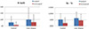
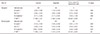
Table 1 shows the analysis on tooth loss rate (N-teeth), change in the number of implants (N-implants), the number of active dental re-treatment during maintenance period (Re-Tx) among all the patients in both control and liver diseasegroup. Independent t-test was applied as a statistical analysis method in this study. Comparing to the control group, patients with liver disease showed higher values on tooth loss rate, change in the number of implant, the number of active dental re-treatment during maintenance period. Statistically significant differences could be found on N-teeth (P = .000) and Re-Tx between control and liver disease group in patients who received non-surgical Pre-Tx (P = .000). Patients with surgical treatment presented higher values on N-teeth, N-implants, Re-Tx, and this finding was identical in both control and liver disease group. The number of visit during maintenance period (F-SPT) was approximately 7 visits per 4 years. Though F-SPT was higher in the control group, it was not statistically significant (Table 1, Fig. 1).
The analysis was performed to find out the possible difference based on severity of liver disease by subdividing patients with viral liver disease from the liver disease group. First, severity of liver disease was subdivided into three grade (carrier stage, active stage of hepatitis, and advanced stage toward liver cancer or cirrhosis). However, it was difficult to differentiate the carrier who received treatment prior to infection from the asymptomatic carrier who received treatment after infection. Thus, classification of carrier into subgroup with same level of severity was considered to be impossible. Therefore, after exclusion of carrier, the analysis was continued only on the patients who could be clearly distinguished depending on severity of liver disease.
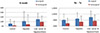
Table 2 represents the analysis of change in the number of teeth (N-teeth), change in the number of implant (N-implants), the number of active dental re-treatment during maintenance period (Re-Tx) and the number of visits during the maintenance period (F-SPT) depending upon severity of liver disease. Other criteria were analyzed using independent samples Krusal-Wallis Test because it could not pass the normality test due to the difference in sample size between each group. F-SPT passed the normality test, so analysis was performed with One-way ANOVA method. As a result, tooth loss rate and the number of active dental re-treatment during maintenance period increased with greater severity of liver disease regardless of received type of treatment before maintenance period. Statistically significant differences could also be found on N-teeth (P = .003) and Re-Tx (P = .044) in patients who received non-surgical Pre-Tx. However, change in N-implants did not show any specific tendency in all the patients regardless of treatment they received before maintenance period. Although F-SPT showed decreasing tendency as severity of liver disease increased, it was not statistically significant (Table 2, Fig. 2).
Among liver disease groups, we tried to find the difference based on drug administration. The type of drug was not categorized. Of the 65 patients in liver disease group who received the surgical treatment, patients taking medication were 20, and patients without medication were 45. No statistically significant difference could be found on the number of tooth loss (P = .661), change in the number of implant (P = .548) and the number of active dental re-treatment during maintenance period (P = .737). Of 165 patients in experimental group who received the non-surgical treatment, patients taking medication were 59, and patients without medication were 106. No statistically significant difference could be found on the number of tooth loss (P = .541), change in the number of implant (P = .100) and the number of active dental re-treatment during maintenance period (P = .361).
In order to find the difference based on the type of hepatitis virus, additional analysis was performed. Since only 2 people had hepatitis A, it was impossible to conduct statistical analysis. Therefore, they were excluded from the analysis. From the analysis based on the 208 hepatitis B patients and 20 hepatitis C patients, no statistically significant difference was found on tooth loss rate (P = .174), change in the number of implant (P = .829) and the number of active dental re-treatment during maintenance period (P = .361).
Difference analysis according to the type of active dental re-treatment during maintenance period (surgical or nonsurgical treatment) could not be observed since 96% of patients with re-treatment received nonsurgical treatment while only few received surgical treatment.
DISCUSSION
Liver disease is one the most common systemic diseases. Main causes of liver diseases are virus infection, alcohol abuse, and disorder in lipid and carbohydrate metabolism. Liver has a broad range of functions in maintaining homeostasis and health and involves in various drug metabolism. Liver disease can present various oral manifestations in the form of mucosal membrane jaundice, increased vulnerability to bruising, gingivitis, bleeding disorder, jaundice, foetor hepaticus, cheilitis, smooth tongue, xerostomia, bruxism, and crusted perioral rash. Especially HCV infection is known to have significant relationship with lichen planus, xerostomia, Sjögren's syndrome, and sialadenitis.15
The most important concerns associated with liver disease in clinical practice are risk of cross-contamination, risk of bleeding disorder, and risk of toxicity caused by altered drug substance metabolism. Since hepatitis C virus can remain stable at room temperature for over 5 days, strict sterilization measure is required.6
From the result of previous studies, it was found that HCV-infected individuals showed higher number of decayed or missing teeth compare to control group along with considerably poor oral health.11 In addition, another research showed that the number of missing teeth in the HBV infected patients was higher than the control group and severe aspect of caries lesion, plaque, calculus, and gingival bleeding followed.12 However, only a few studies tried to find out the transition of oral tissue throughout the maintenance period upon completion of surgery; most of the established researches solely focused on precaution, infection prevention, and procedures that should be considered before treatment. Thus, this study is designed to find out the difference in implant maintenance and relapse of oral disease, which requires active dental re-treatment, between control group and patients with liver disease who are in the maintenance treatment for more than 3 years upon completion of either surgical or non-surgical treatment. From the data, we were to evaluate the influence of liver disease on maintenance of oral health.
In this study, patients without any significant systemic disease were selected as the control group. Our research was designed to evaluate the data solely based on the presence of liver disease. Therefore, patients with other significant systemic disease were excluded from the liver disease group. The study period was 4 years from Jan 1, 2010 to Dec 31, 2013. Every patient enrolled in the study received necessary active dental treatment before Jan 1, 2010 prior to entering the maintenance period. Patients whose data got lost during follow-up were excluded from the experiment. From above standard, after exclusion of 7 patients with non-viral liver disease from the initial liver disease group, research was performed based on liver disease group of 230 patients with viral liver disease.
Type of treatments planned based on patients' oral condition on their first visit is very important to expect the prognosis of future treatment. It could be assumed that patients who were required to be surgically treated had severe dental disease, such as severe periodontitis. On the other hand, patients who needed non-surgically treated were likely to have mild dental disease. Initial oral condition of patients seems to be related with the need for advanced form of dental treatment, such as extraction and placement of implant, even after active dental treatment. Though clinical evaluation data of each patient could not be collected, subjects could be classified into groups with similar level of initial oral condition based on the assumption above. Thus, all the patients were subdivided into two groups (surgical, non-surgical) based type of active dental treatment before starting maintenance period (Pre-Tx).
To decide the type of active dental treatment for patients, initial oral condition of patients and the presence of systemic disease (such as diabetes) that might influence healing were considered. However, since patients with other systemic diseases were excluded in this study, only the initial oral condition of patients became the standard of deciding active dental treatment type. Consequently, presence and severity of liver disease did not affect the determination of dental treatment method.
Analysis of data was made on the number of teeth loss (N-teeth), change in the number of implants (N-implants), the number of active dental re-treatment during maintenance period (Re-Tx) and the number of visits during maintenance period (F-SPT) in all the enrolled patients. As a result, patients with liver disease showed higher values on N-teeth, N-implants, Re-Tx compared to control group regardless of dental treatment received before maintenance period, except for F-SPT. Among those patients, statistically significant differences could only be found on N-teeth (P = .000) and Re-Tx (P = .000) in patients who received nonsurgical Pre-Tx. In the group of patients who received surgical Pre-Tx, higher values on N-teeth, N-implants, Re-Tx was presented in comparison to the group of patients who received non-surgical Pre-Tx; this result was identical in both control and liver disease group.
In our experiment, patients with liver disease were subdivided depending on their grade of severity. After subdivision of the patient group, analysis was done to find out if there was correlations between N-teeth, N-implants, and Re-Tx along with severity of disease. The result showed increasing values in N-teeth and Re-Tx as severity of liver disease aggravates. Since it was difficult to differentiate the carrier who received treatment prior to infection from the asymptomatic carrier who received treatment after infection, carrier factor was ruled out from the analysis. Statistically significant differences could also be found on N-teeth (P = .003) and Re-Tx (P = .044) in patients who received non-surgical Pre-Tx. However, N-implants did not show any specific tendency in any of the patients regardless of the treatment they received before maintenance period. It seemed that, in case of change in the number of implants, comparative analysis had certain limit because the number of implant could be greatly influenced by not only oral tissue condition but also socioeconomic level of patients.
The average number of visit was 7 times in 4 years. It is well known that regularity and frequency of supportive therapy greatly influence the maintenance of healthy oral tissue.1617 In this study, the enrolled patients had similar number of visit (F-SPT) regardless of type of Pre-Tx and presence of liver disease. Thus, it seemed that frequency of supportive therapy did not affect the result of this study.
Data analysis on the type of liver disease was not statistically significant. However, because most of the enrolled patients were HBV infected and there was a huge difference in sampling size (2 HAV, 208 HBV, 20 HCV infected patients), the analysis result could be somewhat questionable. Thus, the analysis using a further reinforced data is required for this reason. Based on the past studies, HCV infection were thought to have the greatest impact on oral disease among other viral liver disease.81112
Difference based on the administration of medication was not statistically significant in our study. However, since detailed investigation on type, duration, and dosage of medication was not available, there was a certain limitation to draw the satisfying result. Therefore, more information on the medication would be required during the visit in order to find out its relationship with oral disease in further study.
Comparative analysis among types of liver disease was unavailable from the data we have because only 7 patients had non-viral liver disease which was not clearly identified in the clinical chart. There are many researches which discovered how alcoholic liver disease affect the oral health.1819 Thus, in further study, we will be able to perform comparative analysis between non-viral disease such as alcoholic liver disease and viral liver disease.
After integration of our result drawn from each criterion, it would be adequate to conclude that presence of liver disease gives malignant effect on maintenance of teeth and healthy oral tissue in patients who were required to receive non-surgical treatment for moderate oral disease. In addition, it is expected that oral tissue will be worsen as severity of liver disease aggravates. Worsen oral condition seems to come from the severe liver disease. However, there are a few more possibilities available. For example, in case of patients with advanced stage of liver disease, they may have possibility to neglect the management of oral hygiene which causes the limitation on maintenance of oral tissue as well. Previous studies found that patients with chronic liver disease had tendency to present poor oral hygiene, which caused higher frequency of tooth loss and carious lesion, presence of plaque and calculus, and gingival bleeding. 12
Thorough intra-oral examination and clinical history review are critical to evaluate the systemic health of patient before dental treatment. If patients have any kind of systemic disease, such as liver disease, which might affect the condition of oral tissue significantly, it is essential to make a consultation with their physicians and evaluate the degree of functional damage of the associated organ. From these procedures, both reasonable and safe dental treatment can be planned with clear judgment on prognosis according to medical condition of patients.6 Liver is the organ with a broad range of function and influence of liver disease might have a greater extent than currently known. Therefore, more careful approach with maintenance treatment will be necessary to treat the patient with liver disease since the disease may have possible malignant effect on the maintenance of oral health.20
CONCLUSION
Study results showed that liver disease might influence the loss of teeth and it was found to cause the relapse of oral disease during the maintenance period in patients with mild to moderate oral disease. A significant positive relationship between aggravation of oral health and severity of liver disease seemed to exist.

 XML Download