

 PDF
PDF ePub
ePub Citation
 Print
Print


INTRODUCTION
Technological developments in materials and devices provide dentists with new and advanced options for indirect prosthetic treatments. Computer-aided design-computer-aided manufacturing (CAD-CAM) technologies continue to advance and offer more options to dentists for manufacturing dental prosthetics. Designing and milling protocols can be performed in a dental laboratory or clinic, reducing treatment time and removing the need for temporary chairside restorations.1 Recently, the entire restoration making process has been implemented in a digital workflow environment, without the need for creating physical models.2
The CAD-CAM block materials for restoration have favorable mechanical and physical properties in comparison with laboratory-processed composites34: remarkable reduction in voids, flaws, and cracks,5 fewer discolorations,6 and higher abrasion resistance.7 Various materials have been used with CAD-CAM machining and commercially provided to dentists: yttria stabilized zirconia (e.g., IPS e.max ZirCAD, Ivoclar Vivadent, Schaan, Lichtenstein),89 feldspathic porcelain (e.g., VITABLOCS Mark II, VITA, Bad Säckingen, Germany),10 glass ceramic (e.g., IPS e.max CAD, Ivoclar Vivadent, Schaan, Lichtenstein),11 and resin composites (e.g., Paradigm MZ100, 3M ESPE, St. Paul, MN, USA).11
The new material Lava Ultimate (3M ESPE, St. Paul, MN, USA), called resin nanoceramic (RNC), is a resin-ceramic composite (primarily ceramic). This material is produced using nanomer and nanocluster fillers with a whole nanoceramic material content of 80 wt%. The nanomers are silica and zirconia with diameters of 20 nm and 4 to 11 nm, respectively.
Nanoclusters have structural integrity that permits a high proportion of ceramic filler to be contained into the blocks, providing great strength, fracture resistance, and wear resistance. The material has excellent polish retention for lasting esthetics and eliminates the need for a firing step after milling.12 The Lava Ultimate material was reported to have better performance than ceramics when applying an ultrathin (e.g. 0.5 mm) restoration.13 This material also showed equivalent fracture resistance to glass ceramics and a fundamental balance similar to enamel structures with a flexural modulus in the identical range as dentin.14 However, at present, few data are accessible in the scientific literature for RNC, in particular regarding resin-bonding protocols.
Resin bonding is an important step for the procedure and longevity of indirect restorations.1516 It is crucial that the adhesive bond is durable to provide high retention,17 prevention of microleakages, and improvement of marginal adaptation.18 A strong resin bond depends on the chemical adhesion between the cement and restoration, and on the micromechanical interlocking produced by surface roughening.19 Current roughening techniques are: (1) grinding,20 (2) abrasion with rotary instruments,2122 (3) air abrasion,23242526 (4) acid etching,27 and (5) a combination of these techniques.
Air abrasion is required for achieving enough bond strength between resins and high-strength ceramics reinforced with either zirconia or alumina.23 Surface modification of alumina has been mostly achieved using a particle size of 50 µm during air abrasion.24 The abrasive process eliminates loose contaminant layers, and the roughened surface supplies some degree of mechanical interlocking with the adhesive. The increased roughness increases the surface area for bonding.25
Silica coating is an air abrasion method also known as Cojet or Rocatec. Restorations are sprayed with alumina particles (around 110 µm) modified with silicic acid,24 resulting in the deposition of a molecular coating of alumina with silicic acid on the bonding surface. The surface is then coated with silane to make it more chemically reactive to the resin.2628
Acid etching with solutions of ammonium bifluoride or hydrofluoric acid (HF) can achieve a suitable surface roughness and texture.23 It was reported that 2.5 - 5% HF solutions applied for 2 - 3 minutes were the most successful.293031 However, the HF etching leaves an amorphous sediment of fluoride on tooth structures, which may negatively affect the bonding.32 Moreover, alumina increases the strength of the ceramic but it is highly chemically resistant and does not etch well.24
Various methods are used to measure bond strength (BS), including shear (SBS), microshear (µSBS), tensile (TBS), and microtensile (µTBS) bond strength tests and pull-out tests; the most common methods are TBS and SBS tests.333435 The advantages of the TBS test are that it uses a small quantity of material and quite even stress distribution can be obtained.35 The advantage of the SBS test is that it is easy to use,3335 but the stresses developing at the bond site are more complicated.3334 Therefore, the reliability of the test is questionable.333435 In contrast, the µTBS test shows more consistent stress distribution during loading and, therefore, higher bond strength values with less cohesive failures can be obtained.353637
Although resin-bonding protocols for silica-based23 and zirconia.1938 ceramics are well known, there are few studies of laboratory-processed composites.39 Studies regarding bond strength and surface treatment of RNC material are rare.16 Therefore, the aim of this study was to evaluate the influence of different surface treatment methods on the microtensile bond strength of resin cement to RNC using a µTBS test.
MATERIALS AND METHODS
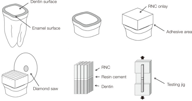
Thirty healthy human molars were collected for this study, obtained and used according to a protocol approved by the Institutional Review Board at Dankook University (IRB No. 1503/003/001). The teeth were stored in 0.1% thymol solution at 4℃ and used within 1 month of extraction. Coronal dentin surfaces were exposed by sectioning occlusal enamel and dentin with a water-cooled diamond saw (METSAW-HS, R&B Inc., Daejeon, Korea). The dentin surfaces were roughened using a wet 600-grit SiC paper (DBG Supplies, Doral, FL, USA) mounted on a disc polishing machine (J-POS2, JISICO, Seoul, Korea) for 30 seconds to produce uniform smear layers. Before the bonding procedure, the dentin surfaces were washed with distilled water and dried with oil-free air.
Thirty onlays (10 × 10 × 5 mm) were cut from CAD-CAM RNC blocks (Lava Ultimate, 3M ESPE, St. Paul, MN, USA) with a diamond saw (METSAW-HS, R&B Inc., Daejeon, Korea). The surfaces to be bonded were polished with a precision lapping machine (SPL-15, Okamoto Machine Tool Works Ltd., Shin-Yokohama, Yokohama, Japan) with diamond pastes of 6 µm and finally 1 µm. The onlays were then ultrasonically cleaned in distilled water for 3 minutes. After air-drying, the RNC onlays were divided into six groups of five onlays each and received one of the surface treatments.
-
1) Group A
-
2) Group AB
Air abrasion was performed as mentioned above followed by a bonding agent. A universal adhesive (Singlebond Universal Adhesive, 3M ESPE, St. Paul, MN, USA) was applied and light cured for 10 seconds with an LED curing lamp (Demi Plus, Kerr, Orange, CA, USA). -
3) Group HB
Surfaces were acid-etched with 4% HF (4% Porcelain Etchant, Bisco Inc., Schaumburg, IL, USA) for 5 minutes. After water washing and air-drying, the Singlebond Universal Adhesive was applied. A group using 4% HF acid etching without universal adhesive was planned. However, a pilot test showed excessively weak bond strengths and these samples were ruled out for µTBS testing. -
4) Group HSB
A silane solution (RelyX Ceramic Primer, 3M ESPE, St. Paul, MN, USA) was applied for 60 seconds before applying the adhesive in the protocol of Group HB. -
5) Group T
Surfaces were air-abraded with 110 µm silica-coated alumina oxide (Rocatec Plus, 3M ESPE, St. Paul, MN, USA) for 15 seconds at 2 bar pressure, 10 mm away from the surface to form a silica layer. -
6) Group TB
After sandblasting with Rocatec Plus the universal adhesive was applied as described above.
To analyze the texture, roughness, and composition of RNC surfaces to be bonded for each group, various surface analyses were undertaken. The group with non-treated surfaces (Group N) and the group with their surfaces treated with 4% HF etching (Group H) were included for a comparison between conditions of before and after surface treatment.
A nano-indenter (TI 750 Ubi, HYSITRON, Minneapolis, MN, USA) was used for measuring the RNC surface roughness, with a 100 nm Berkovich tip with 142.3° angle at a peak force of 2000 µN. The lateral displacement was 20 µm, and the scratch time was 30 seconds. The tests were performed 10 times at different points for statistical analysis.
The surface textures of all samples were examined with FESEM. Observations were performed using an S-4300 instrument (Hitachi, Tokyo, Japan) at 15 - 20 kV, working distance of 16 - 18 mm, and magnifications from ×5000. An integrated EDS system was used at 15 kV to measure the elemental composition of treated RNC surfaces. The test was performed 10 times at different points.
The RelyX resin cement was used for this study and prepared on a mixing pad. The thin layer of cement was applied to the surfaces to be bonded, and each RNC block was luted on the dentin surface while maintaining a continuous pressure of 1 kg over 6 minutes. After curing, the bonded specimens were soaked in distilled water and stored in a laboratory incubator (IB-11E, JEIO TECH, Daejeon, Korea) for 24 hours at 37℃ until the µTBS test was performed.
After storage, the onlays were sectioned in both x and y directions across the bonded interface into beams (2 ± 0.3 × 2 ± 0.3 mm) using a diamond saw (METSAW-HS, R&B Inc., Daejeon, Korea) under constant water cooling. The exact dimensions of the cross-sectional area of the interface were measured by a digital caliper (Absolute Digimatic, Mitutoyu, Tokyo, Japan) to calculate the formal bond strength (in MPa). Thirty samples were selected for each group. For the µTBS tests, each sample was attached with cyanoacrylate resin (Zapit, DVA, Corona, CA, USA) and tested until failure using a microtensile tester (Bisco Inc., Schaumburg, IL, USA) at a speed of 0.5 mm/min. Each specimen was evaluated with a stereomicroscope (SZ-PT, Olympus, Tokyo, Japan), and the failure modes were classified as cohesive (in RNC, dentin, or cement), adhesive (between RNC/cement or dentin/cement), or mixed. The failed surfaces were evaluated with FESEM. The entire protocol of this study is shown in Fig. 1.
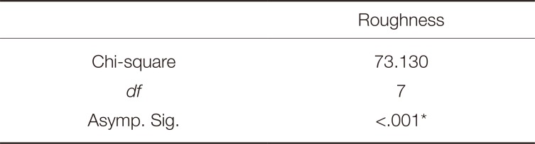
The results of µTBS tests on the samples with different surface treatments were statistically analyzed by one-way ANOVA. Subsequent comparisons were performed using the Tukey HSD test. The roughness and EDS analysis data were analyzed using the Kruskal-Wallis and Mann–Whitney U tests. All statistical tests were performed using the Statistical Package for the Social Science (SPSS v18.0, SPSS Inc., Chicago, IL, USA). P-values less than .05 were considered statistically significant.
RESULTS
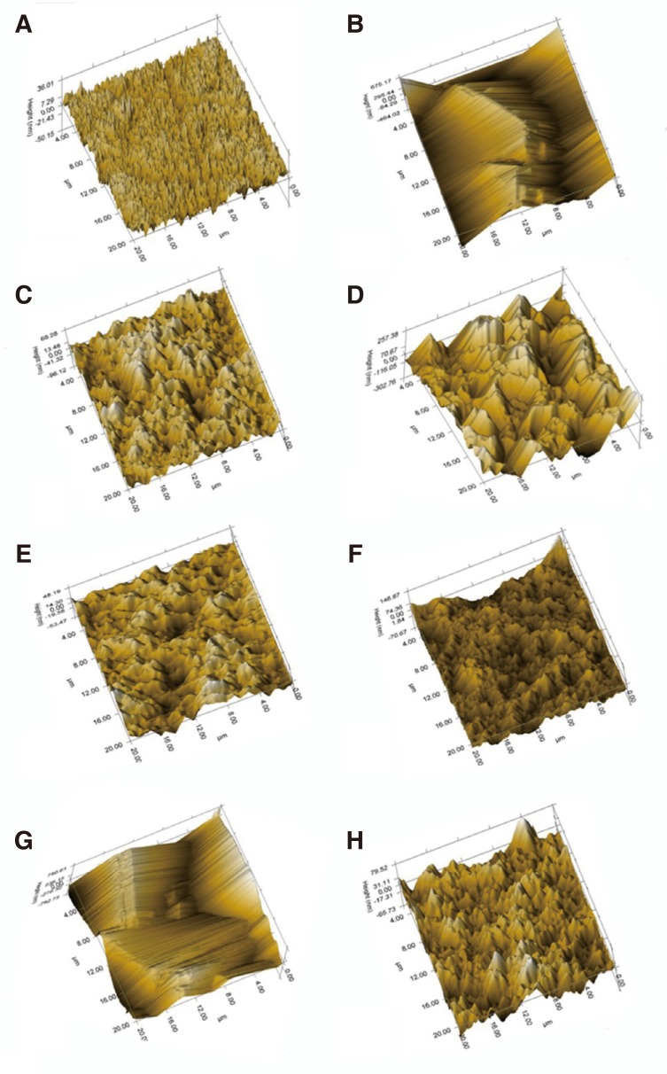
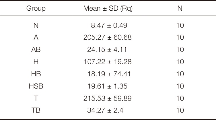
Here, we demonstrated that the nanoscratch test was useful for characterizing the roughness of the RNC samples. The roughness values of the surface of RNC onlays are presented in Table 1. The Kruskal–Wallis test presented significant differences among the experimental groups (Table 2). Group T showed the highest roughness (215.53 ± 60.68 nm) followed by Group A (205.27 ± 60.68 nm). There was no significant difference between Group A and T. However, the surface with 4% HF etching had significantly lower roughness than air-abraded samples, Group A and T (P < .05). Groups AB, HB, HSB, and TB had low roughness values.
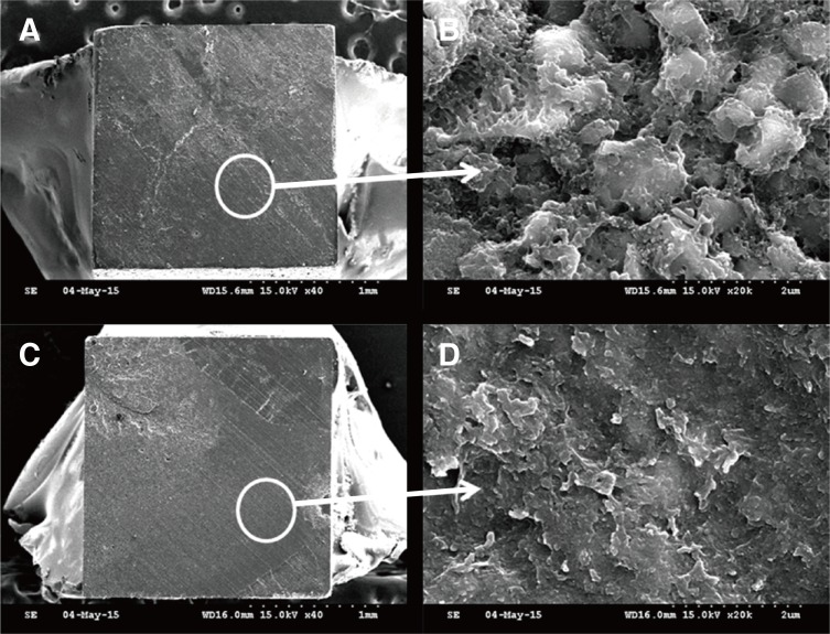
Images of the scratched surfaces of RNC onlays are presented in Fig. 2. The surfaces of Group N samples had an uneven texture that was deeply furrowed. The surface of Group H had a crater-like texture. Group A and T showed a texture with sharp valleys. Group AB, HB, HSB, and TB had similar textures, a smooth wave pattern from the universal adhesive (UA). Regardless of the roughening technique, after applying the UA, the surfaces presented similar texture and roughness.
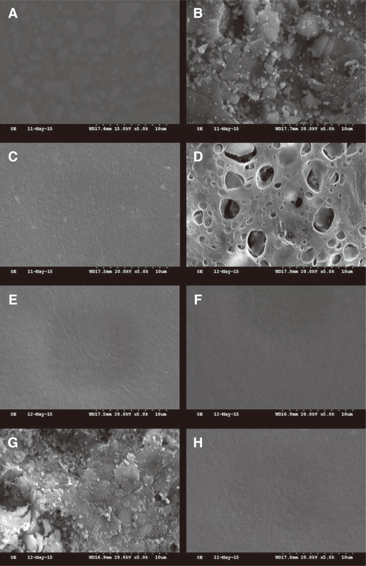
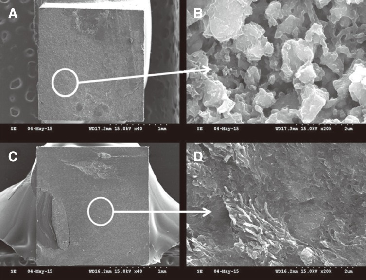
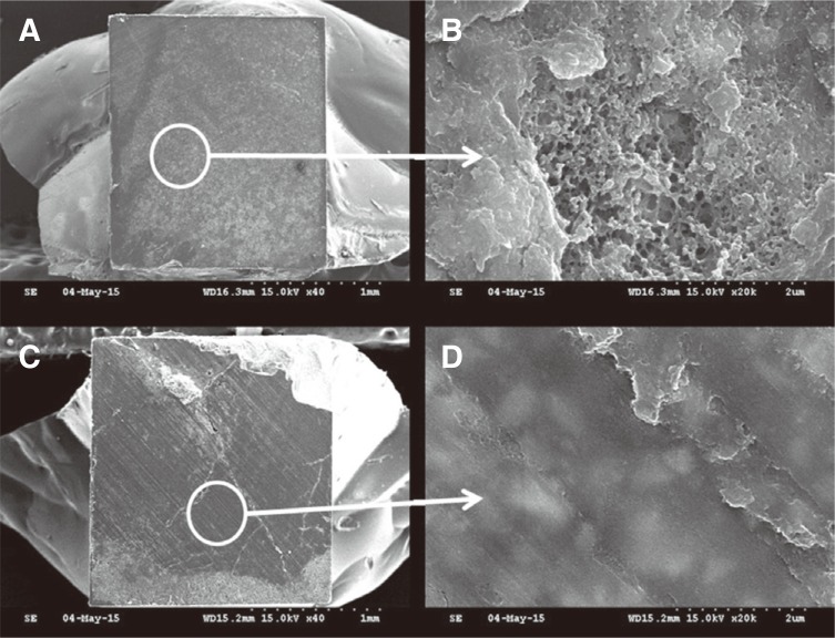
FESEM images of the surface topography of the RNC onlays are shown in Fig. 3. In the photomicrographs of RNC surfaces without any treatment (Group N), there were many fine irregular silica and zirconia fillers in the dense organic resin matrix. Pores in the resin matrix were observed on the surface of Group H. In Group A, rough resin and filler particles were observed. In Group T, the silica coat was visible on the surface. Smooth surfaces caused by the UA were observed in Group AB, HB, HSB, and TB.
After air abrasion, the surface was rougher and irregular particles were mixed in the matrix. After etching with HF, honeycomb-like pores were observed, similar to conventional ceramics. Borges et al.40 assessed the surface topography of different ceramics after treatment with either HF etching or airborne aluminum oxide particle abrasion. They reported highly modified surfaces on IPS Empress after HF etching, and dense pores on the surface were observed. However, for zirconia, there was no change in the superficial structure; many studies have reported that zirconia is not easily etched by HF.38 Swift et al.41 reported a significant decrease in the bond strength after HF etching of a glass-filled hybrid composite. They explained the decrease by the etching effect of HF absorbed in the resin matrix, causing softening and possibly a total dissolution of exposed glass particles. Here, fewer pores were formed after HF etching than for conventional glass ceramics. These samples had an insufficient roughness for resin bonding to RNC. Moreover, RNC includes resin zirconia nanomers in a resin matrix, which may decrease the etching effect and bond strength.
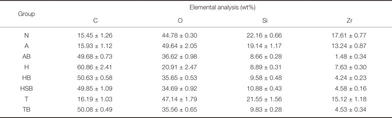
Elemental analysis using EDS showed concentrations of oxygen, carbon, silicon, and zirconium, as shown in Table 3. For the groups without UA, there were significant chemical differences in the surfaces etched with 4% HF for all elements. A decrease in oxygen, silicon, and zirconium ratio and an increase in carbon ratio were observed. However, for samples treated with UA, no significant differences were observed. Moreover, for Group A, a small quantity of aluminum was observed (2.53 wt%), and some remaining fluoride (1.77 wt%) from the HF was detected for Group H samples.
EDS generally has a penetration depth of a few micrometers depending on the material analyzed.4243 Kern and Thompson42 claimed that this depth is appropriate for evaluating chemical changes of a ceramic induced by sandblasting (surface roughening and powder particles mechanically embedded in the ceramic).
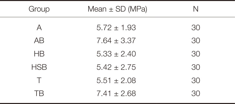
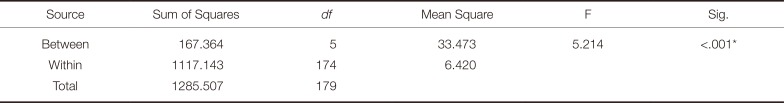
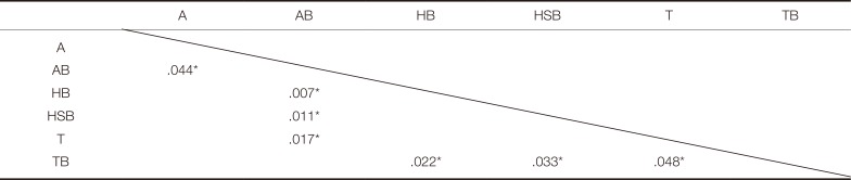
The results of µTBS tests are presented in Table 4. Group AB showed the highest bond strength (7.64 ± 3.37 MPa) followed by Group SB (7.41 ± 2.68 MPa). Group HB had the lowest bond strength (5.33 ± 2.40 MPa). The one-way ANOVA test showed that the differences in bond strength between groups had statistical significance (P < .05) (Table 5). Table 6 presents the results of the Tukey HSD test for the experimental groups.
There were no significant differences among Group A, HB, HSB, and TB or between Group AB and TB. Comparison of Groups A and AB and Groups T and TB showed that the UA increased the bond strength between the RNC and resin cement. However, the silane coupling agent had no effect (there was no significant difference between Groups H and HB). In Group AB, HB, and TB, air abrasion with alumina and the tribological silica coating showed similar bond strengths, but HF etching showed a lower bond strength.
The conventional test methods to evaluate the bond strength are the SBS or TBS test.35 However, as bonding techniques and materials have improved, the bond strength has become sufficiently high to cause cohesive failures in dentin.37 This means that dentin breaks from dentin, leaving the resin–dentin interface intact. Pashley et al.44 reported that the frequency of cohesive failures of dentin can be as high as 80% when the bond strength reaches 25 MPa. Such failures interrupt the measurement of bond strength at the bonding interface and do not mean that the resin-dentin bonds are uniformly stronger than the intrinsic strength of dentin; however, the bond that is stressed is non-uniform and concentrated at the highly focused region where a crack in the dentin is present.37
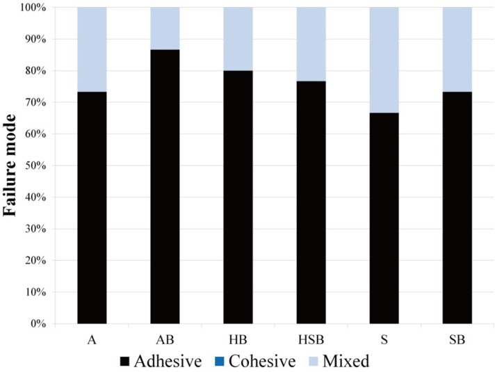
The failure mode distribution is shown in Fig. 4. A high percentage of adhesive failure and non-cohesive failure were observed for all groups. Figures 5, 6, 7 show SEM images of samples after fracture. Figures 5A, 5C, 6C, and 7C show cohesive failures and Figures 6A and 7A illustrate adhesive failures for the RNC surface. Similar to the SEM analysis of the RNC surface, irregular inorganic particles were observed in Group A and detached adhesive and pores were observed in Group HB. The silica coating was also observed in Group T. Group AB, HB, HSB, and TB showed tags and wave patterns caused by the UA.
The µTBS test offers versatility that cannot be achieved using conventional methods. Despite being labor-intensive it has great potential for providing insights into the strength of adhesion.3744 Sano et al.36 mentioned that the µTBS test was useful because small surface areas can be tested, which showed adhesive failures at the bonded interface. Scherrer et al.34 argued that only mixed failures or adhesive failures with small resin segment should be considered for the bond strength computation. Our specimens mostly showed adhesive failure on the RNC surface, and no cohesive failure was observed. Therefore, these results may be useful for the analysis of resin bond strength of RNC.
DISCUSSION
The aim of this study was to evaluate the influence of different surface treatments on the resin bond strength of RNC. It was found that the bond strength between RNC and resin cement can be affected by the specific surface treatment, such as mechanical treatment and application of adhesive material. A strong resin bond depends on micromechanical interlocking and chemical bonding to the surface of the restoration, which requires cleaning and roughening for adequate surface activation.23 Modern surface conditioning methods require airborne particle abrasion of the surface before bonding.25 Here, air abrasion with UA resulted in the highest bond strength. There was no significant difference between air abrasion and a tribological silica coating. Comparing the air-abraded samples, HF etching, and tribological silica coating followed by UA, and HF etched surface with UA showed the lowest bond strengths. Therefore, air abrasion and the tribological silica coating seemed to increase the bond strength more. Imamura et al.45 reported that air abrasion and the Rocatec system were very effective in increasing the bond strength of a laboratory-processed composite resin. However, these methods can induce a high loss of material,42 so excessive use should be avoided.
HF etching can achieve a suitable surface texture and roughness,23 creating a honeycomb-like topography on the ceramic surface that is ideal for micromechanical bonding.40 A chemical reaction between HF and silica in the feldspathic ceramics occurs, forming hexafluorosilicate, which is removed by water.46
Here, we observed that HF etching showed a lower roughness and bond strength than air abrasion and Rocatec treatment. In general, increasing surface roughness through mechanical surface treatment is more effective for increasing the bond strength than chemical bonding,16 which is corroborated by this study.
Here, we showed that there is a need to apply additional adhesive to increase the bond strength between RNC and resin cement. In this study, application of the UA increased the bond strength significantly. Stawarczyk et al.47 reported that when repairing RNC with a direct composite, universal adhesives (Scotchbond Universal and Futurabond U) with phosphoric acid monomers performed better than an adhesive based exclusively on methacrylic monomers, and this could be explained by the presence of 10-methacryloxydecyl dihydrogen phosphate (MDP) monomers in Singlebond Universal. MDP was necessary to achieve a durable resin bond for zirconia.38 Lava Ultimate is composed of resin matrix, silica, and zirconia nanomers, so it can describe the higher bond strength when the UA was used.
There was no significant difference in the bond strength when the silane coupling agent was used in this study. One end of a silane molecule is organically functional, and can be polymerized with an organic matrix (e.g., a methacrylate). The other end is generally composed of alkoxy groups, which can react with a hydroxylated surface, such as porcelain.19 In most studies, silane treatment yielded further increased bond strength.16 In contrast, D'Alcangelo and Vanini48 stated that silane did not have a significant effect on resin bonds, especially HF-etched composite restorations. Here, the silane coupling agent was used with HF etching, and there was no significant effect on the bond strength. The silica coating remained when the tribological silica (Rocatec system) was used. Before forming the silica coat, a roughness increase is achieved by alumina particles modified with silicic acid.24 In this study, there was no significant difference between the bond strengths or roughness values of air-abraded or tribological silica coated samples. Therefore, it seemed that the efficacy of silica coating for RNC was minor.
The manufacturer of Lava Ultimate states that it can be used for inlays, onlays, single/implant crowns, and veneers. Behr et al.49 mentioned that the required tooth and resin bond strength is at least 10 MPa; all samples tested here showed a lower bond strength. Lebon et al.50 investigated the roughness of dental materials, including RNC, after milling. Three commonly used milling tools with average diamond grit sizes of 105, 78, and 43.5 µm were used. They reported that there is a quasilinear correlation between diamond grit size and milled surface roughness. The roughness (Ra) of RNC after milling was approximately 2 to 10 µm. However, in this study, the RNC onlays were polished with diamonds of 1 µm grit and the roughness (Rq) was 8.47 ± 0.49 nm. Therefore, higher surface roughness and resin bond strength are expected in clinical situations. Moreover, when using RNC in certain cases that need more retention or veneers, it will be necessary to use accurate, effective, and systemic surface roughening methods and appropriate adhesives. In addition, more studies are necessary to compare the effects of various surface treatment methods and primers or adhesives on the resin bond strength of RNC.
 XML Download