

 PDF
PDF ePub
ePub Citation
 Print
Print


INTRODUCTION
Adhesively cemented fiber-reinforced composite posts (so-called fiber posts) are commonly used to provide retention and support along with aesthetic appearance for restorations of endodontically treated teeth.12 Low elastic modulus of fiber posts eliminates the need for cementing the length at least equal to the crown length to reduce risk of vertical root fracture.3 However, debonding of the post as the predominant failure mode of the restorations could be associated with the failure of endodontic treatment.13 Establishment of a highly durable bond between the resin cement and root dentin is a critical factor to provide coronal seal and adequate retention.14 This debonding at the adhesive resin-dentin interface is attributed to intrinsic difficulties in relation to homogeneous dentin hybridization in the root canal space.156 Two-step or simplified etch-and-rinse (E&R) adhesives are widely used for coronal dentin bonding; they can be simultaneously applied to cement fiber posts. Etch-and-rinse technique, as the most clinically proven method,6 has been speculated to benefit from dissolving the thick secondary smear layer formed during post space preparation.7 However, E&R adhesives (especially the simplified type) reveal a discrepancy between demineralization depth and monomer penetration.89 The resulting exposed and suboptimally impregnated collagen fibrils along the base of hybrid layer are prone to degeneration by matrix metalloproteinases (MMPs) and cysteine cathepsins over time.81011 This occurrence could be more relevant to intraradicular dentin during post cementation in the narrow and deep root space with limited access and visibility.1213
Dimethyl sulfoxide (DMSO) is known as an amphiphilic and dipolar aprotic solvent that enhances penetration into biologic surfaces in medicine. DMSO molecule has a highly polar S=O group and two hydrophobic CH3 groups. DMSO is completely miscible in all solvents, including most resin monomers contained in adhesive systems.14 DMSO is capable of dissociating the collagen network15 and changing its interfibrillar spaces in the dentin matrix.16 This might be due to suppressing interpeptide hydrogen bonding in the collagen matrix.16 Recently, DMSO was found to be useful in improving and preserving the long-term coronal dentin-adhesive bond strength.16 This positive effect was attributed to improved wetting of collagen by the adhesive. Therefore, this property might be advantageous for adhesive cementation of fiber posts. The aim of this study was to test the null hypothesis that DMSO pretreatment of root dentin has no effect on the bond strength longevity of fiber posts to radicular dentin using an acetone- and an ethanol-based two-step etch-and-rinse adhesive resin cements.
MATERIALS AND METHODS
The research method was approved by the Human Ethics Review Committee of the School of Dentistry, Shiraz University of Medical Sciences. Forty sound human maxillary central incisors with similar size and anatomic shape, and straight roots, without previous endodontic treatments and posts, were selected and stored in 0.5% chloramines-T solution at 4℃ until use for the purpose of the study. The study was conducted following informed consent from patients and the approval of the research protocol by the local Ethics Committee. The absence of cracks and carious lesions in the selected specimens was confirmed using a stereomicroscope and radiographs. The roots were separated from the crowns in a uniform length of 15 mm, using a water-cooled diamond saw (D&Z, Berlin, Germany) at the cementoenamel junction. The roots were endodontically instrumented at a working length of 1 mm from the apex with K-files (Dentsply, Maillefer, Ballaigues, Switzerland) up to #45 with saline solution and 2.5% sodium hypochlorite irrigation. The roots were obturated using AH26 sealer (Dentsply, Caulk, Milford, Germany) and gutta-percha (Aria Dent, Asia Chemi Teb, Tehran, Iran) and coronally sealed using Fuji II LC (GC, Tokyo, Japan) light-cured glass ionomer.
The specimens were stored in water for one week for complete setting. Afterwards, post spaces were prepared to a standardized depth of 10 mm using drills from the respective post manufacturer by the same operator. Cleanliness of the root walls and the remaining 4 mm of gutta-percha at the root end were confirmed by radiographs for the apical seal.
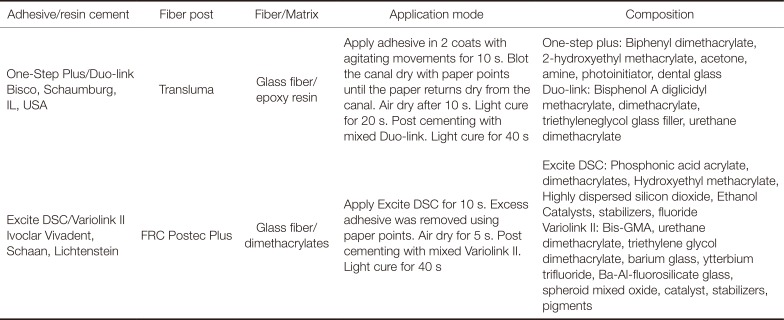
Glass fiber posts from two post/adhesive cement systems (Trasluma Post ISO # 100; Bisco, Schaumburg, IL, USA and FRC Postec Plus #1; Ivoclar Vivadent, Schaan, Lichtenstein) were tried in the canals for a passive fit in the prepared depth. Post surfaces were cleaned with ethanol and air-dried. Then, the Transluma Post surfaces were coated with One-Step Plus and light-cured for 20 seconds; Postec Plus Post surfaces were silanized with Monobond S for 60 seconds and air-dried for 5 seconds according to manufacturers' instructions.
The root canals were acid-etched with 37% phosphoric acid for 15 seconds using an endodontic syringe with endodontic tips, rinsed with water for 30 seconds using a syringe with endodontic needle, and gently dried with an air stream and paper points. The specimens were divided into four groups (n = 10). In the two control groups, the root dentin was treated with 1 mL of distilled water, and in the two experimental groups, the root dentin was treated with 1 mL of 5% DMSO aqueous solution for 60 seconds by light scrubbing method using an endodontic brush. In all the groups, the solutions were delivered into the root canals with a disposal syringe and a 30-gauge needle. The excess water was removed with absorbent paper points to desiccate the root canals (water- and DMSO-wet bonding). During post space preparations and post cementation, the specimens were held in a moist gauze sponge to maintain their moisture content. Two etch-and-rinse resin cements (acetone-based, One-Step Plus/Duo-link, OS/D, Bisco and ethanol-based, Excite DSC/Variolink II, EX/V, Ivoclar Vivadent) were used according to manufacturers' instructions. All the bonding procedures were carried out by the same operator. The composition of the materials and instructions for use are shown in Table 1.
In each group, the respective cement was applied to the post surface and to the post space using elongation tips attached to the automixed tip, supplied by the manufacturer for Duo-link and a #35 lentulo spiral for Variolink II. The post was immediately seated with a slight vibratory motion and held under finger pressure. After removing the excess cement with a microbrush, light polymerization was carried out for 40 seconds by placing the light tip onto the post at 600 mW/cm2 using a light-curing unit (VIP Junior, Bisco) according to manufacturer's instructions. Finally, a tight coronal seal was obtained using Fuji II LC. The specimens were stored in distilled water at 37℃ for one week.
The bonded roots were sectioned into seven 1-mm-thick slices by using a slow-speed cutting machine (Mecatome T201 A, Presi, Grenoble, France). For each root, two slices from each root region (apical, middle, and coronal) were obtained. The first coronal slice was not included.
On half of the root samples from each group (n = 5 roots, 30 slices), push-out test was performed immediately. The other half of the samples were stored in 37℃ distilled water containing 0.4% sodium azide for 1 year before assessing the long-term bond strength.1718
The slices were submitted to a compressive load in a universal testing machine (Zwick, Roell, Ulm, Germany) at 0.5 mm/min on the center of the apical surface of the post in an apico-coronal direction without contact with the root dentin or cement until the shear stress along the bonded interface dislodged the post. The loading was performed using three punch tip diameters (1 mm, 0.8 mm, and 0.7 mm) depending on the diameter of the post in each root region slice.
The load (in N) was divided by the bonded interface area (in mm2). The bond strength was recorded in MPa through the formula π (R + r) [h2 + (R - r)2],0.5 where R and r represent the coronal and the apical post radii, respectively, and h is the thickness of the slice.
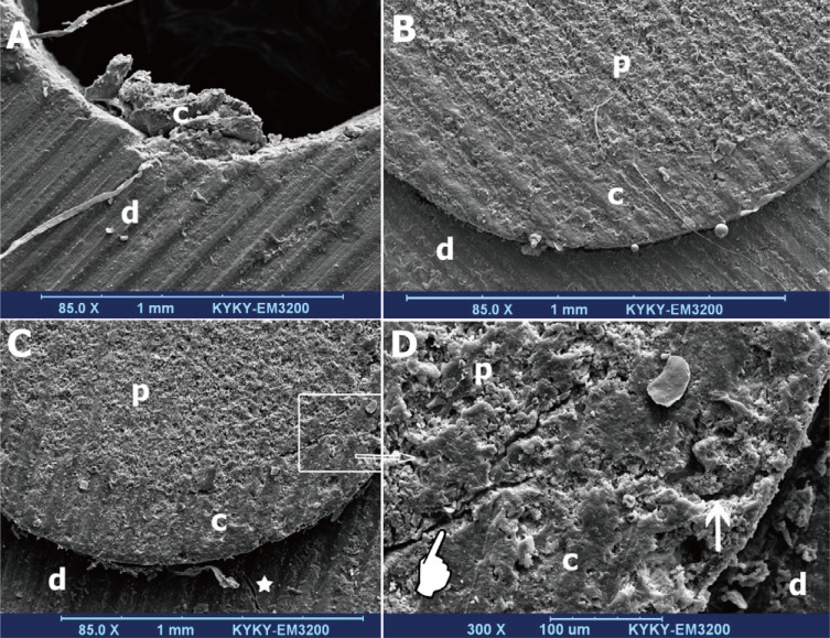
All the debonded specimens were assessed under a stereomicroscope (Carl Zeiss Inc., Oberkochen, Germany) at ×40 and categorized as follows:
Cohesive failure in the dentin
Cohesive failure in the cement or post
Adhesive failure between the cement and the dentin
Adhesive failure between the cement and the post
Mixed failures consisting of a combination of two or more failure modes
The representative specimens of each failure mode were prepared for scanning electron microscopic (SEM, KYKY, EM3200, China) evaluation of the failure pattern as shown in Figure 1.
Data were statistically analyzed in two steps. Initially, a three-way analysis of variance (ANOVA) was performed to evaluate the effects of three main factors (cement type, DMSO treatment, and time) and Student's t-test was used for subgroup analyses. Therefore, the bond strength values of the six slices (in apical, middle, and coronal thirds) originating from the same root were pooled together for each group, and the average bond strengths as the mean values were calculated for the groups (total analysis). Secondly, for each time interval, one-way ANOVA and Tukey tests were used to compare the bond strengths between different root regions in the control and DMSO groups of two cements and these groups were compared in each root region (regional analysis) (α = .05).
RESULTS
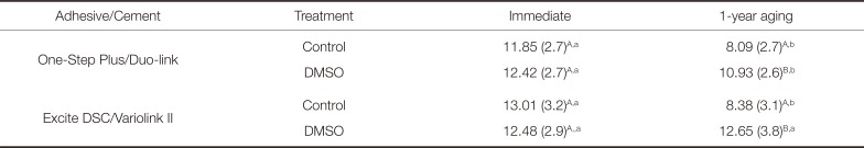
The mean and standard deviations (in MPa) of push-out bond strength (P-OBS) are presented in Table 2. According to the results of three-way ANOVA, the effect of DMSO treatment, time, and the interaction between treatment and time were significant (P < .001). Effect of cement type and interactions among the three factors (cement, DMSO, time), between cement and time, and between cement and DMSO treatment were not significant (P > .05). These results indicated that both cements were similarly affected by the treatment and time, and effects of time and DMSO were dependent on each other.
EX/V exhibited a higher immediate P-OBS compared to that of OS/D and long-term P-OBSs of both cements were similar, but the results were not significant (P > .05). DMSO treatment had no effect on immediate P-OBS of both cements. However, DMSO treatment resulted in a significantly higher P-OBS than the control after aging (P < .001). Aging significantly decreased P-OBS of the control (P < .001); however, P-OBS remained stable when DMSO treatment was applied (subgroup analyses).
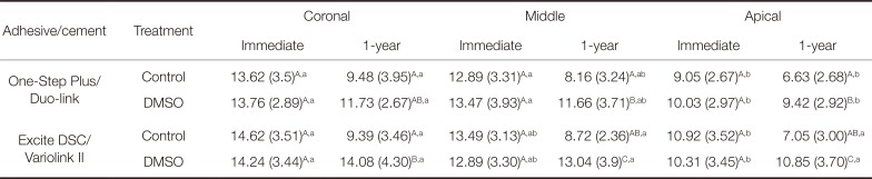
The mean and standard deviation of P-OBS in MPa from different root regions are presented in Table 3. The results of one-way ANOVA and Tukey tests demonstrated progressively decreasing P-OBs at the coronal, middle, and apical regions, respectively, in all the groups. So that, there were significant differences between the coronal and the other two regions (P < .05). However in some groups there was no significant difference between the coronal and middle regions (P > .05). Only in aged groups of EX/V (control and DMSO), the difference was not significant. When analyzed regionally, there were no significant differences in immediate BS between the control and DMSO-treated groups for both cements in the three root regions (P > .05). After aging, the results of comparisons in all the root regions were similar to the results analyzed totally. Only in the coronal region, the difference between the control and DMSO groups of OS/D was not significant (P > .05).
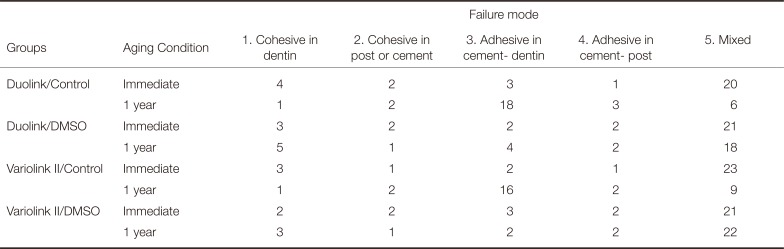
The results of failure modes of the four groups are shown in Table 4. The majority of failures were mixed failures in all the groups, except for aged control groups in which the bond failures were mainly adhesive failures at dentin-cement interface.
DISCUSSION
According to the results of the current study, DMSO treatment of acid-etched root dentin prior to post bonding gave rise to a beneficial effect on the long-term bond strength. In this context, contrary to significantly reduced bond strengths after aging in the control group, DMSO led to a stable bond. Therefore, the tested null hypothesis was rejected.
There is no published study on this issue in relation to fiber post bonding. Only a bond strength study with the use of water storage of microtensile beams showed the positive effect of DMSO on bond durability of a two-step etch-and-rinse adhesive to the control dentin.16
Two etch-and-rinse adhesives used in this study were applied using the water-wet bonding technique. It is difficult to control residual moisture on root dentin after acid etching and rinsing13 because the wall of the narrow root canal space cannot be directly visualized. This wall holds water by surface tension so that displacement of water with the adhesive is difficult.19 In addition, the optimal wetness varies depending on the type of the adhesive solvent;20 acetone-based adhesives need a wetter surface to achieve adequate bond strength.21 Although the ethanol-based adhesive (Excite DSC) used in this study exhibited a higher bond strength compared to the acetone-based one (One-Step Plus), the difference was not statistically significant. These two systems have a number of differences. The post types provided by the manufacturer of the adhesive/cement system are different. Excite DSC is a dual-cured adhesive that has additional chemical polymerization while One-Step Plus is only a light-cured adhesive. Since the accessibility of light in root canal space is problematic, some authors suggested prolonging of the light-curing time for the adhesive.22 Based on this, light-curing One-Step Plus was extended to 20 seconds while the light tip was placed as close as possible to the canal opening. Nevertheless, it was reported that using a post, adhesive and resin cement combination from the same manufacturer could minimize possible incompatibility between the materials and maximize the chemical affinity and potential of each system.23 Despite the differences between the two systems used in this study, when comparing the control group and respective DMSO group, the latter yielded a higher bonding stability for both systems.
Although solvents could facilitate the replacement of water with resin monomers, this replacement is often incomplete in intrafibrillar spaces.24 The residual water causes phase separation of the hydrophilic and hydrophobic components of adhesives, preventing complete monomer penetration into the full depth of demineralized dentin. This is due to the fact that hydrophobic methacrylates are insoluble in water-saturated dentin.2025 The created porous hybrid (hybridoid) layer is responsible for the instability of resin-dentin bonds.92026 In fact, formation of the water-filled channels (nanoleakage) results in water sorption, resin leaching, and hydrolysis through permeable adhesive interface.810 Furthermore, more exposed collagen fibrils may be produced continuously.8 The degradation of the exposed collagens by MMPs is also involved in decreased integrity of the adhesive interface.2728 These factors could explain the observed reduction of bond strength in control groups after aging because sufficient resin impregnation of the intraradicular dentin is clinically more difficult than that of the coronal dentin. The lack of stability of wet bonding method upon aging has been shown in several studies.2930
DMSO as an additional primer on acid-etched root dentin could improve the long-term push-out bond strength of OS/D and EX/V groups in this study. This preservative effect was also supported by failure mode analysis. The number of adhesive failures at the root dentin-cement interface in the aged DMSO-treated groups was less than that in the aged control groups. This finding might be attributed to unique properties of DMSO. The dissociative effect on demineralized dentin collagen was indicated with high concentration of DMSO.16 This effect may not be adequate with low concentration of DMSO used in this study to influence bond durability positively. However, DMSO treatment might result in enhanced collagen wetting (by adhesive) and adhesive penetration into the acid-etched exposed collagen.16 Therefore, resin impregnation of the demineralized dentin could be more complete.
On the other hand, DMSO as a hydrogen bond acceptor is able to form two hydrogen bonds with water molecules. These bonds are stronger than the bond between water molecules.31 As a result, water's self-associative tendency32 may lead to reduced residual water entrapped between the polymeric resin chains.16 This, along with the improved envelopment of collagens following better resin infiltration, might produce a homogeneous and well-polymerized hybrid layer. Mehtälä et al.33 reported that DMSO may significantly decrease dentinal fluid permeation into the hybrid layer during dentin bonding in vitro. The reduced water-filled channels and permeability at the adhesive interface may contribute to the increased long-term integrity of the adhesive interface.25 The correlation between the extent of polymerization and adhesive permeability was previously reported.1734
The DMSO-wet bonding may prevent the possible phase separation of the adhesive components in water-wet bonding technique. The excellent solvent property of DMSO35 could lag behind this assumption. A significant reduction of exposed collagen extension at the base of the hybrid layer was indicated by optical microscopy following 50% DMSO treatment with a water-based etch-and-rinse adhesive.36 Furthermore, MMP inhibitory activity of DMSO of 5% or higher concentrations was demonstrated in zymographic analysis.16 Interactions between the gelatinase binding site and substrate were disrupted by DMSO.37 However, how DMSO functions to improve bonding longevity is not still clear. More investigations should be conducted to understand the real mechanism of DMSO effects on bonding interface in coronal and intraradicular dentin and to determine its optimal concentration for maximal beneficial effects on the adhesive interface.
The use of DMSO treatment as an extra step in the complex bonding procedures in root canal space may be considered a disadvantage. To solve this, DMSO could be possibly incorporated into adhesive compositions because it is able to solvate commonly used adhesive monomers16 and is fully miscible in all the solvents.14 The compatibility of this combination in short- and long-term bond strength tests should be evaluated in future studies.
Sectioning of microtensile specimens before bond strength testing and water storage of microbeams was considered as the accelerating aging of bonded interface. This is due to rapid water diffusion through the small surface area of the adhesive interface, resulting in rapid degradation process.38 Although this rapid degradation may be supposed to lack clinical relevance, similar patterns of hybrid layer degradation have been demonstrated in vivo from the base of adhesive bonded cavities.39 Similarly, sectioning the tested bonded roots was performed in this study before water storage. This long-term water storage of root microslices prior to submitting to dislodging forces during the push-out test cannot closely mimic clinical aging conditions. However, this experimental set-up was previously designed to examine the role of MMP inhibitor agents in bond stability of fiber posts to root dentin.171840 Furthermore, direct exposure of micro-specimens was used to detect the effect of DMSO on bond stability in coronal dentin.16 The push-out test used in this study could provide a true shear stress parallel to the adhesive interface, resulting in a better estimation of the bond strength than the conventional shear test. The other advantage of push-out test is that it is more dependable than the microtensile test for bonded posts.41
Following post cementation, the final restoration was placed and subjected to complex forces, low intermittent functional forces from different directions, and temperature changes.42 In this in vitro study, the intraoral conditions were not fully simulated. A single load testing on microslices of bonded root without thermomechanical cycling was used. These were considered as the limitations of the current study. Additionally, two adhesive/cement systems were tested; therefore, the current finding cannot be generalized to all the systems.
 XML Download