

 PDF
PDF ePub
ePub Citation
 Print
Print


INTRODUCTION
The use of titanium (Ti) implants is considered to be a suitable treatment option for fully or partially edentulous patients.1 In the context of titanium implants, surface microstructure of suspensions and pockets affect the cell spreading, morphology and its alignment. Roughness of the surface and surface chemistry (titanium hydride) contribute to the high compatibility of such implants.2
To improve physical and chemical properties of Ti implants and achieve faster osseointegration, the Ti surface must be modified appropriately. Many studies suggested that, in comparison with conventional sandblasted with large grit and acid-etched (SLA) implants, modified SLA (MSLA) implants yielded more successful bone formation at the initial stage of healing, resulting in significantly higher levels of bone-to-implant contact.3456 The immunostimulatory effect of implant surfaces on the pro-inflammatory cytokine gene expression profile of human macrophages has been shown to be attenuated by surface hydrophilicity.3 However, the differentiation rate of osteoblasts is enhanced on hydrophilic surfaces. Indeed, it is known that surface roughness and hydrophilicity are two important factors that affect osteoblast behavior; the factors target osteoblast proliferation and the initial stages of osteoblast differentiation via the PI3K(Phosphoinositide 3-kinase)/Akt signaling pathway.4 Modified SLA surfaces with smaller contact angles and higher surface energies have been suggested to enhance osteogenic properties when compared with conventional SLA surfaces.4 Lai et al.5 have indicated that cell attachment is greater on surfaces with a higher surface energy. Osteoclasts are formed via monocyte fusion, so monocytes are essential for osteoclast differentiation. According to the study conducted by Bang and colleagues, the enviroment surrounding a MSLA surface results in the limited monocyte adhesion to MSLA surface. Moreover, the surface properties of MSLA were found to inhibit osteoclast growth and differentiation.6 On the other hand, higher surface free energy (SFE) on MSLA surfaces is thought to enhance hydrophilicity, and this increased hydrophilicity and SFE of Ti implant surfaces may in turn accelerate the initial healing at the biomaterial/biosystem interface.7
Various studies have documented the capacity of laser wavelengths and laser properties to influence implant surfaces.891011 The ultraviolet (UV) irradiation from a YAG (yttrium aluminum garnet) laser set to a wavelength of 355 nm has been shown to confer high hydrophilicity on titanium oxide, an effect that has been attributed to the transformation of Ti4+ into Ti3+.8 New bone apposition has also been observed on a laser-treated and acid-etched (LAT) surface during the initial stages of bone regeneration.9 Another study showed that cell attachment on surfaces subjected to a single dose (3 J/cm2) or multiple doses (1.5 or 3 J/cm2) of laser treatment was higher than that on those not subjected to laser treatment. Nonetheless, in the initial cell attachment levels, no differences were found between surfaces treated with a single dose and those treated with multiple doses.10 Low-level laser therapy (LLLT) has been shown to promote initial stages of the destructive construction process at the implant-soft tissue interface.11
There are various articles that use several parameters, including cell attachment, cell morphology, RUNX2 (Runt-Related Transcription Factor), OPN (Osteopontin) and OCN (Osteocalcin) testing as markers for osteogenic activity. Another parameter, measure of ALP activity, has proven to be a reliable and adequate marker as well. Numerous studies have reported the modification of implant surfaces; however, few studies have been conducted to compare the effects of LT(LAT) or MSLA on bone-implant bonding. Therefore, the aim of this study was to compare the cellular activity between MSLA, LT, and LAT Ti-surfaces in order to determine the most favorable surface modification for successful osseointegration in the early healing period.
MATERIALS AND METHODS
A total of 84 Ti6Al4V disks (diameter: 15 mm, thickness: 2 mm) were manufactured from commercially pure Ti (grade 23, ASTM F136, Galimplant S.L., Spain) and were standardized by polishing with 600-grit silicon carbide abrasive paper to produce smooth surfaces. Specimens were washed with 70% ethanol, rinsed three times with distilled water, and dried. Ti disks were divided into three groups: MSLA (sand-blasted with large grit, acid-etched, and immersed in 0.9% NaCl), LT (laser treatment), LAT (laser-treated and acid-etched).
To prepare MSLA surface, Ti specimens were sandblasted with Al2O3 particles and then acid-etched for approximately 40 min in a boiled solution (60℃) containing a mixture of 18% HCl and 49% H2SO4.9 Prior to use, modified Ti specimens were rinsed once with 100% isopropanol and twice with distilled water in an ultrasonic bath (5 min each).12 Samples were autoclaved and immersed in 0.9% NaCl for 7 days before being used in experiments.7 An Nd:YAG laser (Jenoptic Laser Optik) with a pulse repetition frequency of 15 kHz and a pulse width of 2 µsec was applied for Ti surface ablations. The equipment was operated at a sustained wavelength of 1064 nm and a power output of 10 W. LAT surface was treated by laser and acidetched for approximately 40 minutes in a boiled solution (60℃) containing a mixture of 18% HCl and 49% H2SO4. The 84 prepared Ti disks were divided into three groups and tested for their effects on differentiation.
The mouse calvaria-derived preosteoblast cell line MC3T3-E1 subclone 4 was obtained from the American Type Cell Culture Collection (ATCC, Manassas, VA, USA). Cells were cultured in alpha-minimum essential medium (Invitrogen-Gibco, Carlsbad, CA, USA) supplemented with 10% fetal bovine serum (Invitrogen-Gibco, USA, Carlsbad, CA, USA) and 100 U/ml penicillin solution (Invitrogen-Gibco, Carlsbad, CA, USA) at 37℃ in a humidified atmosphere of 95% air and 5% CO2. The cells were grown to confluence and culture medium was replaced every 2-3 days. After rinsing the confluent cultures twice with phosphate buffered saline (PBS, pH 7.4; Gibco, USA), cells were harvested with 0.25% trypsin-EDTA treatment (Invitrogen-Gibco, Carlsbad, CA, USA) for 2 min followed by centrifugation. MC3T3-E1 cells were considered normal osteoblasts. The experiment was conducted using cells with passage number of 10-15.
ALP activity was measured using a commercially available kit (SensolytepNPP alkaline phosphatase, AnaSpec, Fremont, CA, USA).13 Cells seeded onto Ti disks at a density of 4 × 10⁴ cells/well in 24-well plates were cultured for 7 days, 14 days, and 21 days. Culture medium was removed, and cells were washed twice with cold PBS before detachment by scraping in accutase. Cells were then lysed in a solution of Triton X-100 and 1 × assay buffer. After centrifugation, the cell layer was suspended before removal of the lysis solution. Lysed cells were transferred to 96-well plates, and a colorimetric alkaline phosphatase substrate (50 µl) was added to every well. The plate was shaken for 10 min at 37℃ in a 5% CO2 humidified atmosphere, and absorbance readings (405 nm) were taken using an ELISA Ultra Microplate reader (n = 7).
Samples were mounted on aluminum specimen holders and coated with gold-palladium using a sputter coater (Bio-Rad SC510 Watford, UK). Coated samples were assessed using scanning electron microscopy (SEM, TM-100, Hitachi, Tokyo, Japan) and photographed using a magnification of ×500.
Energy dispersive analysis of X-ray spectroscopy (EDAX: Horiba EX -300, Japan) was used to determine the element analysis of the specimens in this study.
Surface roughness was evaluated by a contact profilometer (URFPAK-SV; Mitutoyo, Kawasaki, Japan) and characterized by two parameters: mean roughness (in Ra) and maximum peak-to-valley height (in Rt).
Data was analyzed using SPSS17.00 (SPSS Inc., Chicago, IL, USA) and presented as mean ± standard deviation. Statistical significance among control group and test groups, and respective response from 7 days, 14 days, and 21 days in every group were evaluated by one-way ANOVA with the Tukey HSD post-hoc test. Differences with P < .05 were considered statistically significant.
RESULTS
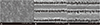
SEM photographs of MSLA, LT and LAT treated surfaces are shown in Fig. 1. Results showed that LT and LAT treated surfaces were rougher than MSLA surfaces.
EDS photographs of MSLA, LT and LAT surfaces are shown in Fig. 2. The less carbon and more oxygen elements were performed on LT, LAT than MSLA specimens.
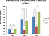
Differentiation of MC3T3-E1 cells was quantified using ALP as a marker for osteoblast maturation. Both MSLA and LAT surface showed a gradual increase of ALP synthesis in cells. Total ALP expression in MSLA, LT and LAT surface-adherent cells was found to be highest at 21 days (P < .05). Furthermore, ALP expression levels at 7, 14, and 21 days were significantly higher on MSLA and LAT surfaces when compared to LT (P < .05).
Roughness values for specimens in control (MSLA) and test groups (LT and LAT) are shown in Fig. 3 and Table 1. Ra and Rt values of MSLA specimens were 1.31 µm and 19.41 µm, respectively. For LT surfaces, Ra and Rt values were 7.48 µm and 89.68 µm, higher than those obtained for the control group, suggesting that LT is rougher than MSLA surfaces. Additionally, the Ra and Rt values of LAT surface were higher than those of MSLA and LT (9.93 µm and 93.47 µm, respectively); therefore, of the three surfaces examined, LAT surfaces were roughest.
DISCUSSION
Measurement of surface wettability is a typical method by which the hydrophilicity of material surface is evaluated. Contact angle is quantified in the evaluation of wettability. The angle is governed thermodynamically by the solid-vapor, solid-liquid, and liquid-vapor surfaces and the interfacial energy of an ideal surface by roughness and chemical heterogeneity of a real surface.7 The SLA surface showed highest contact angle of approximately 90°, whereas surface-modified SLA substrates which is hydrophilic had a contact angle of 0°. By soaking the surface-treated implant in saline for more than 2 weeks, the surface changed from hydrophobic to hydrophilic, which shortened the time of fixation and increased the initial stability of implants. This result is similar to the result by the study of Rupp et al.15
Modified SLA surfaces can retain the desired hydrophilicity even under harsh drying procedures, whereas SLA surfaces stored in a NaCl solution will reduce the initial hydrophilicity after vacuum treatment.7 Alfarsi et al.3 showed that surface hydrophilicity affected biological events involved in the initial stages of the bone healing process. They found that biomaterials with hydrophilic surfaces inhibited processes by which macrophages formed foreign-body giant cells. In this scenario, hydrophilicity conferred anti-inflammatory properties on surface-modified biomaterials. Alfarsi's findings further suggested that modulating of the response of proinflammatory cytokines was a significant biological factor by which improved and/or faster wound healing around hydrophilic titanium dental implants could be achieved. Bang et al.6 found that, on modified SLA surfaces, the osteogenic effort was promoted and bone resorption was inhibited via the inhibition of osteoclastic differentiation. Osteoclasts are formed via the fusion of monocytes, and therefore the abundance of monocytes is essential for osteoclast differentiation. In modified SLA surface, monocyte attachment is reduced, and the osteoclastic differentiation process is thus generally inhibited.7 Therefore, modification of implants as processing procedure is a technique widely used, and it was included in this experiement.
Usually, the osseointegration between titanium implants is evaluated using animal models with histomorphometrical or biomechanical measurements. Since not many studies have been conducted regarding osseointegration of implant and accordingly, few animal models are available to refer to, osteoblasts are considered very important in the osseointegration. The osteoblastic cell response to titanium surface was applied to mimic the initial level of osseointegration in vivo. The MC3T3-E1 is one of the most widely used cell line for the studies on osteoblastic cell response. The cell line expresses high ALP activity with mineralization of the extracellular matrix in vitro. Consequently, MC3T3-E1 is a good candidate for evaluating the proliferation, adhesion, and differentiation of osteoblasts on titanium surfaces.16
ALP activity is considered an initial marker of osteogenic differentiation.13 Elementary progenitor cells do not express ALP activity. However, following osteoblastic differentiation up to a defined number of cell divisions, cells ultimately express a mature osteoblast phenotype, i.e, a postmitotic, osteogenic cell type with ALP activity.17 The ALP enzyme is active in all cell membranes and is present at higher levels in osteoblasts to allow for matrix mineralization.18 In this study, cell adhesion on MSLA and LAT surface was higher than that on LT surfaces after 7, 14 and 21 days in culture, and cell adhesion on MSLA was lower than that on LAT surface. These findings suggest that MSLA and LAT surfaces provide outstanding and stable conditions for osteoblast proliferation at every time point included in this study (Fig. 4). While Coombe et al.19 repeatedly reported that laser irradiation did not show a significant effect on the ALP activity, Cho et al.20 found that implant surfaces treated with laser show higher removal torque than turned implant surfaces.21 Francisley et al. suggested that laser-treated surface had regular cavities similar to honeycomb and showed excellent osseintegration.22 The reason was that primary stability of implants was initially gained from mechanical fixation in the peripheral bone and then from bone remodeling, which occurred during the 2nd and 4th weeks after implantation. Increasing roughness on macro-surface can enhance the implant anchorage and stability. Removal torque value is determined by tissue, which consists of vein and biomaterials, and therefore removal torque value cannot be a critical index of osseointegration.
Usually, cells grown on rough surfaces exhibit an approximately cuboidal shape while cells grown on smoother surfaces appear more flattened.18 Cell adhesion to the surface reflects the first interaction between cells and biomaterials and is influenced by surface chemical and topographical characteristics. The topography of LT is much rougher than that of MSLA, and the roughness is not fit for proliferation22 (Fig. 1, Fig. 3, Table 1). Conversely, on smooth surfaces, the cells exhibited high proliferation rate; however, alkaline phosphatase and osteocalcin production was little, suggesting the loss of differentiated osteoblastic phenotype cells. Surface roughness can change osteoblast proliferation, differentiation, and matrix production; however, various kinds of osteoblasts may perform differently on surface.23 Osteoblasts are sensitive to surface roughness and show reduced proliferation and high phenotypic differentiation on rough surfaces.24 Microscale alterations, such as mechanical interlocking and nanoscale alteration, can help the phenotypes of osteoblast and adhesion ability, and subsequently bone anchoring and biomechanical stability of implants in bone.12
When comparing ALP expression of cells adhered to MSLA and LAT surfaces, the values were similar even though LAT expression was a little higher than that of MSLA. It is suggested that, although pure laser treatment cannot develop the ability of collecting osteoblast cells, addition of acid etching to laser surface is an excellent method to activate osteoblast cells. Acid etching (A) to titanium surfaces can create hydrophilic surfaces and increase SFE, which, in turn, accelerate initial healing reactions between biomaterial/bio system interface.7 The EDS results suggested that the amount of oxygen and carbon elements on both LT and LAT surfaces were higher than that on MSLA surfaces (Fig. 2). The oxygen element on LT couldn't play an important role in ALP activity.
Though the oxygen element on LT did not show any significant effect on ALP activity, LAT surfaced showed better result than LT surface. It is possible that the hydrophicility, conferred by acid treatment as a critical factor, affected ALP activity but not the oxygen element on the surface.
This study focused on the in vitro characterization of the effects of surface chemistry on cell differentiation of osteoblast cells. The limitation of this study is the lack of use of animal models for in vivo studies, which are necessary for comparing the osseointegrative properties of the four different treated surfaces like smooth Ti6Al4V, MSLA, LT, and LAT. Furthermore, the cell attachment, OPN, OCN, RUNX2, extracelluar calcium deposition assay, and Alizarin red staining assay should be investigated in future.
CONCLUSION
Within the limitation of this study, our findings allowed us to conclude that ALP activity of osteoblasts cultured on modified SLA and LAT surfaces were of a significantly higher quality and abundance than those cultured on LT surfaces. LAT and MSLA treatments showed better results than LT, therefore both of them were recommended as the ideal treatment to Ti implant surfaces.


 XML Download