

 PDF
PDF ePub
ePub Citation
 Print
Print


INTRODUCTION
World Health Organization (WHO) and the National Institute on Ageing (NIA) reported that tooth loss could be a risk for Alzheimer's disease.1234 The basal ganglia, limbic system, thalamus, and cerebral cortex control the mastication and are linked with the centers of mastication, deglutition, and respiration.56 Inhibition of masticatory movement linked with the mastication center and deglutition center can cause a sudden deterioration of brain function.789 Masticatory efficacy could be improved by several ways in edentulous patients, from conventional complete denture to implant supported prosthesis.1011 The effect of complete denture on restoring patients' mastication has been sufficiently evaluated by different methods, but the role of balanced complete dentures in the functional improvement of brain function has not been elucidated. This pilot study aims to know complete denture's influence on the brain and cognitive functions in edentulous patients.
Go to : 
MATERIALS AND METHODS
The protocol of the study was approved by the Institutional Ethical committee of SRM University, Chennai, India (SRMU/M &HS/SRMDC/2012/MDS-PG student /205). The patients of age group between 50 and 60 years were informed of the procedure and their consents were obtained (Fig. 1). The patients selected were edentulous for a year and they were treated with complete denture for the first time. Patients participated in this study had class 1 ridges and adequate interarch spaces. A comprehensive examination excluded patients with symptoms of temporomandibular disorders, xerostomia, orofacial motor disorders, severe oral manifestations of systemic diseases, and psychological or psychiatric conditions that could influence their response to treatment.
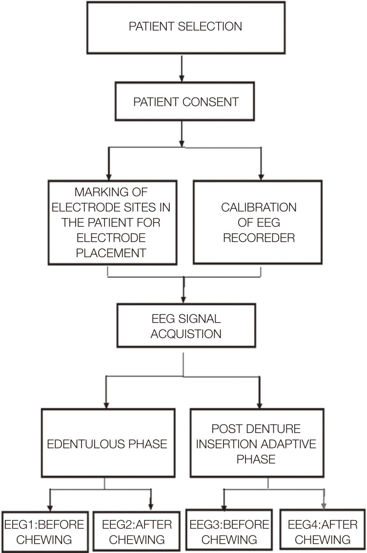
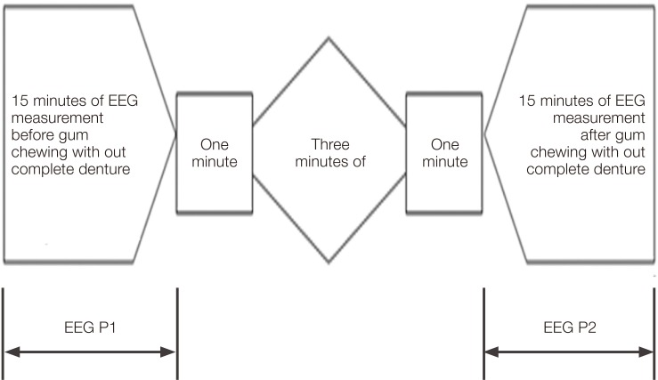
The electroencephalogram (EEG) signals were used to check the brain activity.12 Patients were instructed to take adequate sleep the night before and were restricted from consuming caffeine at least eight hours before the EEG analysis. The electrode placement for EEG was done in accordance to the international federation in electroencephalography and clinical neurophysiology as a 10-20 electrode placement system.1314 EEG recordings were obtained in two settings: before chewing paraffin gum (Saliva-Check kit, GC Corporation, Japan) and after three minutes of chewing gum with one-minute resting intervals between each EEG recording. The recordings were made for the total of fifteen minutes. The signals were sampled at 256 samples per second with 16-bit resolution and with fifteen minutes of digitized signal duration. This data was saved to be analyzed and compared with the EEG records two months after denture placement (Fig. 2, Fig. 3 and Fig. 4).
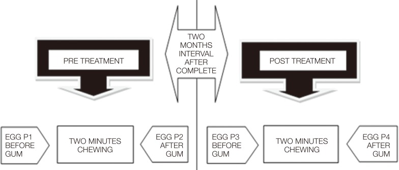
Complete dentures were fabricated with balanced occlusion for all the patients with maximized comfort. After two months of denture usage, the EEG recordings made before and after chewing were similar to pre-treatment EEG results. Both the pre-treatment and post-treatment EEG data were analyzed for differences in power spectral density (PSD) with gum chewing.
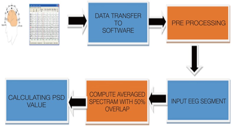
The algorithm was tested using a single channel (C3-P3). The alpha waves of the greatest amplitude were recorded. The data was pre-processed by computing average spectrum with 50% overlap in the epoch signal. EEG signal baseline wanders were viewed in the LAB VIEW platform (National Instrument, India). The recordings were corrected for artifacts and the signal amplitude was quantified to micro-volts.15 A digital low pass finite element filter (FIR) filtered the EEG signal by Hamming window technique. The order of the filter was 40 and the cut off frequency was 32 Hz. Flatness without a ripple in the pass band was desirable in the analysis of EEG signals. The filtered EEG segments were chosen for analysis.
Averaged spectrum and power measure were calculated. The power spectrum of the signal was computed using Fast Fourier Transformation (FFT). The equation for FFT is given as a formula16:
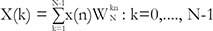

where one value of 'k' has N complex multiplications, since 'k' = 0, 1, …, N-1. The multiplication of x(n) and wkn was done for N times, since n = 0 to N-1.
Spectral analysis is the function of power over frequency. In medicine, a spectral analysis of various signals measured from a patient, such as electrocardiogram (ECG) or electroencephalogram (EEG) signal, provides useful information for diagnosis. A random signal usually has finite average power and is characterized by an average power spectral density as in equation:
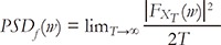
where F (w) XT represents the FFT output and T is the total duration of the input signal (Fig. 5). The power spectral density of alpha waves calculated from the data was obtained (Table 1). The Kolmogorov-Smirnov test was used to check the normal distribution of the values and the results were statistically analyzed using a paired t-test.
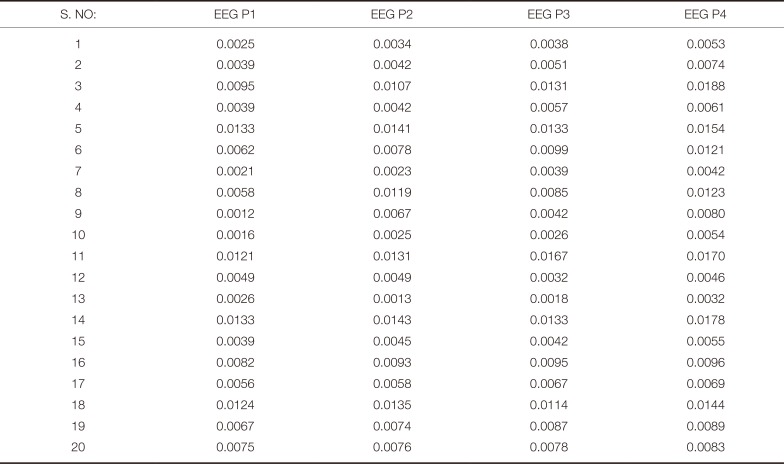
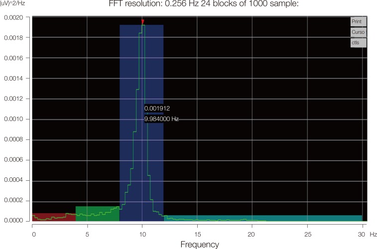
Table 1
Power spectral density (relative units)
![]()
Go to :
RESULT
The mean values were grouped as edentulous phase before chewing (EEG p1-0.006), edentulous phase after chewing (EEG p2-0.007), post denture insertion adaptive phase before chewing (EEG p3-0.007), and post denture insertion adaptive phase after chewing (EEG p4-0.009). The values were statistically analyzed using a paired t-test, which showed a significant increase in alpha waves PSD values after chewing in both edentulous and post denture adaptive phase. The alpha waves PSD related to before-chewing values increased in post denture adaptive phase (P < .05). (Table 2, Fig. 6)

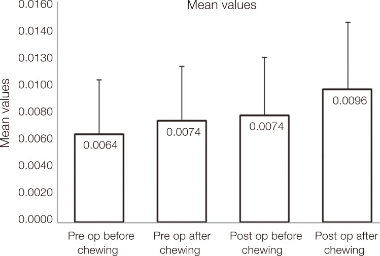 | Fig. 6Mean values of edentulous phase and post denture insertion adaptive phase before and after chewing.
|
Table 2
Paired samples t-test to compare the mean value
![]()
Go to :
DISCUSSION
The sensory information transmitted through the trigeminal nerve from the masticatory muscles, temporomandibular joint, and residual mucous membrane increased when subjects chewed. This transmission was unique and different from the control mechanisms involved in the movements of the arms and legs. Otsuka et al.16 proved the relationship between malocclusion and the limbic circuits. Narita et al.17 and Hirano et al.18 estimated that gum chewing was more effective in stimulating brain activity than other treatments. Yamazaki et al.19 demonstrated that, in both mature and old animals, the spatial memory declined as the number of hippocampal neurons decreased from impairment in hippocampal function due to early tooth loss. Hippocampal dysfunction caused impairment in episodic memory such as learning new information and retrieving information.20 Like hippocampus, other brain areas, such as the prefrontal cortex (PFC), the striatum, and the cerebellum, play an important role in executive abilities like performing multiple tasks simultaneously, set-shifting, and inhibition. These areas are sensitive to ageing.2122 Morokuma23 and Klimesch24 showed a positive relationship between alpha waves and cognitive performance as well as the speed of processing information. With this concept, brain activity was evaluated by studying power spectral density of alpha waves in edentulous patients before and after denture insertion. PSD values were calculated and a comparison was made for the EEG taken in two phases - edentulous phase and post denture insertion adaptive phase - before and after chewing paraffin gum.
The power spectral density of alpha waves increased in both edentulous phase and post denture adaptive phase after chewing. The masticatory stimulation travels from the masticatory muscles to the hypothalamus via trigeminal nerve. This mechanism is believed to involve a wide area of the brain and gum chewing is thought to be effective in stimulating the brain activity.25 The study result did not synchronize with the studies performed by Masumoto et al.,26 which stated no significant differences in EEG frequencies between a control and post gum condition. Masumoto et al.27 contradicted their earlier study with the findings indicating that chewing flavorless and odorless gum base led to increased alpha and theta activity.
Comparison of PSD values before chewing between edentulous phase and post denture insertion adaption phase showed that post denture insertion adaptive phase PSD value increased and influenced the brain function. The results concluded that dentures showed eminent improvement in brain function.
The limitation of this study is that the brain function can potentially be affected by other factors involving the subject, including his/her living environment, hospital visits, conversations with the attending physician, or the treatment administered. There is also a concern over the potential effect of circadian variability of EEG on the measurement sequence of brain function. These factors have to be standardized in future studies.
Go to :
 XML Download