

 PDF
PDF ePub
ePub Citation
 Print
Print


INTRODUCTION
The restoration of an endodontically treated tooth is a subject that has been evaluated and discussed widely in the dental literature.12
One of the functions of a post-core is to distribute the applied forces along the root to avoid fracture.34 Recent reports suggested that the rigidity of the post should be equal or close to that of the root to distribute any occlusal force evenly along the long axis of the tooth.56 The traditional custom cast or prefabricated posts are rigid in nature, similar to all-ceramic and zirconia posts. This rigidity may become a potential risk for a subsequent root fracture. More recently, fiber reinforced composite (FRC) posts placed into the root canal have been introduced as an alternative to previously employed materials.78 The biomechanical properties of FRC posts have been reported to be close to those of dentin.9 In addition, posts made of carbon/graphite fibers,10 glass, polyethylene or silica fibers (which are white or translucent) can be used where there is a higher esthetic demand.511 The use of such materials also allows an increased transmission of light within the root.12
There are different FRC posts currently in use in clinical dentistry. According to the manufacturer; Snowlight composite posts are made of unidirectional, longitudinal glass fibers for strength, and the fibers are embedded in a resin matrix for shock absorption. The use of zirconia glass fiber gives Snowlight posts a higher resistance to acid and alkali agents, a better resistance to hydrolysis in wet environments, and also a better resistance to fatigue when compared to common glass fibers. The special components of the Snowlight resin matrix, together with the zirconia glass fiber, make Snowlight posts translucent. The cylindro-conical, metal free FRC Postec root canal posts consist of glass fibers and a composite matrix containing dimethacrylate and ytterbium fluoride. The use of leno-woven polyethylene ribbon (Ribbond Bondable Reinforcement Ribbon) reinforced composite resin as a post in the restoration of extensively damaged teeth has also become established as an alternative treatment technique in clinical dentistry.1314 A review of literature shows that there has been a substantial number of in vitro studies dealing with the resin luting agents,1516171819 the post design,2202122 the length of the post,23242526 and the mechanical properties of the posts.272829303132333435363738 In addition, many studies30313233343536373839 have been conducted to compare the mean fracture resistances of different fiber-reinforced post-core systems, but there has not yet been a report in the literature about the fracture resistances of the different sizes of fiber post with all-ceramic crowns. Therefore, the purpose of this in vitro study was to determine the fracture resistance and the mode of fracture of endodontically treated canınes teeth restored with different sizes of fiber post and all-ceramic crowns.
Go to : 
MATERIALS AND METHODS
Two types of fiber post systems ((1) Postec, Ivoclar-Vivadent, Schaan, Liechtenstein and (2) Snowlight, Abrasive Technology, Lewis Center, OH, USA) in two different sizes (thick and thin), and two different thicknesses (2 and 3 mm) of polyethylene fiber ribbon (Connect KerrHawe, Bioggio, Switzerland) were chosen for evaluation (Table 1). 48 recently extracted, unrestored, and non-carious human maxillary canine teeth with similar root sizes were selected. All of the teeth were kept in a thymol solution for 24 hours for disinfection. Tissue remnants and debris were removed with a scaler before the teeth were brushed with pumice and placed in distilled water until use. Root lengths were measured from the cementoenamel junctions (CEJs) on the facial surface, and mesiodistal widths were measured from between the proximal surfaces at the CEJs.
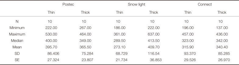
Table 1
Mean, median, and standard deviations of the fracture resistance values of the all the materials tested (in N)
![]()
All roots were instrumented with a 40 K file until 1 mm short of the apex and irrigated with sodium hypochlorite. The canal was dried with paper points, filled with zinc oxide eugenol sealer (Endomethasone, Septodont, Cedex, France), and obturated with a no. 4 gutta percha master cone (Gutta Percha Points, DiaDent, Canada) and three medium-fine cones using the lateral condensation technique. The endodontic treatment was completed with the use of a water-cooled diamond rotary cutting instrument (806 314 2837 014, Komet, GEBR. Brasseler GmbH & Co. KG, Lemgo, Germany) to remove the coronal parts of all the specimens by sectioning perpendicular to their long axes at 2 mm above the CEJs to provide a ferrule effect. The specimens were divided into 6 groups (n = 8). The post spaces in all of the specimens were then sequentially drilled. The Postec thin and thick (Ivoclar-Vivadent, Schaan, Liechtenstein) specimens were drilled with a no. 1 (0.8 mm) and no. 3 (1.0 mm) drill, respectively. The Snowlight thin and thick (Carbotech, Gages, France) specimens were drilled with a no. 12 and no. 16 drill, respectively, according to the manufacturers' instructions. The polyethylene fiber ribbon specimens were drilled using a Gates Glidden bur (Gates Drill, Mani Inc., Tochigi, Japan). At least one third of gutta-percha was left apically to seal the root apex. With the help of a silicone stopper on the drill, all the roots were uniformly drilled down to the same length of remaining gutta-percha.
Post cementation procedures for the fiber reinforced post groups were as fallows; the walls of the root canals were washed with 5% sodium hypochlorite solution. After a drying procedure with paper points, 37% phosphoric acid solution was used to etch the root canal wall and remaining tooth surface for 15 seconds before the specimens were washed thoroughly for 30 seconds. The fiber posts were then washed with 75% alcohol solution and air dried. Resin cement (Variolink, Ivoclar Vivadent, Liechtenstein) was used for cementation. Syntac Primer (Variolink, Ivoclar Vivadent, Liechtenstein), Syntac Adhesive (Variolink, Ivoclar Vivadent, Liechtenstein), and Heliobond (Variolink, Ivoclar Vivadent, Liechtenstein) were separately applied to the dentine in accordance with the manufacturer's directions. The fiber posts were coated with Variolink resin cement (Variolink, Ivoclar Vivadent, Liechtenstein) and then inserted into the canal under rotation without using a lentulo spiral. The posts were polymerized for 40 seconds from each surface (a total of 200 seconds) with a wide-tipped prismatic light-polymerizing unit (Optilux VLC 401, at 420 mW/cm2; Kerr Corp, Orange County, CA, USA) in accordance with the manufacturer's directions.
Post cementation procedures for polyetylene fiber ribbon-reinforced resin composite post-core buidup restorations were as follows: polethylene fiber ribbon (Connect KerrHawe, Bioggio, Switzerland) at two different thicknesses (3 mm and 2 mm) was used. The root canal filling was removed down to the apical one third by using the Gates Glidden bur (no. 1 to no. 4 for thin specimens and no. 1 to no. 6 for thick specimens) and then washed with 5% sodium hypochlorite solution. After drying with paper points, 37% phosphoric acid solution was used to etch the root canal wall and remaining tooth surface for 15 seconds before the specimens were washed thoroughly for 30 seconds. Any excess water was then eliminated, leaving the dentine moist. Variolink resin cement was used for luting the polyethylene fiber ribbon, and Syntac Primer, Syntac Adhesive, and Heliobond were applied to the dentine separately in accordance with the manufacturer's directions. A piece of fiber ribbon 5 - 6 mm longer than the prepared root canal length was cut off and embedded in mixed Variolink resin cement. The root canal was then filled with the resin cement. The fiber-resin combination was carefully placed into the canal with the use of titanium nitride coated instruments (Brilliant Esthetic Line Composite Instrument, Coltène/Whaledent Inc., Cuyahoga Falls, OH, USA), leaving a loop formed by 2 - 3 mm of ribbon above the occlusal surface of the root. The combined fiber ribbon and luting resin was light cured for 40 seconds with the light-polymerizing unit. To fabricate a core, the exposed ribbon loop was incrementally filled and covered with composite resin (Tetric Ceram; Ivoclar Vivadent), and light cured for 20 seconds from each surface.
A fine particle hybrid composite (Tetric Ceram; Ivoclar Vivadent) was used to fabricate core build-up for all the group. The light-cured composite was applied to the post in 1 - 2 mm thick increments. Each increment was carefully adapted to the post surface and light cured separately for 20 secondsfrom all surfaces according to the manufacturer's instructions. The total preparation height was 7 mm coronal to the facial cementoenamel junction.40
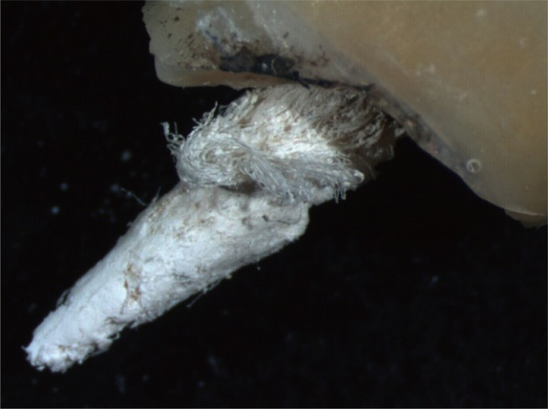
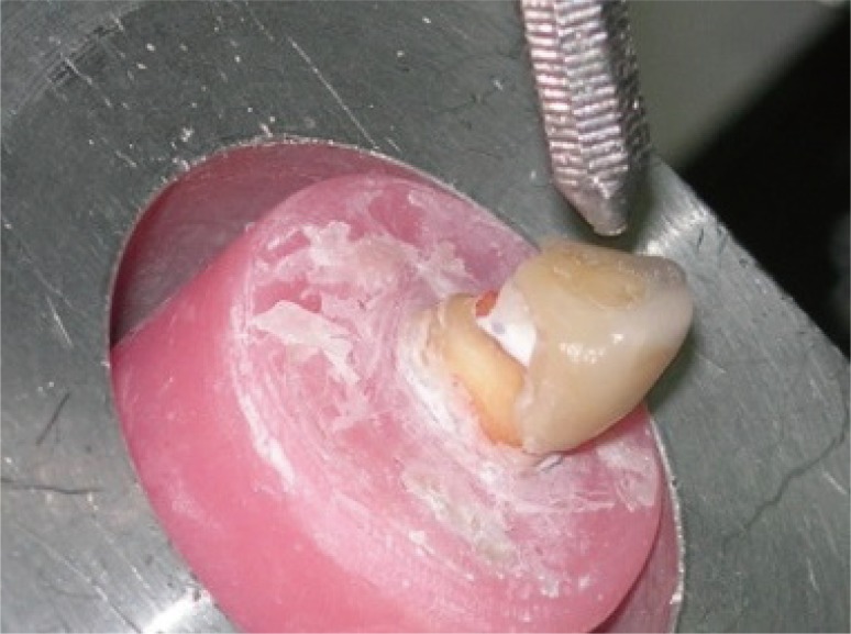
The roots of each tooth were embedded in autopolymerizing acrylic resin (Meliodent, #64713278 Heraeus Kulzer, Hanau, Germany), which was extended from the bottom to 2 mm below the cementoenamel junction. The preparation of the teeth was done using a parallelometer (Kavo EWL Type 990, Leutkirch, Germany) to ensure standardization. Medium and coarse diamond burs (Accurata, G+K Mahnhardt Dental, Thurmansbang, Germany) were used for tooth preparation. A 1 - 1.2 mm circumferential "chamfer" finish line and a 1.5 mm occlusal reduction were prepared. 2.0 mm of coronal tooth structure and the tooth/core junction was left to enhance fracture resistance. The core preparation was completed with a circumferential deep chamfer finish line. The finish lines for all specimens were placed at the CEJ level. Impressions were made with an addition silicon impression material (Affinis, Coltene, Whaledent, OH, USA). Full ceramic crowns (Empress II, Ivoclar, Vivadent, Amherst, NY, USA) were fabricated. The complete seating and marginal adaptation of each crown was checked, and all the crowns were cemented according to the manufacturer's instructions by using Variolink II high viscosity resin cement (Ivoclar Vivadent). Excess cement was removed with a brush. The restorations were photo polymerized for 40 seconds from all surfaces. Each specimen was placed in a custom apparatus that allowed the specimens to be positioned at 130 degrees to the buccal/lingual long axis (Fig. 1). The teeth were subjected to a compressive load in a universal test machine (Lloyd LRX; Lloyd Instruments, Fareham, Hants, England) using material testing software (Nexygen version 2.0; Lloyd Instruments). Shear bond strength at failure was determined by loading each specimen at a crosshead speed of 1 mm/min until fracture. The load was measured in Newtons (N) (Table 1). The mode of failure was recorded after the test using a stereomicroscope at ×20 magnification (Leica Cambridge Ltd., Cambridge, England). Failure was defined according to four groups: fracture of the all-ceramic crown, fracture of the post, fracture of the root, and post dislodgement (Fig. 3, Fig. 4, Fig. 5, and Fig. 6). All continuous random variables were normally distributed, and then results belonging to continuous random variables were analyzed statistically by a One-Way ANOVA test to compare the means of independent groups. When P < .05 was found, Tukey Kramer Multiple Comparisons post test was used to compare all pairs of groups. Fisher exact test was used to compare the nominal data related to the failure type. P value less than 0.05 was considered as significant. Data analysis was done by NCSS 2007 and Graphpad version 3.05.


Go to :
RESULTS
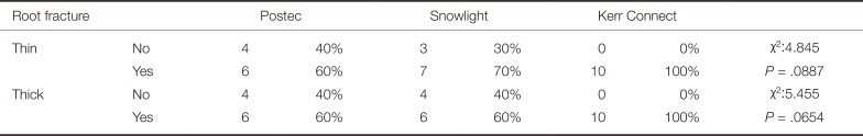
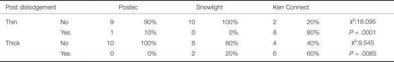
The mean, median, and standard deviations values of the tested specimen groups, and the mean fracture resistance of the groups were shown in Table 1. The One-Way ANOVA test results and the Tukey Kramer Multiple Comparisons test results are shown in Table 2 and Table 3, respectively. The failure type and distribution in percentages are shown in Table 4, Table 5, Table 6, and Table 7.
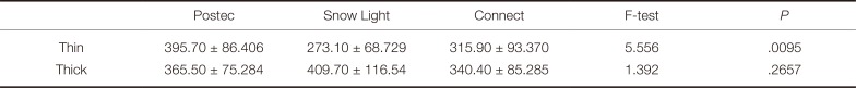

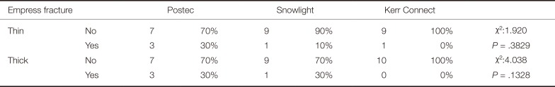
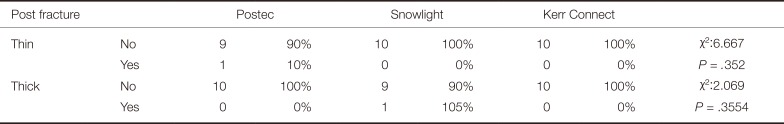
Table 2
Comparison of fracture resistance of the tested specimen groups
| Postec | Snow Light | Connect | F-test | P | |
|---|---|---|---|---|---|
| Thin | 395.70 ± 86.406 | 273.10 ± 68.729 | 315.90 ± 93.370 | 5.556 | .0095 |
| Thick | 365.50 ± 75.284 | 409.70 ± 116.54 | 340.40 ± 85.285 | 1.392 | .2657 |
![]()
Table 3
Tukey Kramer multiple comparison test results for the thin specimens
| Tukey Kramer multiple comparison test | Thin |
|---|---|
| Postec / Snow light | P < .01 |
| Postec / Connect | P > .05 |
| Snow light / Connect | P > .05 |
![]()
Table 4
Empress fracture's failure results
| Empress fracture | Postec | Snowlight | Kerr Connect | |||||
|---|---|---|---|---|---|---|---|---|
| Thin | No | 7 | 70% | 9 | 90% | 9 | 100% | χ2:1.920 |
| Yes | 3 | 30% | 1 | 10% | 1 | 0% | P = .3829 | |
| Thick | No | 7 | 70% | 9 | 70% | 10 | 100% | χ2:4.038 |
| Yes | 3 | 30% | 1 | 30% | 0 | 0% | P = .1328 | |
![]()
Table 5
Post fracture's failure results
| Post fracture | Postec | Snowlight | Kerr Connect | |||||
|---|---|---|---|---|---|---|---|---|
| Thin | No | 9 | 90% | 10 | 100% | 10 | 100% | χ2:6.667 |
| Yes | 1 | 10% | 0 | 0% | 0 | 0% | P = .352 | |
| Thick | No | 10 | 100% | 9 | 90% | 10 | 100% | χ2:2.069 |
| Yes | 0 | 0% | 1 | 105% | 0 | 0% | P = .3554 | |
![]()
Statistically significant differences were observed among the Postec, Snowlight, and Kerr Connect thin specimens. The mean fracture resistance values were 395.70 N, 273.10 N, 315.90 N for Postec, Snowlight, and Kerr Connect thin specimens, respectively (P < .0095). The Postec results were found to be significantly higher than the others. No statistical difference was observed among the thick specimens (365.50 N, 409.70 N, and 340.40 N, respectively) (P < .2657). The mean fracture resistance values of the Snowlight thick samples was found to be higher than those of the Snowlight thin samples.
The all-ceramic crown fracture failure results indicated that the percentage ratio of the thin and thick specimens of Postec and Kerr Connect materials were the same, 30% and 0% respectively. No all-ceramic crown fractures were seen in the Kerr Connect specimens, whereas 30% of the fractures were seen in the Postec specimens. 10% of the thin and 30% of the thick specimens of the Snowlight all-ceramic specimens were fractured. The fractures always occurred around the cemento-enamel junction at the palatinal side (Fig. 2 and Fig. 3, Table 4).
No post fractures were observed in the thin Snowlight and Kerr Connect specimens or in the thick Postec and Kerr Connect specimens. The percentage ratios of the thin Postec and thick Snowlight specimens were 10% (Fig. 4, Table 5).
Among the common failure types of the specimens, the worst was observed to be the root fracture failure. The percentage ratios of the thin Postec, Snowlight, and Kerr Connect were high, and the ratios were 60%, 70%, and 100%, respectively. The thick specimen percentage ratios were 60%, 60%, and 100%, respectively (Fig. 5, Table 6).
The highest post dislodgement failure result (80%) was obtained from the thin Kerr Connect specimens. A statistically significant difference was found between the thin specimens of all 3 post systems tested (P = .001). No post dislodgement was observed for the thin Snowlight specimens. No statistically significant difference was observed for the thick specimens of each material tested (P = .069). The percentage ratios of the thick Postec, Snowlight, and Kerr Connect specimens according to the post dislodgement factor were 0%, 20%, and 60%, respectively (Fig. 6, Table 7).
Go to :
DISCUSSION
Post and core materials should be both esthetically compatible with the full porcelain crown and biologically compatible with the surrounding tissues. The use of conventional custom cast posts with full porcelain crown has been noted to compromise esthetics due to the gray tint of the underlying metal. This cosmetic concern has led to the development of more aesthetic posts. The use of fiber post systems and polyethylene fiber ribbon reinforced composite resin as post-core restoration materials14 satisfied the aesthetic demands of all ceramic restorations and also provided a level of strength to the composite core material replacing the lost tooth structure.
Cementation plays a significant role in enhancing retention, distributing stress, and sealing irregularities between the tooth and the post.12 The effect of the cement on post retention is influenced by the strength and adherence of the cement.19 Mendoza et al.16 evaluated the ability of cemented posts to reinforce structurally weak teeth and investigated the effect of cements, reporting that dentine bonding of the cements increased the roots' resistance to fracture. It was reported that resin luting agents showed good adhesion to both carbon fiber and glass fiber posts.17 In the present study, all specimens were cemented with the same adhesive cement, so the bonding effect of the tested post systems was more constant by eliminating cement effectiveness as a parameter.
The primary reason for using a post is to retain a core as a substitute for the missing coronal tooth structure. Therefore the post head design is important.20 The amalgam and composite resin core has been reported to help the distribution of stress to the surface underneath the core.21 Although composite resin cores have a significantly higher failure rate compared to amalgam when used with metal posts under simulated chewing conditions,41 resin cores have esthetic advantage. Sadek et al.22 reported that, to build up the core, low-viscosity, highly filled core materials and hybrid composites are better alternatives to flowable composites. In this study, a fine particle hybrid composite was used to fabricate core build-up for all groups.
Although it is claimed that additional root canal preparation for Postec and Snowlight posts is minimal, radicular dentin requires considerable preparation to achieve the best level of retention, and this may weaken the root structure. On the other hand, residual radicular dentin can be preserved during polyethylene fiber ribbon-reinforced resin post-core restoration since there is no need for additional preparation of the root canal. This reduces the risk of perforations and optimizes the remaining root dentin to resist fracturing.14 Our results support this claim since ribbon post dislodgement, rather than root or post fracture, was observed under compressive load.
The length of the post influences stress distribution in the root and thereby affects its resistance to fracture.20 An increase in the success rate of the restoration of endodontically treated teeth has been noted when the length of the post is greater than or equal to the crown length.2123 A higher failure rate is seen when the post length is too short.23 Therefore, at least one third of gutta-percha was left apically to seal the root apex while preparing the post spaces of all the specimens in the present study by keeping posts at an optimum and equal length.
Assif et al.29 indicated that a complete crown with a 2-mm ferrule on a sound tooth structure distributed the forces to the root and post complex. In addition, Pereira et al.30 reported that a ferrule length of 2 mm significantly increased the resistance of endodontically treated teeth. In the present study, 2.0 mm of coronal tooth structure was left in tooth/core junction to enhance the fracture resistance for all the crown preparations. So, all-ceramic crowns were trasferred the distribution of forces to the root and post complex of each specimens.
Regarding the results of this study, no statistically significant difference was found between the thin and thick posts of the Postec system or the polyethylene fiber ribbon (Connect) used as post material. The mean fracture resistance was 395.70 N and 365.50 N, respectively, for the thin and thick Postec systems and 315.90 N and 340.40 N, respectively, for the thin and thick polyethylene fiber ribbon (Connect). In contrast, the fracture resistance of the thin and thick posts of the Snowlight systems was statistically different. The mean fracture resistances for the thin and thick posts were 273.10 N and 409.70 N, respectively. Although the overall fracture resistance of the thick post specimens was higher than that of the thin posts, the difference was not statistically significant. There has not been any information related to the effect of post thickness on fracture resistance in the literatures. For this reason, the results of our study can only be compared with the mean fracture resistance results of former studies.31323334
Cormier et al.31 reported a 180 N mean for the failure loads of glass fiber posts and FibreKor posts, which is lower than the mean value of the results of this study. Al-Wahadni et al.32 recorded a 393 N mean fracture load for teeth restored with glass fiber posts, which is consistent with the findings of the present study. However, Stricker et al.33 reported a 672.6 N of mean failure load for teeth restored using glass fiber posts (FRC) with 2 mm ferrules, which is much higher than the present study. This difference could be related to the methodology of the Stricter et al. study, as the loading from a computer-controlled masticator might have increased the success rate. Akkayan34 reported a 99.84 kg mean fracture resistance value for the glass fiber ER Dentin Post, which is the highest value currently recorded in the literatures.
The mean fracture resistance of the all the groups tested in this study was higher than the maximum physiological forces acting on the teeth intraorally. Tan et al.35 reported that the maximum forces on anterior teeth varied, but the amount was almost always below 200 N. Lyons and Baxendale42 observed that the mean force applied to a maxillary canine was 215 N. In the presence of parafunctional loading, the authors noted that this force increased to 254.8 N, and the maximum forces were between 343 N and 362.6 N. In this study, the mean fracture resistance obtained from maxillary canine teeth with 2 mm ferrules, restored with fiber post systems and covered with all-ceramic crowns, was greater than the mean actual functional and parafunctional forces applied to maxillary canines.
The failure type was classified into 4 categories: fracture of Empress 2 crowns, fracture of post, fracture of root, and dislodgement of post. No statistically significant difference was observed among all the tested groups when the failure types were considered. However, a statistically significant difference was found between the thin samples of all 3 tested post systems, and the highest post dislodgement distribution was obtained in the Kerr Connect system. Although this result conflicted with that of the in vivo study by Turker et al.,14 this might be considered an advantage with regard to the protection of the root structure under excessive load. Even though laboratory fracture strength tests cannot fully mimic intra-oral loading conditions, they offer a controlled environment for preparing and testing specimens, thus allowing a comparable evaluation of the variables under investigation.28
Root fractures occurred at the cervical portion of the root for all the tested materials. Eskitascioglu et al.36 evaluated fiber composite laminate (FLC) post core restoration by using a finite elemental stress-analysis method and concluded that using FLC post cores distributed stress to the cervical 1/3 region of the tooth and the supporting bone structure, which agrees with the result of this study.36 In another study, Sirimai et al.27 added polyethylene woven fiber ribbon to the post and core restoration and reported that, with this procedure, there was a significant reduction in the incidence of vertical root fractures. In the present study, no vertical root fracture was observed in the groups using polyethylene fiber ribbon as post material. Giovani et al.37 and de Oliveria et al.38 reported fractures at the cervical region of longer glass-fiber posts (8 and 10 mm) and also fractures of the restorative material or the coronal structure of the prefabricated carbon fiber post. The results of the present study are therefore supported by those of Sirimai,27 Giovani,37 and de Oliveria et al.38.
Marchionatti et al.43 reported that the elastomeric materials used to stimulate the periodontal ligament did not affect fracture or bond strength of the teeth restored with fiber posts and composite cores in fracture test. So that, in the present study no artificial periodontal ligament was used to stimulate the clinical condition.
Although the methodology of the presented study could not exactly simulate the oral conditions, the in vitro comparison of fiber reinforced posts in different sizes offered information that may be of use in post system selection. Another limitation of this study is the absence of thermal cycling of the specimens and fatigue loading. In vitro studies including thermal cycling, fatigue loading of specimens, and the fracture resistance of endodontically treated teeth can be done in the future. In addition, a longitudinal clinical study should be done to compare the clinical survival of fiber reinforced post systems.
Go to :
CONCLUSION
In the light of the results of this study, although the Snowlight thick posts were the strongest of the 3 post-core systems, the overall fracture resistance of the Postec posts was found to be greater than that of the Snowlight posts. In terms of optimizing the fracture resistance of restored teeth, fiber post size selection should be done according to the forces that applied to them.
Go to :
 XML Download