

 PDF
PDF ePub
ePub Citation
 Print
Print


INTRODUCTION
Metal ceramic crowns and fixed dental prosthesis have been widely used in clinical practices because of their good mechanical properties and acceptable aesthetics,12 regardless of the development of all ceramic crowns.34 Noble metal ceramic alloys have been frequently used for the fabrications of metal ceramic restorations. The advantages of noble metal ceramic alloys include excellent biocompatibility, sufficient mechanical properties and excellent bonding capacity with the veneered porcelain.567 However, because of the soaring price of noble metal alloys in the last decade, the non-precious metal alloys have been attracting considerable attention in clinical practices. The non-precious metal ceramic alloys, including nickel-chromium (Ni-Cr), cobalt-chromium (Co-Cr) alloys, commercially pure titanium (cpTi), and Ti alloys are the alternatives to noble metal ceramic alloys. Although cpTi and Ti alloys possess excellent biocompatibility, corrosion resistance, and mechanical properties, they require special casting machines and investment materials due to their high melting temperature.89 Furthermore, cpTi and Ti alloys show some difficulty in porcelain fusing.101112 When Co-Cr alloys is compared to Ni-Cr, the use of more biocompatible Co-Cr alloys is suggested due to the hazardous effect of Ni-Cr alloys.131415161718
Appropriate oxidation of metal surface is required for the stable bonding between metal alloy and porcelain.192021 However, non-precious metal alloys are readily oxidized and thick oxidized layer is formed on the alloy surface during porcelain firing,1322 and excessively oxidized layer leads to the complications in bonding between alloy and porcelain by yielding the facture through the thick oxidized layer.1322 In order to resolve this problem, there have been attempts to modify alloy components2324 and metal surface treatment, including degasification,25 firing temperature of opaque layer26 and employment of air-borne particle abrasion,1427 have been made. Among these endeavors, applications of metal conditioners1928 have also been attempted. These metal conditioners are employed with the intension of improving the aesthetics of the porcelain by masking dark oxide color of non-noble metal frameworks and also increasing the bonding of porcelain to noble metal alloys,1028 cpTi, Ti alloys,1011 and Ni-Cr alloys.1122 However, the reports on the effectiveness of metal conditioners to Co-Cr alloys are very limited,22 and the effectiveness of metal conditioner to Co-Cr alloys is yet unknown.
Therefore, studies on the efficacy of metal conditioners for Co-Cr alloys are desired. A metal conditioner for non-noble metal ceramic alloy has been developed recently. This conditioner improves the bonding of porcelain to non-precious metal ceramic alloy by controlling the formation of oxidized layer and by increasing the wettability of alloy surface.
The purpose of this study was to evaluate the efficacy of a novel metal conditioner for a Co-Cr alloy, and to compare its efficacy to that of traditional metal conditioner available for non-precious metal alloy. The null hypothesis of the study was that the metal conditioners were not effective for the improvement of the bonding of porcelain to a Co-Cr alloy.
MATERIALS AND METHODS
Materials used in this study are presented in Table 1. For the metal ceramic alloy, Co-Cr alloy (Heraenium Pw, Heraeus-Kulzer GmbH, Hanau, Germany) was used. For the metal conditioners, a novel conditioner for non-precious metal alloy (HeraCeram NP primer, Heraeus-Kulzer GmbH) and a traditional conditioner for all noble and non-precious PFM alloys (Initial IN-Metalbond, GC Europe N.V., Leuven, Belgium) were employed. Low fusing feldspathic porcelain (Heraceram Opaque and Dentin, Heraeus-Kulzer GmbH) was used as veneering porcelain.
Disk-shaped specimens (2.5 × 10.0 mm) were cast with Co-Cr alloy according to the manufacturer's instruction, and used as adherend materials. The bonding surfaces of the adherend were polished with a 240, 400, and 600-grit silicon carbide paper (Carbimet Paper Discs; Buehler, Lake Bluff, IL, USA) under ample water coolant. Then the bonding surface was airborne-particle abraded using 110 µm alumina particles (Korox110, Bego, Bremen Germany) at 0.4 MPa pressure for 10 seconds with a 5 mm nozzle-to-alloy distance, followed by drying with filtered air.
Procedures for fabrication of bonding specimens are presented in Fig. 1. A 30 µm-thick masking tape (Mending Tape; Kokuyo Co., Ltd., Osaka, Japan) with a 5 mm diameter opening was placed on the airborne-particle abraded surface to restrict the porcelain firing areas, and a thin layer of either of the metal conditioners was applied. The masking tape was removed and the metal conditioner was fired in the furnace (KDF Master Spirit 120, Denken Co., Ltd., Kyoto, Japan), according to the firing schedule presented in Table 2. Another sheet of masking tape was placed to fit the opening into the baked metal conditioner, then the second firing of each metal conditioner was achieved in the same manner (Fig. 1A).
After the firing of each metal conditioner, the masking tape was placed so that the opening is fit into the baked metal conditioner, the opaque porcelain paste was applied onto the baked metal conditioner. The tape was carefully removed and opaque porcelain was fired in the furnace, according to the firing schedule shown in Table 2. These procedures were repeated to layer the opaque porcelain with two increments (Fig. 1B).
After the firing of opaque porcelain, dentin porcelain powder was mixed with purified water. Additional dentin porcelain slurry was layered on the opaque porcelain, and was fired in the furnace according to the firing schedule (Table 2). Dentin porcelain was fired into 1.5 mm in height by repeating the procedure twice (Fig. 1C). Then a Teflon® tube (5 mm inner diameter and 3 mm in height) was fixed so as to cover the baked porcelain and additional dentin porcelain was placed in the Teflon tube. The Teflon® tube was carefully removed and dentin porcelain was fired in the furnace (Fig. 1D), according to the firing schedule (Table 2). Finally, the porcelain was subjected to glazing procedure, according to the schedule shown in Table 2, to complete the specimen fabrication (Fig. 1E). Specimens without metal conditioner were also fabricated and used as controls. Eight specimens were fabricated for each of three groups, including two metal conditioner groups and a control group.
Apart from bonding specimens, specimens for scanning electron microscopic (SEM) observation were fabricated. Rectangular specimens (3 × 10 mm and 0.5 mm in thickness) were cast from Co-Cr alloy, and porcelain-firing surfaces were prepared according to the procedures as same as the bonding specimens. Then the metal conditioner and porcelain were fired into 1.5 mm height, according to the fabrication procedures of bonding specimens. Specimen without metal conditioner was also fabricated. Specimens were invested with translucent auto-polymerizing resin (Rigolac 2004, Showa Denko K.K., Tokyo, Japan), and SEM specimen with 1 mm thickness was sliced off from the center of rectangular specimen using a precision saw (IsoMet Low Speed Saw, Buehler, Lake Bluff, IL, USA), and carefully polished using 2000, 4000, and 8000-grit lapping film (3M™ Lapping Film, Sumitomo 3M Ltd., Tokyo, Japan) under ample water coolant.
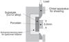
Bonding specimens were embedded into an auto-polymerizing resin and mounted on a shear-testing device. Shear bond test was performed using universal testing machine with 5.0-kN load cell (AGS-5kNG, Shimadzu Corp., Kyoto, Japan) with a 1.0 mm/min crosshead speed until failure occurred. A cross-sectional view of shear testing is illustrated in Fig. 2. A unibevel-chisel apparatus made from stainless steel was used to produce a shear force adjacent and parallel to the bonding interface. The shear bond strength (MPa) was determined by dividing the maximum load (N) by the bonding areas (mm2). The data were statistically analyzed by a 1-way analysis of variance (ANOVA) and Bonferroni/Dunn test to identify the significant differences among the groups (α = 0.05). Statistical analyses were performed using IBM SPSS Statistics software, version 22 (IBM Corporation, New York, NY, USA).
The deboned surface of each specimen was analyzed with a stereo and zoom microscope (SteREO Discovery. V12, Carl Zeiss Microscopy GmbH, Jena, Germany) at ×40 magnification and images of both failed Co-Cr alloy and porcelain surfaces were captured using CCD camera (Axio Cam ERc5s, Carl Zeiss Microscopy GmbH) and digital image processing software (AxioVision 4.8, Carl Zeiss Microscopy GmbH). Failure modes were categorized as cohesive failure of porcelain (CP), cohesive failure of metal conditioner (CM), adhesive failure at the alloy-porcelain interface (AP), adhesive failure at alloy-metal conditioner interface (AM), or mixed failure of cohesive and adhesive failures (MF).
SEM specimens were then gold-coated with an ionsputtering device (Fine Coat Ion Sputter JFC-1100, JEOL Ltd., Tokyo, Japan) and observed with an SEM (JSM-5510LV, JEOL Ltd., Tokyo, Japan).
Then, energy dispersive spectrometer (EDS) analysis was performed for each specimen to determine the chemical elements of metal conditioners. Failed porcelain surface of each shear specimen were evaluated to determine the failure modes by using the SEM equipped with energy dispersive X-ray analyzer (JED-2300BU, JEOL Ltd., Tokyo, Japan).
RESULTS
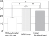
The results of the shear bond strength tests of each group and statistical analysis among groups are presented in Fig. 3. Two groups that used metal conditioners showed significantly higher shear bond strengths than the group that did not use metal conditioner (P < .0001). For the comparison between two metal conditioner groups, the group using NP primer showed significantly higher shear bond strength than the group using Initial IN-Metalbond (P = .0153).
The SEM observation for the sectional view of bonding interface for non-metal conditioner specimen revealed loose contact between porcelain and Co-Cr alloy surface and a gap formation at alloy-porcelain interface (Fig. 4A). On the other hand, Initial IN-Metalbond-applied and NPPrimer-applied specimens (Fig. 4B and Fig. 4C, respectively) showed hermetic contacts at both the alloy-metal conditioner interface and metal conditioner-porcelain interfaces. From the results of EDS analysis, NP-Primer mainly contained silicon (Si). In addition, sodium (Na), zirconium (Zr), aluminum (Al), potassium (K) and gold (Au) were also detected. On the other hand, Initial In-Metalbond mainly contained titanium (Ti) and Si. Na, Al, and K were also detected in Initial In-Metalbond.
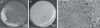
Distribution of failure modes in each group are presented in Table 3. For the comparisons of failed surfaces, all the specimens showed mixed failure, which included cohesive failure of Co-Cr alloy, cohesive failure of porcelain and cohesive failure of metal conditioner. Non-metal conditioner specimens showed remnant of porcelain on the Co-Cr alloy surface (Fig. 5A) and numerous numbers of black spots scattered on the failed porcelain surface (Fig. 5B). SEM observation showed porous porcelain surface (Fig. 5C), and cobalt (Co) and chromium (Cr) were detected by EDS analysis. For the Initial IN-Metalbond-applied specimens, cohesive failure of both porcelain and metal conditioner were observed on the alloy surface (Fig. 6A) and black spots scattered on the failed porcelain surface (Fig. 6B). SEM observation showed entrapment of air-bubbles on metal conditioner surface (Fig. 6C), and Co and Cr were detected by EDS analysis. Regarding the NP-Primer-applied specimens, cohesive failure of porcelain and metal conditioner were observed on the alloy surface (Fig. 7A) and black spots scattered on the failed porcelain surface (Fig. 7B). SEM observation showed entrapment of air-bubbles on metal conditioner surface (Fig. 7C), and Co and Cr were detected by EDS analysis.
DISCUSSION
The null hypothesis of the study was rejected, because both of the metal conditioners employed in this study significantly improved the bonding of porcelain to the Co-Cr alloy. Metal-ceramic bonding is usually established by the development of a suitable oxide layer on the substrate metal surface.192021 In noble metal alloys, an addition of small amount of base metal develops an optimal oxide layer to achieve bonding with porcelain during porcelain firing, while an excessive oxidized layer forms and diminishes the bonding in non-precious metal alloys.1322 Metal conditioners have been used to overcome this problem for mainly non-precious metal ceramic alloy. They react with metal oxides and form a new interface which seals the alloy surface and protects from further oxidation, thus prevent the production of a thick oxidized layer and make opaque porcelain fuse well.101122 Hermatic contents at both the alloy-metal conditioner interface and metal conditioner-porcelain interface for both metal conditioner applied groups (Fig. 4B and Fig. 4C) must have proved effect. However, there are some limitation in affinity of metal conditioners to metal substrate for the improvement of metal-ceramic bonding.2229 The other possible functions of metal conditioner, the improvement of esthetics by regulating color change of porcelain during subsequent firing cycles, have also been mentioned.22
Both of metal conditioner employed in this study revealed remarkable effect, and shear bond strength of metal conditioner-applied groups (30.1 MPa for NP-Primer group, 27.1 MPa for Initial IN-Metalbond group) were significantly higher compared to that of non-metal conditioner group (17.2 MPa). These shear bond strength values range between those of gold alloy and porcelain (24.5 MPa) reported by Saito et al.30 which employed identical specimen configuration. Therefore, metal conditioners employed in this study must be effective for the improvement of the bonding between Co-Cr alloy and porcelain.
Components of both metal conditioners are not announced. From the results of EDS analysis (Table 4), NP-Primer mainly contains Si, and small amount of Au was also detected. Ceramic-containing metal conditioners perform a function of absorbing excessive oxides that are formed on the alloy surface during porcelain firing.10 As the Au-containing bonding agent reduces the interfacial stress by improving the compatibility between porcelain and alloy,3132 it may lead to the increased bond strength of porcelain to alloy. Si-containing metal conditioners perform a function of absorbing excessive oxides that are formed on the alloy surface during porcelain firing.1029 On the other hand, Initial In-Metalbond mainly contains Ti and Si. Ti-containing bonding agent acts as oxygen scavengers protecting the alloy surface from progressive accumulation of excessive oxidation layer with firing cycles,293233 and it may cause the increase in porcelain-metal bond strength. The difference in the main component of both bonding agents must have led to the differences in bond strength.
Regarding the failure mode for both groups, Initial In-Metalbond applied group showed cohesive failure of both porcelain and metal conditioner on the alloy surface, and cohesive failure of alloy on the opposing failed porcelain surface (Fig. 6). Cohesive failures of porcelain on the alloy surface and of Co-Cr alloy on the failed porcelain surface (Fig. 7) were both observed in the NP-Primer-applied specimens. Both metal conditioner-applied groups revealed similar failure mode. Therefore, it is assumed that mechanical properties of Initial In-Metalbond, whose main component is Ti, must be inferior to those of NP-Primer. Differences in mechanical properties between the metal conditioners must have led to the difference in bond strength between the groups.
It is difficult to compare the bonding results of this study with other studies because of differences in test methods. Various test methods including pull through test,28 shear test,1218223034 three-point flexural test,167192025 and four-point flexural test1011 have been employed to evaluate the bond strength between metal and porcelain. Among them, high reliability of shear test has been mentioned based on its minimal experimental variables and less residual stress.12 Shear test with flat interface mostly directs the tension to the bonding interface, and the result is not influenced by the difference in elastic modulus of a metal as seen in flexural tests.24 Therefore, this study employed shear test for the measurement of bond strength between metal and porcelain.
Airborne-particle abrasion of alloy surface is commonly performed prior to porcelain firing.12152530343536 Lombardo et al.15 have reported the efficacy of airborne-particle abrasion with 50 µm alumina particles for the improvement of shear bond strength of porcelain to Co-Cr alloy compared to roughening with a tungsten bur. This effect was produced by the increased surface roughness and the resultant micromechanical retention of porcelain, and by the enhancement of wettability using airborne-particle abrasion.33 Furthermore, Külünk et al.34 have compared the effects of particle size for airborne abrasion and concluded that higher bond strength was obtained using 110 µm alumina particles for both Co-Cr and Ni-Cr alloys. Therefore, airborneparticle abrasion using 110 µm alumina particles must have been adequate as a surface treatment method.
Metal-ceramic crowns are exposed to chemical, thermal and mechanical stresses under intraoral conditions; however, this in vitro design did not sufficiently simulate these clinical conditions. Therefore, further investigations combining thermal cycling, which could weaken the metal-ceramic bonding,101237 need to be employed to evaluate the bonding under more closely simulated clinical conditions.


 XML Download