

 PDF
PDF ePub
ePub Citation
 Print
Print


INTRODUCTION
Even though many types of resins have been brought out into dental treatments for the construction of complete and partial dentures PMMA is the most preferred material in spite of its low strength.1,2 Dentures' physical and mechanical properties could be affected by the type of denture base material and its polymerization method, as well as the distinction in processing techniques.3 Some deficiencies in mechanical properties and many factors that weaken the denture may cause fracture failures. The addition of metal wires, different types of fibers and cross linking agents or generating chemical modifications by adding rubber graft copolymer into the PMMA, may improve the mechanical properties of denture base resins and makes it strong against the fracture failure.1
The addition of copolymer have been used to strengthen the acrylic resin dentures.4 By blending two different polymers having different physical properties, in different volume ratios, a new material with tunable properties may be formed.5 As a result of the miscibility of the two polymers incompatible with each other, both homopolymer chains of the resulting copolymer chains have structures. Provided that the system combines mixable mixture, an intimate mixture or mix-up of (at least) two polymer chains occur in the blend.6 The final features of the material then count on the interactive manner of the two diverse polymer chains at different volume fractions. The physical features which might be enhanced contain effect, thermal and chemical struggle, adhesion, modulus, and elongation at break.5 Copolymerization mechanism provides polymer system arranges, bringing about various favorable modifications such as a rise in polymerization rate, superior mechanical and physical features, and a decline in water solubility when compared to that of linear polymers.7 It is possible to gain structure resistance by addition block or by a random copolymers to polymer blend that is immiscible. It has been reported that adding styrene and a methyl methacrylate copolymer to the same components can enhance the mechanical properties of the structure.8
Numerous types of monomers have been added to methyl methacrylate (MMA) and made ready for copolymer formation of PMMA. Fluoroalkyl, butyl acrylate, butadiene styrene, butyl methacrylate, ethyl methacrylate, IBMA, tertbutyl methacrylate, tetramethyldisiloxane, carbonyl, and phenyl methacrylate monomers have been applied with MMA to yield a adjusted copolymer formation but conflicting results have been reported.4,9,10 Also, HEMA was used to develop the some mechanical properties of PMMA.11
Attempts to modify the composition of PMMA material led to the development of high impact strength acrylic denture bases. This is a graft copolymer of butadiene styrene rubber with PMMA. However, these materials showed poor flexural strength compared to conventional acrylic resins.12 Butyl methacrylate is used as solvent, adhesive, shutter in the resin and application of dental properties. Strong interactions are constituted between the surfaces by the combination and distribution of butyl methacrylate and HEMA in the polymer matrix.13 Isaksson reported that HEMA and ethylene glycol dimethacrylate could be used for dental and other acrylic products in low concentrations.14 Yoshii performed a study about cytotoxicity of acrylate and methacrylate. The results showed that acrylate is more toxic than methacrylate and also reported that dimethacrylates which have 14 or less oxyethylene chains showed similar toxic findings, while dimethacrylate having 23 oxyethylene chain showed less toxic findings.15
Biocompatibility can be characterized as the adoption or refusal of synthetic material by the surrounding tissues and whole of body.16 Many studies have been reported the cytotoxicity of miscellaneous types of acrylic resins. According to these studies the cytotoxicity of acrylic resins depends on their polymer to monomer ratio,17 storage time, water immersion,18 polymerization cycle,17,19 and polymerization methods.20
The aim of this study is to obtain a resin material with high strength properties by developing a chemical structure that has both hard and soft segments in the same molecule by using IBMA and HEMA and investigate the cytotoxic effect.
MATERIALS AND METHODS
Two PMMA-based acrylic resins-a conventional (Paladent 20, Heraeus Kulzer GmbH & Co. KG, Hanau, Germany) and a injection-molded resin (Palaxpress, Heraeus Kulzer GmbH & Co. KG, Hanau, Germany) and also two different methacrylate monomers-isobutyl-methacrylate (IBMA), (Fluka, Sigma Aldrich GmbH, Germany) and 2-hydroxyethyl-methacrylate (HEMA) (Merck Schuchardt OHG, München, Germany) were used.
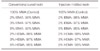
This study had 14 experimental groups; two of them were control groups (Paladent20 and Palaxpress). The specimens of copolymer groups, which were generated by adding various concentrations of IBMA and HEMA monomers to the Paladent and Palaxpress' monomers', and the control groups are given in Table 1. A Teflon mold was prepared to fabricate wax duplicates of flexural strength test resin specimens. 98 (n=7), wax rectangular-shaped specimens were prepared with dimensions of 65 × 10 × 2.5 mm (complying with ADA specification no.12)21 for three-point flexure test and 98 wax specimens were prepared in 1 mm diameter and 1.5 mm thickness for agar-overlay test.
IBMA and HEMA monomers were added to the monomers of Paladent 20 and the Palaxpress acrylic resin with a millimeter syringe at various content percentages of 2%, 3%, and 5% per volume and were bottled separately.
The conventional heat-polymerized resin specimens were prepared by using denture base resin material according to recommendation of the manufacturer. The polymer/monomer ratio 20/7 g/mL and the mixing time 30 seconds was set for the control groups. In the copolymer groups, the IBMA and HEMA monomer mixtures, which were added to monomers of Paladent 20 acrylic resin and bottled at various content percentages of 2%, 3%, and 5% by volume maintained a polymer/monomer ratio of 20/7 g/mL and a 30 seconds mixing time, as with the control group. The hydraulic pressing apparatus (Emmevi SPa, Italy) was used at 8000 kPa to compress the flasks for 5 minutes. Conventional heat-polymerized resin specimens were processed in a thermally controlled water bath (KaVo EWL 5501, KaVo Electrotechnisches Werk GmbH, Germany). Subsequently the flasks were put into cold water for heated up to 100℃ and boiled for 45 minutes.
The injection-molded resin specimens were prepared by using denture base resin material according to recommendation of the manufacturer. The polymer/monomer ratio 30/15 g/mL was set for the control groups. In the copolymer groups, the IBMA and HEMA monomer mixtures, which were added to the monomers of the Palaxpress acrylic resin and were bottled at various content percentages of 2%, 3%, and 5% by volume, maintained a polymer/ monomer ratio 30/15 g/mL. The specimens of Palaxpress for the injection-molded resin were polymerized at 55℃ for 30 minutes under 2 bar pressure.
Acrylic resin specimens were bench-cooled before deflasking. Afterwards all the specimens were grounded with 600-grit size silicon carbide paper (Mırka, Finland). Before the test procedure the storage of specimens in distilled water at 37℃ for 50 ± 2 hours was carried out.
A three-point bending test was conducted on the universal testing machine (Lloyd NK 5, Lloyd Instruments Ltd., Fareham, Hampshire, UK) according to the ISO 1567:199922 specifications for denture base polymers. The specimens were then placed on the standard three-point bending jig with circular supports 50 mm apart and the test performed using a 5 mm/min crosshead speed until there was a failure to determine the flexural strength.
The flexure strength of each resin, N/mm2 (δ), was calculated from the equation: δ = 3fL/2bh2, and the elasticity modulus, N/mm2 (E), of each resin was determined from the equation : E = L3m/4bh3d where F is the load, (N) at a given point in Newton (N), L is the distance between the support span (mm), b is the breadth of specimen (mm), h is the height of specimen (mm), and d is the maximum flexure.
Data were statistically analyzed by using SPSS 12 statistical software (SPSS, Inc., Chicago, IL, USA). In order to evaluate the effects of acrylic resin copolymers with different concentrations and polymerization methods, mean values and standard deviations of test groups were analyzed by Kruskall-Wallis test. Then pairwise comparisons were performed between significant groups by Mann-Whitney U test.
The cytotoxicity test was carried out by an agar overlay test. Specimens were performed in sterile with ethylene oxide gas. Each test group consists of 7 test specimens. Mouse fibroblast cell (L929) suspensions at 2.5 × 10⁵ cells density were seeded in each 35 × 15 mm petri dish. The used culture medium was Dulbecco's Modified Eagle Medium (1% L-glutamine, 1% penicillin-streptomycin mixture, 4% fetal bovine serum).
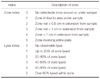
After twenty-four hours of incubation (5% CO2/ 95% air, 37℃), following up a confluent cell layer creation, suspend of the medium was carried out. Afterwards, 10 mL of medium including 3% agarose was replaced. Subsequent to solidifying the agarose, plated acrylic resin specimens on the agar were incubated for 24 hours at 37℃ (5% CO2/ 95% air). Twenty-four hours later, 0.01% neutral red added to the cells and staining was carried 20 minutes at 37℃. After incubation, the cytotoxicity of the test specimens was determined by assessing microscopically the cell lysis and zone index under a light microscope. The results that assessment with respect to the zone and lysis index, was shown in Table 2.23
Thin transparent tablets, which were formed from the control and copolymer resins samples, were triturationed through KBr (potassium bromide) under the 200 bar. Also the MMA, HEMA, and IBMA monomers were added drop wise into purged air cells, and the FTIR spectra of the samples were registered by using Unicam Mattson 1000 FTIR spectrometer (Cambridge, UK).
RESULTS
Table 3 summarizes the results of the determined flexural strength of three concentrations of two different monomers polymerized by two-polymerization process. For conventional heat-polymerized resin, 2% HEMA groups showed the highest flexural strength (102.10 ± 9.14 MPa), the control groups showed the lowest flexural strength (87.60 ± 1.69 MPa). There were significant interactions between the control and 2% IBMA, 5% IBMA, and 2% HEMA (p<.05), and there were no significant interactions between all others groups for the conventional heat-polymerized resin (P>.05).
For the injection-molded resin, the 2% IBMA groups showed the highest flexural strength (92.78 ± 3.41 MPa), and the 2% HEMA groups showed the lowest flexural strength (91.82 ± 8.64 MPa). There were significant interactions between the 2% IBMA group and all the other groups: the 3% IBMA group and the 2% IBMA, 5% IBMA, 2% HEMA, 3% HEMA, and 5% HEMA groups; the 5% IBMA group and the 2% HEMA group and the 3%HEMA groups, the control and the 2% HEMA, 3% HEMA, and 5% HEMA groups (p<.05). There was no significant interactions among all the other groups for injection-molded resins (P>.05). For both polymerization types, the flexural strength of the control groups showed no significant effect (P>.05) while there were significant interactions between the groups of the same monomer concentrations (p<.05).
Table 4 summarizes the results of the determined elasticity modulus of, three different concentrations of two different monomers in a two-polymerization process. In conventional heat-polymerized resin, the 3% HEMA group showed the highest (2944.40 ± 238.52 MPa) and the control group showed the lowest (2498.5 ± 278.57 MPa) elasticity modulus. There were no significant interactions between the copolymer groups in the conventional heat-polymerized resin (P>.05). In the injection-molded resins, the 3% IBMA group showed the highest (2938.71 ± 126.42 MPa) and the 2% HEMA group showed the lowest (2100.24 ± 255.94 MPa) elasticity modulus. There were significant interactions between the control, the 2% and 3% IBMA groups and the 5% IBMA, and the 2% HEMA groups. There was also significant interaction between the 2% HEMA and the 5% HEMA groups (p<.05). When the same copolymer groups were compared for polymerization methods, there were significant interactions between the control, the 3% IBMA, the 5% IBMA and the 2% HEMA groups (p<.05).
Results of the agar overlay test are shown in Table 5. Sterile drying paper discs impregnated with the phenol, the positive control, induced a zone/lysis index of 4-5, sterile drying paper impregnated with the DMEM, the negative control induced a zone index 0-0. 5% HEMA caused lysis or inhibition zone in conventional cured resin showed 1/1. All the specimens except 2% IBMA caused lysis or inhibition zone in injectional molded resin showed 1/1.
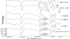
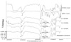
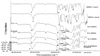
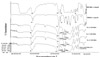
The copolymer synthesis was approved by FTIR spectroscopy. Fig. 1 shows the FTIR spectrum of Paladent 20 control, 2% IBMA, 3% IBMA, 5% IBMA, MMA monomer and IBMA monomer; Fig. 2 shows the FTIR spectrum of Paladent 20 control, 2% HEMA, 3% HEMA, 5% HEMA copolymers, MMA, and HEMA monomers; Fig. 3 shows the FTIR spectrum of Palaxpress control, 2% IBMA, 3% IBMA, 5% IBMA, MMA monomer, and IBMA monomer; Fig. 4 shows the FTIR spectrum of Palaxpress control, 2% HEMA, 3% HEMA, 5% HEMA, MMA monomer, and HEMA monomer; 'a' indicates the C-H; 'b' indicates the C=O ; 'c' and 'd' indicate C=C bonds. Bending of C=C which were observed in the MMA, IBMA and HEMA monomers, were not observed in both the control and the resin structure of the MMA-IBMA and MMA-HEMA copolymers. These findings are evidence of forming MMA-IBMA and MMA-HEMA copolymers as a result of polymerization by fragmentation of the double bonds between MMA, IBMA, HEMA monomers. When we look at the areas of peak amplitude wavelengths resulting from copolymerization, (approximately 1740-1050 cm-1) conventional heat-polymerized resins have higher peaks of amplitude than the injection-molded resins.
DISCUSSION
Incorporation of monomers into polymeric dental materials has been a rising concern.1,10 PMMA is most widely used material to fabricate the complete dentures. Because of its low strength, several studies were carried out to test the physical and mechanical features of dentures, the addition of reinforcement material, the injection-molded, microwave polymerization process, and chemical modification.24,25 The studies which include the chemical strengthening of acrylic resins have mainly included the preparation of copolymers to be used as a filling phase within the structure formed by the resin matrix.2,5,26 In this study, copolymer structure was formed without damaging chemical structure matrix of PMMA, by adding products which have similar characteristics of that structure. The scope of the present study is to obtain a resin material with high strength properties by developing a chemical structure that has both hard and soft segments in the same molecule. Transverse strength is related to efficiency subsequent formation of short chain of polymers with low molecular weight.27 The flexural test is able to provide beneficial data for comparison of experimental denture base materials, where a stress of this type is used to the denture during mastication.28 This study has several limitations and direct extrapolation of results to other studies is not possible but it is possible to comment on the chemical structure.
Doğan et al.9 observed the effect of different monomers on tensile and flexural strengths of PMMA denture base resin using a liquid unit as a homopolymer, and discovered that MMA had higher flexural and tensile strength than IBMA, tert-butyl methacrylate, and ethyl methacrylate. The higher flexural strengths gathered from the current study for IBMA are not in agreement with those obtained from Doğan et al. This may be due to the use of IBMA as a copolymer instead of a homopolymer.
Cho et al.29 added butyl methacrylate to the methyl methacrylate-functionalized portion of a copolymer of a structure for developing PMMA. They investigated, interfacial adhesion between the matrix structures, consisting of PMMA, butyl methacrylate, to define the rubber phase. It was concluded that van der Walls attraction wasn't too weak for the toughening of the phase that separated multiphase polymers for a low strain rate test; nevertheless, adequate toughness in the high strain rate test and good interfacial adhesion are crucial to obtain an irrespective strain. In the current study, the reason for choice of two different monomers has terminated by the methacrylate functional group. However, such copolymer structures have two different alkyl groups whereas not one rubber phase.
Johnson and Jones30 added 25%, 50%, 75% butyl methacrylate and ethyl methacrylate into MMA and comonomer structure was obtained, and bulk polymerization was undertaken. According to their investigation, the elastic modulus decreased linearly with increasing concentrations of ethyl methacrylate or butyl methacrylate. In the current study, increasing the concentration of IBMA resulted in a decreasing elasticity modulus. The 2% IBMA groups showed the maximum flexural modulus (2848.6 ± 197.84), the 5% IBMA group showed the lowest flexural modulus (2712.16 ± 349.13). And the 3% HEMA group showing the highest flexural modulus (2944.40 ± 238.52 MPa) at a higher concentration resulted in a decreased modulus of elasticity for the conventionally cured resin. The injection-molded resin of the 3% IBMA group showed the highest elasticity modulus and in all other concentrations showed a lower elasticity modulus than the control group.
Clarke31 stated that the flexibility may increase using alkyl group that have higher numbers of carbon, such as (-CH2CH3, -CH2CH2CH3, -CH2CH2CH2CH3) on the main chain of PMMA instead of CH3- group. We can also explain why two different monomer types were used, which have higher number of carbons than methyl methacrylate.
In the present study for the conventional cured resin's flexural strength of the experimental groups of both IBMA and HEMA, the 2% concentration showed the highest flexural strength; also when concentration was increased, the flexural strength value decreased. Group of the injection-molded resin flexural strength of the IBMA group, the 2% concentration showed the highest value; also, when the concentration increased the flexural strength value decreased. But in the HEMA group, when the concentration increased, the transverse resistance value did not change.
In the flexural experiments, some groups have higher flexural strengths than the control group. This could be explained by the strong van der Walls attraction between MMA-IBMA and MMA-HEMA. Having a higher number of carbons of IBMA and HEMA than MMA could improve the flexibility of the copolymer structure and increase the durability Mechanical features of polymers might be highly affected by the side groups bound by the polymer backbone. An increase of the steric difficult formations on the backbone may lead to the polymer to be more inflexible.9 In respect of MMA's chemical structure, IBMA and HEMA have bigger groups31 which performed a copolymerization effect during the polymerization MMA. By the increasing concentration of IBMA and HEMA, larger molecules had taken part in the chemical structure and flexural strength of new product had been decreased32 Increasing the concentration of IBMA resulted in decreasing the flexural strength of our examples, which might be due to having a large volume molecule and the steric hindrance of using monomers.
Rodford12 also studied the impact of copolymerization with butadiene styrene monomer rate of 6-14% on the mechanical features of PMMA-based denture resins and announced an increase in the impact strength and a decrease in elastic modulus on all groups, which made the clinical use of this copolymer impossible. It should be considered that structure of the copolymer obtained in the present study is completely different from that used in Rodford's study. Rodford's copolymer structure consists of macromer beads which embedded in a matrix of poly methyl methacrylate. In our study when the rates of methacrylate monomers that have different alkyl groups that we used in order to form copolymer, in copolymer structure (as a last product) was calculated; it was seen that addition of 2% monomer was resulted that in 0.7% value, 3% monomer was resulted that in 1% value, 5% monomer was resulted that in 1.7% value. Cerveny et al.33 reported that, if the structure is modified by using fillers in this way, filler type, size, shape and concentration affect the mechanical properties. There is no similar study in literature as present study which was formed copolymer structure by chemical modification, but study of Rodford, which was different from present study to form copolymerization, had higher concentration (6-14%) than present study (0.7-1.7) in the copolymer structure (as a last product). Because of having steric hindrance and large volume molecule of using monomers31,32 and also advantage of not damaging main structure, we preferred lower concentration as 2-5%.
Other important factors that affect the mechanical behavior are the distribution of fillers in the matrix structure and adhesive bond forces between the phases. In the present study copolymer structures were not formed by chemically generated phase separation. The specimens were obtained by polymerization which was done with the powder of PMMA and monomer mixtures, through to investigate the effect of different alkyl groups which were varying proportions in copolymer structure, by this way the copolymers which have different chain than PMMA.
In the current study resins fabricated with different polymerization methods showed different transverse strength in the same concentrations according to the polymerization methods. The copolymer synthesis was approved by FTIR spectroscopy. When compared with the conventional and injection-molded resins, areas of peak amplitude wavelengths resulting from copolymerization approximately 1740-1050 cm-1, are lower in the injection-molded resin than in the conventional one. Resins' copolymerization, fabricated by the injection-molded method at 55℃, did not completely occur when compared with conventional heat polymerized method which was fabricated at 100℃, because 55℃ was not enough to complete the copolymerization and to extend the copolymer chain length. These situations were reflected in the physical properties of the copolymer resin materials and describes the difference between the transverse strength values of the conventional and injection-molded methods.
Several authors have reported biological reaction to acrylic resin materials.34,35 To date, there are not any articles in the literature involving the copolymerization by chemical modification like the current study, it is not likely to compare the results. Hence, in the present study cytotoxicity effect of polymerization techniques and varying concentration of monomers which could cause cytotoxicity have been compared.
The cytotoxicity test has been performed as a crucial screening test to diagnose the behavior of cells in the presence of biomaterials. Although there are numerous test approaches for evaluating the cytotoxicity of biomaterials, the agar overlay test is well set up for assessing the cytocompatibility of biomaterials,36 it was therefore selected for use in the present study.
The polymerization technique is a significant criterion in the cytotoxicity of denture base acrylic resins.37,38 The injection molding technique enables oriented control of the polymerization process owing to the flask design. A sustained flow of new material from the sprue balances for the polymerization shrinkage and develops a more precise denture compared to that developed by the compression molding method.39 However, Phoenix et al. stated that injection-molded acrylic resin mostly needs a greater monomer content to enhance flow characteristics and ease of the mold cavity. They reported that this can frequently cause additional unreacted monomer within a polymerized acrylic resin.40 The cytotoxic effect of heat-cured, chemically-activated, microwave-activated and injection-molded acrylic resins were reported by Sipahi. Cytotoxic effects of these materials were observed, chemically-activated, injection-molded, and heat-cured microwave-activated, respectively.41 On the other hand, the findings of Ergun, who investigated cytotoxic effect of auto polymerized, injection-molded, heatand microwave-polymerized resins by using agar-overlay test on primary human gingival fibroblasts culture medium, revealed that none of the resin had cytotoxic effect.42
In the current study, the mixtures were obtained according to the manufacturers' polymer/monomer ratio and control groups of conventional cured resin and injection-molded resins have no cytotoxic effect. It was in agreement with those obtained from Ergun et al.42
Whichever method was used to begin the polymerization of denture base resins, the monomers may not be converted to the polymer definitely and some unreacted monomers, named residual MMA monomers, are remained in the denture base resins.43 It is assumed that unreacted compound or products which released from resin cause cytotoxicity.18 MMA, formaldehyde, benzoyl peroxide, hydroxyquinone, methacrylic acid, dibutyl phthalate, phenyl benzoate, phenyl salicylate, dicyclohexyl phthalate have potential to cause cytotoxity in the denture base resins.18,38,44 Release of this kind of materials cause cytotoxicity however the main reason is residual monomer.45
Most of the authors reported that amount of the residual monomer in the acrylic resin depends on; type of acrylic resin, polymerization method, polymerization time, thickness of resin and powder-liquid ratio.46 To reduce the residual monomer content, powder-liquid ratio and avoidance of the low temperature or short time polymerization should be considered.47,48 Dogan reported that increasing in the polymerization temperature and time causes a decrease in the residual monomer. If the temperature rises much the molecules move fast and polymerization reaction is more completed.45
According to the information given above, it can be reported that the resins polymerized by conventional method, in 100℃ for 45 minutes lead to appear less residual monomer through the high temperature and long polymerization cycle. However in injection molded resins polymerized in 55℃ for 30 minutes, the temperature was not enough to complete the copolymerization of the added monomers and caused the release of residual monomers. It is thought that the increase in the lysis and zone index was caused by this manner. As the polymerization process is included in converting monomer to polymer, many authors debated that enough polymerization is a crucial factor to maximize the physical features and biocompatibility of acrylic denture base resins.19,49 Residual monomer behaves as a plasticizer in the polymer matrix, causes porosity, affects physical and mechanical properties of the acrylic resins.49
Also, it is thought that incomplete copolymerization may cause residual monomer. This state inspires porosity49 and supports that porosity and strength inversely proportional.50 These situations were reflected in the physical properties and cytotoxicity of the copolymer resin materials and describes the difference between the transverse strength values and cytotoxicity of the conventional and injection-molded methods.


 XML Download