

 PDF
PDF ePub
ePub Citation
 Print
Print


INTRODUCTION
Titanium (Ti) has been used as a practical biomaterial for dental implants in edentulous areas. Many studies have demonstrated that microgrooved rather than smooth titanium surfaces promote better adhesion of gingival fibroblasts and osteoblastic cells, enhancing soft tissue sealing and an osteogenesis reaction.123 In our previous study, we showed that 60-µm-wide and 10-µm-deep etched microgrooves were the most effective surface topography for promoting osteoblast maturation and inducing differential gene expression in human bone marrow-derived mesenchymal stem cells (MSCs).1
Because undifferentiated MSCs are osteoblastic precursors cells producing bone marrow, their capability for forming bone and undergoing differentiation into osteoblasts is important for estimating the interaction between the bone and implant surface during osseointegration. MSCs cultured on nanoscale topography of Ti surfaces have been demonstrated to express significantly higher levels of alkaline phosphatase (ALP) activity and type I collagen, extracellular matrix mineralization, and bone matrix protein production than those cultured on smooth Ti surfaces.45 In our previous study using osteogenic cultures of primary human cells at day 14, it was found that the osteoblast marker gene expression, except for type I collagen α1 (COL1A1), was significantly higher on 60-µm-wide and 10-µm-deep etched microgrooves than it was on smooth surface.6
Fibronectin, a plasma protein that is one of the major molecules mediating cell adhesion, was found to bind specifically to collagen and gelatin.7 In vitro, fibronectin is primarily adsorbed to titanium surfaces and promotes fibroblast adhesion and activity8 through increased focal contact formation9; however, in vivo, dermal attachment around transcutaneous devices is not affected by adsorbed fibronectin.10 It is suggested that adsorbed fibronectin can be dissipated during implant insertion due to physical abrasion or the presence of other competing serum proteins. To enhance dermal attachment of fibronectin for intraosseous transcutaneous titanium implants in vivo, a more predictable chemical attachment other than simple adsorption to Ti is required. Silanization causes covalent bonds between proteins and metals with a silane complex, improving cell adhesion and activity.11 The bioactivity of fibronectin conjugated on silanized Ti alloy was determined by analysis on cell morphology, area, immunolocalization of focal contacts, and metabolism.12 Several studies have reported that microgrooved Ti surfaces precisely modulate endogenous fibronectin expression at the transcriptional and post-transcriptional levels, as well as the amount of fibronectin assembled in the extracellular matrix.1314
In our previous study, etched microgrooves on Ti substrates also enhanced cell proliferation and altered the expression of various genes including fibronectin.4 Such surface treatment appears to increase the degree to which exogenous human plasma fibronectin is adsorbed onto the Ti substrates.2 These data suggest a need to determine whether an organic coating of fibronectin (such as conjugated fibronectin) on silanized microgrooved Ti12 would further enhance the overall cell response.
In this study, our aim was to determine the effect of fibronectin (FN)-conjugated microgrooved Ti on the osteoblast differentiation and osteoblast marker gene expression in MSCs. For this, we performed multiple tests, including the ALP activity assay, extracellular calcium deposition assay, and quantitative real-time PCR.
MATERIALS AND METHODS
Grade 2 commercially pure Ti sheets (TSM-TECH, Ulsan, Korea) with 0.14-mm thickness were polished using a cloth wheel (Yougar Enterprise, Incheon, Korea) at 1,800 rpm on an angle grinder (GWS 20-230) (Robert Bosch, Stuttgart, Germany) to produce a polished surface with a roughness average (Ra) of 0.1 µm. These sheets were washed using acetone and acted as the smooth Ti surface control (non-etched control, NE0). Subsequent acid etching was performed for 10 seconds using 1% hydrofluoric acid (HF), and these constituted a second control group (etched control, E0). As described in our previous study,15 we used photolithography to create truncated, V-shaped microgrooves 15-, 30-, or 60-µm wide and 3.5- or 10-µm deep on Ti surfaces. Subsequent acid etching was performed for 2 seconds using 1% HF on the overall surface of the microgrooved Ti substrates (E15/3.5, E30/10, and E60/10) (Table 1).
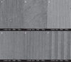
In every experiment, the fabricated Ti substrates were washed three times for 30 minutes in an ultrasonic device with sterile distilled water. After another triple wash using distilled water, Ti substrates were dried at room temperature overnight before use. The surfaces of the five Ti substrates (NE0, E0, E15/3.5, E30/10, and E60/10) were examined using field emission scanning electron microscopy (S-800 FESEM) (Hitachi, Tokyo, Japan) (Fig. 1).
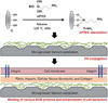
Human serum fibronectin was conjugated on Ti surfaces that had been functionalized (silanized) with 3-amino-propyltriethoxysilane (APTES) (Sigma-Aldrich, St. Louis, MO, USA) (Fig. 2). Functionalized amine was produced in anhydrous toluene by adding 1 mL of APTES to 9 mL toluene solution per well of Ti substrates (silanization). The reaction was maintained at 120℃ in a nitrogen atmosphere with a reflux condenser for 24 hours. After the reaction, the Ti substrates were washed three times with methanol to eliminate silane remnant. The silanized Ti substrates were dried with a vacuum pump for 24 hours and then washed three times for 20 minutes each time using distilled water in an ultrasonic device.
Human plasma fibronectin (human pFN) (Sigma-Aldrich) was conjugated on the surface of the silanized Ti substrates in a 10 µg/mL concentration and maintained for 24 hours at 4℃. After that, the pFN-conjugated Ti substrates were washed with distilled water to remove residual pFN and were dried at room temperature for 45 minutes. As a result, the FN-conjugated non-etched control Ti substrates (NE0FN), the FN-conjugated acid-etched control Ti substrates (E0FN), and the FN-conjugated microgrooved Ti substrates (E15/3.5FN, E30/10FN, and E60/10FN) were produced (Table 1).
Human bone marrow-derived MSCs were purchased from Lonza (Walkersville, MD, USA). The cells were cultured in specific growth medium (MSC GMTM) (Lonza) at 37℃ in 5% CO2. Cells were maintained in Dulbecco's modified Eagle's medium (DMEM; WelGene, Daegu, Korea), containing 10% fetal bovine serum (FBS; Sigma-Aldrich, St. Louis, MO, USA) and 1% antibiotic-antimycotic solution at 37℃ in 5% CO2. MSCs at passages 3 to 5 were used.
MSCs were seeded on 24-well Ti substrates (NE0, E0, E15/3.5, E30/10, and E60/10) and FN-conjugated Ti substrates (NE0FN, E0FN, E15/3.5FN, E30/10FN, and E60/10FN) at 4×10⁴ cells/well and were cultured for 2 days for confluence. Cells were then cultured in osteogenic media, which consisted of DMEM (WelGene) supplemented with 10% FBS (Sigma-Aldrich), with 50 µg/mL of α-ascorbic acid (Sigma-Aldrich), 10 mM of β-glycerophosphate (Sigma-Aldrich), and 100 mM of dexamethasone (Sigma-Aldrich), at 37℃ in 5% CO2 for 7 and 14 days to determine ALP activity. The ALP activity was assayed according to the procedure described previously.2 The reaction suspensions were transferred to 96-well plates to be monitored at 405 nm by a microplate reader (Bio-Rad, Hercules, CA, USA). Measurements were compared using p-nitrophenol standards to be normalized to total protein.
MSCs were seeded on 24-well Ti substrates of all 10 groups at 4×10⁴ cells/well and were cultured for 2 days for confluence. Cells were cultured in osteogenic media at 37℃ in 5% CO2 for 21 days and were washed in phosphate buffered solution (PBS) (Gibco BRL, Grand Island, NY, USA). Ti substrates with remaining calcium deposits were incubated with 0.5 N hydrogen chloride at 4℃ overnight. After centrifugation, the resultant amount of calcium was quantified by Calcium LiquiColor (Stanbio Laboratory, Boerne, TX, USA). The reaction suspensions were transferred to 96-well plates to be monitored at 650 nm by a microplate reader (Bio-Rad, Hercules, CA, USA).
The expression of osteoblast marker genes in MSCs was analyzed using quantitative real-time PCR. The genes were COL1A1 (Hs00164004_m1), ALP (Hs01029144_m1), CBFA1/RUNX2 (Hs01047978_m1) , SP7/OSX (Hs01866874_s1), BGLAP/OC (Hs00609452_g1), SPP1/OPN (Hs00959010_m1), bone sialoprotein II (IBSP/BSP2; Hs00173720_m1), and osteonectin (SPARC/ON, Hs00234160_m1). MSCs were seeded on the 24-well Ti substrates of all groups at 4×10⁴ cells/well and cultured for 2 days for confluence and were subsequently cultured for 14 days in osteogenic media at 37℃ in 5% CO2. The quantitative real-time PCR was performed as in our previous study.6 The relative-fold-change expression levels were analyzed by normalizing the values to the internal control, glyceraldehyde 3-phosphate dehydrogenase.
The ALP activity assay, extracellular calcium deposition assay, and quantitative real-time PCR analyses of MSCs were repeated independently and simultaneously five times. One-way analysis of variance (ANOVA) was used to compare the mean values of the results for the substrate groups under Tukey's multiple comparison tests. Pearson's correlation analysis was performed to determine the correlations. Multiple regression analysis was used to determine the influential factor genes on osteoblastic differentiation. The SPSS 18.0 was used for statistical analyses.
RESULTS
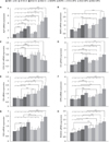
MSCs' ALP activities were tested after 7 and 14 days of osteogenic culturing. These time points were selected from previously reported of MSCs' peak ALP activity after 14 days of osteogenic culturing.16 The extracellular calcium deposition assay was conducted after 21 days of osteogenic culturing. MSCs showed the highest level of ALP activity on both day 7 and day 14 (Fig. 3), with the highest calcium level found to be on E60/10FN relative to the other groups of Ti surfaces (Fig. 4). On the extracellular calcium deposition assay, every Ti surface group with conjugated fibronectin had significantly higher levels of calcium compared with the Ti groups that had the same microstructure but without conjugated fibronectin at 21 days. When compared with NE0 or E0, E60/10FN showed a significant increase (up to more than four-fold) in calcium deposition at 21 days (Fig. 4)
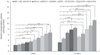
In our previous study, it was shown that the relative COL1A1 gene expression was distinctly patterned when compared to other osteoblast marker genes analyzed at day 14.6 Thus, we chose day 14 as the critical time point during MSCs' osteoblast differentiation on microgrooved Ti substrates. The expression levels of BSP2, ALP, RUNX2, OSX, OC, OPN, and ON were higher on E60/10FN than on any other Ti substrate (Fig. 5). However, the difference in the expression levels of BSP2 mRNA was not statistically significant between E60/10FN and E30/10FN, whereas the levels of ALP, RUNX2, OSX, OC, OPN, and ON were significantly different compared to other groups. Only COL1A1 showed a lower level of gene expressed on E60/10FN versus NE0. This result corresponds to those in our previous study, in which E60/10FN caused significantly lower expression levels of COL1A mRNA than any of the Ti substrates.6 Fig. 5 provides detailed results of the relative protein levels in MSCs.
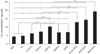
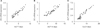
Using the representative gene expression as independent variables, we performed a Pearson's correlation analysis. Correlations were significant between the expression levels of each of the eight mRNAs and MSC differentiation. Multiple regression analyses showed that expression of ON, OC, ALP, COL, and OSX had significant influence on MSC differentiation at 21 days on the Ti substrates with various topographies and fibronectin conjugation (Table 2). These were determined as the influential factor genes that enhanced MSCs' osteoblast differentiation in response to FN-conjugated microgrooved Ti substrates, with ALP apparently being the most influential one. From the stepwise method, ON, OC, and ALP were determined as the most influential factor for ALP 7 days, ALP 14 days, and Ca 21 days, respectively, and the scatter plots for each correlation result are presented in Fig. 6.
DISCUSSION
The results of a series of tests, including the ALP and extracellular calcium deposition assays, quantitative real-time PCR, Pearson's correlation, and multiple regression analysis, suggest that FN-conjugated microgrooved Ti substrates promote osteoblast differentiation in MSCs. Indeed, more than three-fold increase in ALP activity at 14 days and more than four-fold increase in calcium concentration at 21 days were observed in the MSCs on E60/10FN compared with those on NE0 or E0 (Fig. 3 and Fig. 4). The conjugation of fibronectin on Ti significantly increased osteoblast differentiation in MSCs when compared with the non-conjugated Ti substrates. Also, the extracellular calcium deposition assay revealed an approximately two-fold increase in calcium concentration in MSCs on E60/10FN compared with E60/10. The addition of both conjugated fibronectin and microgrooves on Ti substrates enhanced osteoblast differentiation in MSCs synergistically when compared with that achieved on the control substrates. The E60/10FN substrates used in this study provided optimal environment for the promotion of a variety of cellular responses in vitro; therefore, an optimal microgroove structure and the secondary submicroscale of etched topography with conjugated fibronectin on Ti substrates can exert the strongest impact on osteoblast differentiation in MSCs.
Earlier studies indicated that microgrooved Ti regulates the amount of fibronectin that assembles in the extracellular matrix.14 In addition, etched microgrooves and ridges on the Ti surface exclusively control endogenous fibronectin expression and appear to increase the degree to which exogenous human plasma fibronectin is adsorbed onto Ti substrates.2 Other studies suggest that a fibronectin coating on Ti improves a longevity of the outcome of transcutaneous implants by promoting soft-tissue attachment and preventing infection or epithelial downgrowth.17 MSCs were allowed for limited spreading but enhanced osteoblast differentiation on microgrooved substrates.18 Moreover, they displayed high sensitivity to substrate differences, and in appropriate-for-surface topography studies, differentiated more rapidly into osteoblasts, and required less time for matrix maturation and mineralization, as compared with human periodontal ligament cells.4 Microgrooves that are narrower than 10 µm induce changes in cell morphology and alter specific gene expression.1419 On the other hand, sufficient surface area of truncated, V-shaped, etched microgrooves with 30- or 60- µm width induce fibroblast crawling or migration, and the proliferation of cells is significantly enhanced.4 However in this study, ALP activity and calcium concentration were higher on E60/10 than on E30/10 and on any other surface groups in the MSC cultures (Fig. 3 and Fig. 4). Taken together with our result in the present study, cells on FN-conjugated microgrooved Ti substrates with microgrooves of sufficient dimension are expected to secure the peri-implant environment and regulate strong and rapid osseointegration.
Based on the osteoblast marker gene expression in human bone marrow MSCs after 14 days of osteogenic culture on Ti substrates having various surface topographies and fibronectin conjugation by quantitative real-time PCR, we observed significant upregulation of ALP, RUNX2, OSX, OC, and OPN genes in MSCs at day 14 of osteogenic culture on E60/10FN. We also observed significant downregulation of COL1A1 gene expression in MSCs on E60/10FN. This result corresponds with our previous study,6 and indicates a presence of mechanism for preventing excessive collagen deposition and for inducing effective mineralization.20 The 14-day time point culture (Fig. 5) was chosen for gene expression analysis based on previous reports showing that the nanotopography upregulates the expression of genes responsible for the sequential osteoblast differentiation progress in MSCs on Ti.212223 Several studies suggest that MSCs at day 14 represent an immature osteoblast-dominant phase between pre-osteoblast and mature osteoblast.2425 Although our results contradict previous reports that showed a decrease in OPN on dual acidetched Ti substrates,26 they coincide with the findings in other studies showing that upregulation of ON, RUNX2, and OPN gene expression correlated with the increase in adhesion and proliferation on Ti grooved surfaces.272829 Expression patterns of RUNX2 and OPN were similar during osteoblast differentiation and de novo bone formation,29 due to the known fact that RUNX2 is a transcription factor activating the expression of OPN,2830 and also to the reported possibility that RUNX2 may maintain the expression levels of OPN in immature osteoblasts with ultimate downregulation.31 The multiple stepwise regression analysis showed that expression of ON, ALP, COL, and OSX had the most significant influence on MSC differentiation at 21 days. OC and ALP were influential genes at 14 days, and ON was important at 7 days on Ti substrates with various topographies and fibronectin conjugation (Table 2). These results demonstrated the influence of several marker genes on MSC osteoblast differentiation. Scatter-plot results showed significant correlations between the expressed genes which are found to be the factors with the greatest influence on osteoblast differentiation and relevant assays (Fig. 6).
CONCLUSION
The conjugation of FN by silanization on Ti with 60-µm-wide/10-µm-deep etched microgrooves significantly enhanced the ALP activity and osteoblast differentiation in human MSCs. Likewise, induction of osteoblast marker gene expression caused by etched microgrooves with conjugated FN is indicative of the positive effect of such surface modification on the acceleration of the osseointegration for oral and orthopedic Ti implants. Further study is essential to explore the application of other nanoscale and submicroscale topographies in various methods of cellular assays and to perform a various timelines of analysis on osteoblast marker gene expression.


 XML Download