

 PDF
PDF ePub
ePub Citation
 Print
Print


INTRODUCTION
Dental implants are a reasonable treatment for single-tooth restoration. The high success rate of implant osseointegration has been well documented,123 and the maintenance and longevity of implants supporting the restoration are also important issues. However, biological and mechanical complications, such as peri-implantitis, screw loosening, and fracture of various parts of an implant, remain of concern.45 These complications do not always lead to the loss of an implant, but they can compromise oral function. To minimize these complications, various new implant surfaces and implant-abutment connection configurations have been developed.
Recent attention has focused on the conical internal connection (CIC, internal friction type), in which the conical interface provides a tight junction through the friction between the implant and abutment, and not through a screw.6 The CIC was developed to reduce the incidence of mechanical complications. An in vitro study of this connection showed that it is biomechanically superior compared with the external and internal hexagonal connections, and that it may provide better long-term stability in clinical applications.7
Certain CIC-type implants also have as their design a switched platform. Gardener, Lazzara, and Porter introduced the platform-switching concept, by which a larger-diameter implant is combined with a narrower abutment, which results in movement of the implant abutment gap away from the implant shoulder.89 In clinical studies, the platform-switched implant maintained interimplant bone height and reduced crestal bone loss.1011
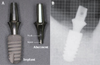
The Ankylos® implant (Dentsply-Friadent GmbH, Mannheim, Germany) comprises a rough progressive thread and a cervical collar, with a conical implant-abutment connection for the switched-platform connection (Fig. 1). This implant is thought to have some advantages, including a lower incidence of mechanical failure, by improving the interface and superior strength and stability because of the reduced micro movement of this system.1213 These advantages have been supported by some clinical studies in Western countries or large multicenter studies under the manufacture request.14151617 The reported results show that the Ankylos implant system offers a higher long-term survival rate and much lower incidence of mechanical complications, including the rare occurrence of fractures of the implant system, which has been reported as nearly zero or <1%, and a low rate of screw loosening (1.3%).12141718
Further clinical studies are needed in other environments and groups of patients to determine whether implant survival and complications differ according to ethnicity, which is associated with differences in the anatomy of the dental arch, dietary patterns, and clenching force. There are few data on the outcomes of the Ankylos implant in Asian populations,19 and no clinical study has analyzed the clinical outcomes of the Ankylos implant in the Korean population. Considering the hard and stiff texture of Korean food and different diet habits, we reasoned that differences in mechanical force and accumulated fatigue may affect the outcomes and mechanical complications of restorations using the Ankylos implant in the Korean population.
The aim of this study was to evaluate the clinical outcomes of the single-tooth Ankylos® implant over an 8-year period in Koreans. We assessed the cumulative survival rate (CSR), causes of failure, and complications. We performed a detailed analysis of the mechanical complications and identified the predisposing factors related to it.
MATERIALS AND METHODS
This retrospective clinical study was conducted in dental clinics at an urban university hospital. The study protocol was reviewed and approved by the institutional review board at our medical center (IRB No. 2013-i007). The inclusion criteria for our study subjects were adult patients who had received an Ankylos® implant for single-tooth restoration from October 2005 to October 2012 (8 years). The exclusion criteria included any underlying uncontrolled disease or health condition; e.g., severe liver or renal disease, complicated diabetes mellitus, or history of recent radiotherapy in the head and neck area or active chemotherapy.
Insertion of the Ankylos® implant system was performed by 6 experienced faculty dentists (4 oromaxillofacial surgeons, 2 prosthodontists) according to the recommended protocol from the manufacturer. All placements of Ankylos® implants were performed in two stages. Following a healing period of 3 - 6 months, the submerged implant was uncovered, and the prosthetic procedures were performed. A 2-piece balance abutment was connected to the implant and tightened with a force of 15 N and restored to cementation type. After insertion of the definitive prosthesis, periapical radiographs were taken to confirm the fit. All patients were asked to return to the clinic for routine follow-up visits every 6 - 12 months or at any time if they had pain or discomfort from the implant. Any clinical problems such as implant failure, screw loosening, fracture, or any other complaint were checked and recorded in an electronic medical recording system.
Data were collected from the patients' electronic medical records. The baseline data about the patients and implants at the time of implant placement were collected as follows: 1) age and sex, 2) date of implant placement, 3) clinical data about the bone graft, and 4) implant diameter, length, and location. Further data about clinical problems such as infection, fracture, screw loosening, other functional problems, and related information such as the date, type of implant, restoration position, and preceding significant events were also collected.
The two main outcomes of interest were survival of the implants and mechanical complications after osseointegration. Variables related to the survival of implants included the 8-year CSR and the occurrence of implant failure, which was defined as any case of implant removal because of early osseointegration failure, removal because of serious marginal bone loss, or an implant or abutment fracture that could not be restored. The time (in months) between the date of completion of the implant restoration and the last follow-up or implant failure was measured to calculate the survival duration of the implants. Mechanical complications included screw loosening and any type of fracture of the implant system (implant fixture, abutment, screw, and crown), and the number of occurrences and cumulative fracture rate (CFR) were analyzed. Other factors were analyzed to identify the predisposing factors for the main outcomes: 1) sex (male/female); 2) age group (young-aged, 18 - 39 years; middle-aged, 40 - 65 years; and old-aged, >65 years);20 3) bony graft (yes/no); 4) diameter of the implant (3.5 mm, 4.5 mm, or 5.5 mm); 5) length of the implant (8 mm, 9.5 mm, 11 mm, or 14 mm); 6) position (anterior, premolar, or molar); and 7) arch (mandible or maxilla).
Kaplan-Meier analysis was used to estimate the CSR and CFR, and the Breslow (generalized Wilcoxon) test was used to compare the cumulative rates between groups. To compare variables between two groups, a chi-square test or a Mann-Whitney rank-sum test was used. Two-sided P-values < 0.05 were considered significant. The data were analyzed using SPSS statistical software (version 20, SPSS Inc., Chicago, IL, USA).
RESULTS
During the study period, 450 Ankylos implants from 257 patients were included. Of these patients, 133 were men (51.8%) and 124 (48.2%) were women. The patients' ages at the time of implant placement ranged from 16 to 81 years (mean age, 47.8 ± 14.3). The mean duration of follow-up of the implants was 63.5 ± 16.0 months. Table 1 shows the baseline data for sex, age, and the characteristics of the installed Ankylos implants such as bone graft, implant length, diameter, location, arch, and loading time.
The 8-year CSR of the implant was 96.9%. Thirteen (2.9%) fixtures failed during this time. Three failed because of early osseointegration failure at 2, 4, and 6 months of the loading period. Six fixtures were removed because of marginal bone loss with peri-implant inflammation at 17, 31, 35, 40, 42, and 58 months of the loading period. Four implants failed because the fractured parts of the abutment and screw could not be retrieved. Analysis of CSR according to the patient and implant factors showed no significant differences between groups (Table 1).
Of the 450 implants, 10 instances of abutment screw loosening (2.2%) and 10 of significant mechanical fractures (2.2%) occurred. There were no implant fractures, and 10 abutment fractures occurred. All abutment fractures were located in the neck portion, and concurrent screw fractures were observed in all cases of abutment fractures (Fig. 2). In the Kaplan-Meier analysis, the estimated 5-year CFR was 1.6%, and the 8-year CFR was 2.8%. The median time of fracture occurrence was 35.5 (28.3 to 78.8) months after loading. Eight abutment fractures occurred in the first 5 years of the postloading period, and 4 resulted in implant failure because of difficulty in removing the fractured part within the fixture. Two abutment fractures occurred after 5 years, and these could be restored with a new abutment (Table 2). In the 10 abutment/screw fractures, 4 exhibited screw loosening before the abutment fracture occurred, and 1 case of bruxism was reported (Table 3).
Analysis of the rates of mechanical complications grouped according to the patient or implant factors showed no significant differences in the occurrence of screw loosening (Table 2). However, in cases of abutment/screw fracture, the middle-aged group had a higher 8-year CFR (5.3%) compared with the young and old groups. The molar position had the highest 8-year CFR (5.8%) compared with the anterior or premolar position. The CFR was significantly higher for the Ankylos implant of 4.5 mm or 5.5 mm in diameter compared with that of 3.5 mm in diameter (Table 2).
DISCUSSION
This is the first clinical study to evaluate the clinical outcomes of the insertion of the Ankylos implant in the Korean population, and the first to report on the details of mechanical complications in patients treated by Ankylos implant for single-tooth restoration. We tried to identify the clinical factors predisposing patients to implant failure and mechanical complications, which have not been reported in detail by previous studies. We found a 96.8% 8-year CSR, which is similar to the rate reported by other studies. Ten fractures (2.2%) were observed, 4 of which resulted in implant failure. All fractures were concentrated on the neck of the abutment and had concurrent screw fractures. The predisposing factors for fractures were the molar position, middle-aged patients, and a large implant diameter.
Now, Ankylos implant is accepted as the suitable option for single-tooth replacement. In this study of Koreans, the 96.8% of overall CSR was similar with that survival outcome in previous reports.12131417 Special clinical reports published by the Ankylos Implant Clinical Research Group in 2004 reported a 97.5% 5-year CSR for the Ankylos implant. A recent large study of 12,737 Ankylos implants in Germany reported a 97.3% 5-year CSR. However, there was a key difference in the clinical outcomes between our study and previous studies. Despite the lack of a significant difference in the results of survival rate, a higher frequency of fracture and related implant failure occurred in our study.
The previously held general concept about the Ankylos implant is that mechanical complications, including screw loosening and fracture, are rare complications.12131718 Because of the tightness from the rough progressive thread and the cervical collar with a platform-switched connection, the Ankylos implant enables appropriate loading transmission when functional loading starts.12 This provides a mechanical advantage in shifting the stress concentration area away from the cervical bone implant. The dense and hard soft tissue in the loading portions also contributes to the implant stability. The specific design of the Ankylos implant provides mechanical stability and is expected to reduce the frequency of mechanical complications. A clinical study by Döring et al.12 that analyzed the clinical outcomes of 264 single-tooth Ankylos implants for 8 years found no fractures during the healing period, and rare screw loosening (1.3%) was reported in another clinical study.18
In our study, 10 cases of screw loosening (2.2%) and 10 (2.2%) of abutment fractures were observed. Screw loosening is known as the most frequent mechanical complication, and its rate varies widely from 1% to 45%. Although the incidence of 2.2% in our study is higher than the 1.3% in a previous study, it is not as high as that reported for other implant systems which and somewhat lesser rate. The most interesting finding of our study is that the occurrence of fractures was higher than in a previous study, and all fractures occurred in the neck of the abutment and screw, and that these fractures occurred after long post loading periods.
From the geometric point of view, the Ankylos implant has a mechanical advantage in shifting the stress concentration area away from the cervical bone-implant interface, which is an advantage in avoiding overload of the implant fixture.6 By contrast, greater stress can cause overload to the abutment or abutment screw, as shown by numerous in vitro studies. The study by Pessoa et al.,21 which mapped the stress distribution in internal conical interface implants, demonstrated greater stress on the conical joint between the abutment and implant. In another study by Quaresma et al.,22 lower von Mises stress was furnished on the alveolar bone prosthesis, but greater von Mises stress was observed on the neck portion of the abutment-prosthesis complex in the conical implant. The laboratory study by Freitas-Junior et al.23 focused on the reliability and failure/fracture patterns in internal conical implant designs. According to the fatigue testing in that study, the internal conical design gave a b value (also called the Weibull shape factor, which indicates a change in the failure rate over time) of > 1. This means that a fracture is caused by accumulated damage and usually occurs in a later phase after the accumulation of loading fatigue. These in vitro studies emphasize that there is a consistent feature of mechanical fractures arising from accumulated fatigue in the internal conical implant, and that the main feature of such fractures is the abutment-implant connection involving a screw.
Another important finding about fractures in our study is that some cases of abutment/screw fractures resulted in implant failure. In our study, 4 implants (0.9%) failed because of abutment/screw fracture, and this rate is higher than that reported by a German study in 2013 (30 of 12,737; 0.23%).17 For restoration of a fractured implant system, the fractured part within the implant fixture should be removed. However, retrieval of the fractured parts of the abutment/screw is a significant challenge. The abutment of the Ankylos implant provides a high frictional retention at the implant-abutment junction because of the 4-degree tapered connection of the abutment. However, in cases of abutment/screw fractures, it is too difficult to remove the fractured element because the fractured part can be stuck tightly. It may be difficult to assess and manipulate within the sub-gingival environment, which is a narrow and deep space containing saliva, blood, and exudate. Thus, abutment/screw fracture may always have a latent risk of implant failure because of the inability to retrieve fractured parts within the implant body.24
There is no clear explanation of why the incidence of fracture was higher in our study than in previous studies. Accumulated metal fatigue from mechanical overload is thought to be the main cause of fractures of implant systems.25 Metal fatigues can be influenced by various factors, such as the implant diameter, implant-prosthesis structural design, ocular force magnitude, and marginal bone loss.526 Greater overloading stress or accumulated fatigue loading may be closely associated with the higher incidence of fracture in our study. In particular, the coarseness of Korean foods may have contributed to the higher incidence of mechanical overloading and subsequent abutment/screw fractures in our study.27
We identified some predisposing factors for abutment/neck fracture. In our study abutment/screw fractures occurred most often in middle-aged people (40 - 65 years) and in restored teeth at a specific anatomical position (the molar position) and with a large-diameter implant. We have found no clinical or other reports on the relationship between fracture risk and patient age. It is possible that different ingestion habits in middle-aged Korean from other age groups may be associated with over-loading fatigue to implant systems and higher risk of abutment fractures. The molar is fractured more frequently than anterior or premolar teeth because the molar region is subjected to greater masticatory forces.26 The axial force might reach 120 N and an increased bending overload may affect metal fatigue,28 and more frequent fractures were occurred in molar position. Implant diameter was identified as another factor related to the rate of implant fracture.29 In cases of implant body fracture, a larger diameter may provide greater resistance to overloading force and may then have a lower incidence of fracture.5262830 However, we found the opposite with the Ankylos implant: large-diameter (4.5 mm or 5.5 mm) implants had a significantly higher CFR at the abutment/screw compared with small-diameter (3.5 mm) implants. In contrast to the case of implant body fractures, the implant-abutment joints may exhibit a greater resistance to vertical overload in small-diameter implants. Unfortunately, to the best our knowledge there was no definite studies which explain the causal relationship between the diameter of implant and the abutment fracture, and further study may be necessary in future.
This study has some limitations. First, its retrospective design may have introduced selection and measuring biases during data collection and assessment of the contributing factors. Second, this study was based on data from only one hospital and included a relatively small sample size, which limits the ability to generalize the results. However, considering the lack of data dealing with the mechanical complications, our results should help in establishing the treatment options for use of the Ankylos implant system.
CONCLUSION
Single-tooth restoration with Ankylos implants showed suitable survival in this study of Korean patients. However, more frequent rate of abutment fractures (2.2%) were observed compared with a previous study (0.23%). Middle-aged patients, the molar position, and a large implant diameter were associated with a high incidence of abutment fracture. And 40% of abutment fractures resulted in implant failures because of failed extraction of broken fragments.

 XML Download